10 January 2008 - Case #107
All cases are archived on our website. To view them sorted by case number, diagnosis or category, visit our main Case of the Month page. To subscribe or unsubscribe to Case of the Month or our other email lists, click here.
This case was contributed by Dr. Mowafak Hamodat, Eastern Health of Newfoundland and Labrador, St. John's, Canada.

ARUP Laboratories First Annual Winter Update in Clinical and Laboratory Medicine is scheduled for March 3-7, 2008 at The Canyons, in Park City, Utah.
This 22.5 hour review and update in the areas of clinical chemistry, immunology, microbiology, and molecular medicine is intended to improve knowledge about the pathogenesis and clinical manifestations of a wide variety of metabolic infectious, immunologic, and genetic disorders along with the selection, performance, and interpretation of clinical laboratory tests.
This course will provide a forum for the exchange of ideas among clinicians, clinical and anatomic pathologists, and laboratory scientists on new developments diagnosing these disorders. Ample discussion time will be provided to address controversial issues.Advertisement
Case #107
Clinical history:
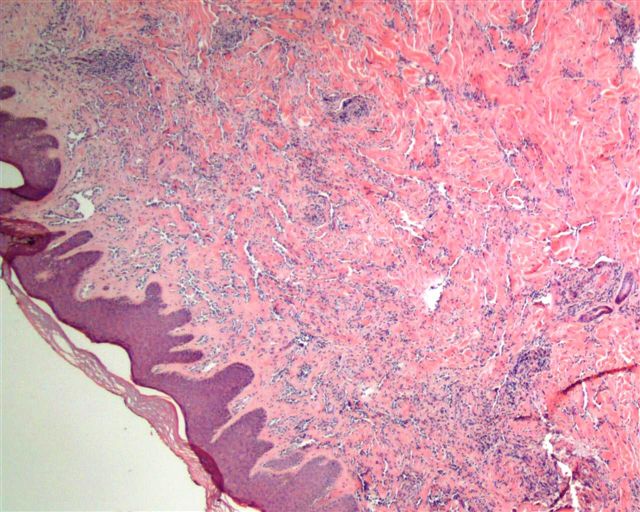
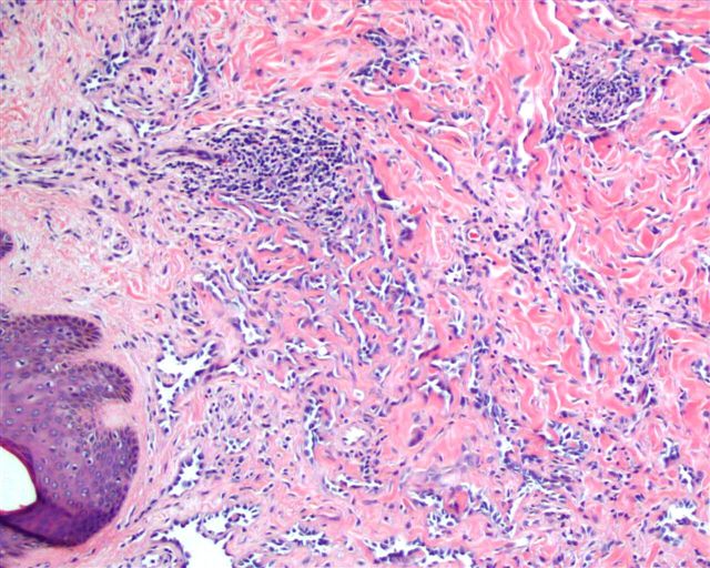
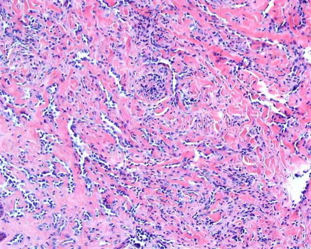
A 37 year old man had a skin lesion on the back, which was excised.
Microscopic images:
What is your diagnosis?
Diagnosis: Retiform hemangioendothelioma
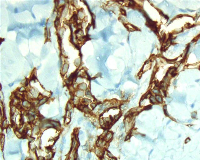
Immunostains:
Discussion:
Retiform hemangioendothelioma is a low grade variant of angiosarcoma that usually occurs in the distal extremities of young individuals. Weiss and Goldblum use the term hobnail hemangioendothelioma for retiform and Dabska type tumors, which they believe to be closely related (Goldblum: Enzinger and Weiss's Soft Tissue Tumors, 5th Edition, 2007).
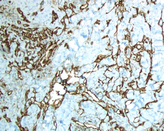
Microscopically, the reticular dermis and subcutaneous tissue have a retiform (net-like, similar to rete testis) pattern of blood vessels that disperse through the tissue and is highlighted by endothelial markers. The vessels are lined by monomorphic hobnail endothelial cells with scant cytoplasm and rounded, naked type nuclei. There often is a prominent lymphocytic infiltrate, as seen focally in this case. There are no epithelioid areas or cytoplasmic vacuoles (Am J Surg Pathol 1994;18:115). They are rarely multiple (Am J Dermatopathol 1996;18:606).
The endothelial cells are immunoreactive for CD34 (strong), CD31 and vWF. They are negative for keratin.
The differential diagnosis includes angiosarcoma, which may focally have low grade features but also exhibits areas of marked atypia and pleomorphism. Angiosarcomas also dissect between individual collagen bundles and have mitotic activity. Hobnail hemangioma is a smaller, more superficial and more localized lesion, with papillary dermal vessels that disappear into the reticular dermis.
Treatment is wide local excision. These tumors are considered to have intermediate malignancy, with frequent recurrence (particularly if a wide excision was not done) but only rare metastases and no tumor related deaths.
All cases are archived on our website. To view them sorted by case number, diagnosis or category, visit our main Case of the Month page. To subscribe or unsubscribe to Case of the Month or our other email lists, click here.
This case was contributed by Dr. Mowafak Hamodat, Eastern Health of Newfoundland and Labrador, St. John's, Canada.

ARUP Laboratories First Annual Winter Update in Clinical and Laboratory Medicine is scheduled for March 3-7, 2008 at The Canyons, in Park City, Utah.
This 22.5 hour review and update in the areas of clinical chemistry, immunology, microbiology, and molecular medicine is intended to improve knowledge about the pathogenesis and clinical manifestations of a wide variety of metabolic infectious, immunologic, and genetic disorders along with the selection, performance, and interpretation of clinical laboratory tests.
This course will provide a forum for the exchange of ideas among clinicians, clinical and anatomic pathologists, and laboratory scientists on new developments diagnosing these disorders. Ample discussion time will be provided to address controversial issues.
Website news:
(1) We recently completed an extensive update of the Leukemia-Acute chapter, which includes all AML and ALL diagnoses under the WHO classification, plus non-WHO entities that have been described in the literature. There are over 350 image links and 130 references. We are currently updating a separate chapter on Myelodysplasia / Myeloproliferative disorders.
Visit and follow our Blog to see recent updates to the website.
Case #107
Clinical history:
A 37 year old man had a skin lesion on the back, which was excised.
Microscopic images:
What is your diagnosis?
Click here for diagnosis and discussion:
Diagnosis: Retiform hemangioendothelioma
Immunostains:
Discussion:
Retiform hemangioendothelioma is a low grade variant of angiosarcoma that usually occurs in the distal extremities of young individuals. Weiss and Goldblum use the term hobnail hemangioendothelioma for retiform and Dabska type tumors, which they believe to be closely related (Goldblum: Enzinger and Weiss's Soft Tissue Tumors, 5th Edition, 2007).
Microscopically, the reticular dermis and subcutaneous tissue have a retiform (net-like, similar to rete testis) pattern of blood vessels that disperse through the tissue and is highlighted by endothelial markers. The vessels are lined by monomorphic hobnail endothelial cells with scant cytoplasm and rounded, naked type nuclei. There often is a prominent lymphocytic infiltrate, as seen focally in this case. There are no epithelioid areas or cytoplasmic vacuoles (Am J Surg Pathol 1994;18:115). They are rarely multiple (Am J Dermatopathol 1996;18:606).
The endothelial cells are immunoreactive for CD34 (strong), CD31 and vWF. They are negative for keratin.
The differential diagnosis includes angiosarcoma, which may focally have low grade features but also exhibits areas of marked atypia and pleomorphism. Angiosarcomas also dissect between individual collagen bundles and have mitotic activity. Hobnail hemangioma is a smaller, more superficial and more localized lesion, with papillary dermal vessels that disappear into the reticular dermis.
Treatment is wide local excision. These tumors are considered to have intermediate malignancy, with frequent recurrence (particularly if a wide excision was not done) but only rare metastases and no tumor related deaths.

