24 May 2007 - Case #85
All cases are archived on our website. To view them sorted by case number, diagnosis or category, visit our main Case of the Month page. To subscribe or unsubscribe to Case of the Month or our other email lists, click here.
This case was contributed by Dr. Ankur Sangoi, Stanford University, Stanford, California (USA).

Vision BioSystems advancing the science of histology
Vision BioSystems is advancing the science of histology. By uniting superior reagents with precision automation we improve quality, increase productivity and create diagnostic confidence.
The Vision BioSystems product range includes the revolutionary Bond system that delivers brilliant IHC and ISH staining; the Peloris rapid tissue processor that performs faster than any other instrument; and the Novocastra range of antibodies that are renowned for their sensitivity, specificity and consistency.
Vision BioSystems is a global business with over 20 years of histology experience. With expertise in histological science, laboratory automation and advanced applications support, Vision BioSystems is the ideal histology partner.
Advertisement
Case #85
Clinical history:
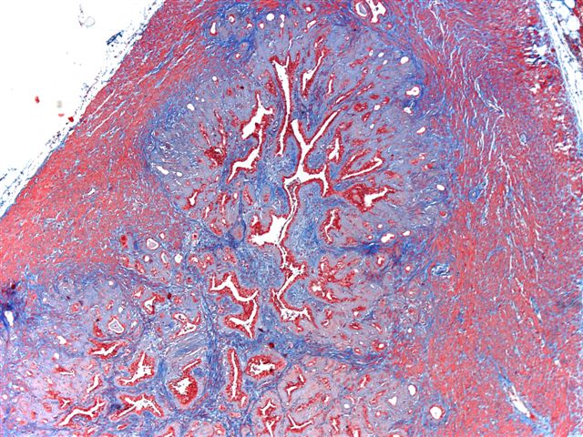
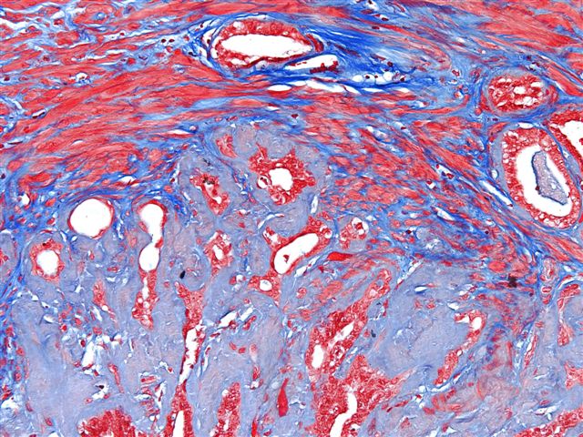
A 66 year old man had a prostate biopsy showing adenocarcinoma (Gleason 3+3) in 1 of 12 cores. He had a radical prostatectomy.
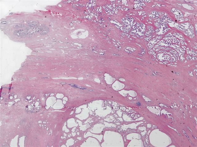
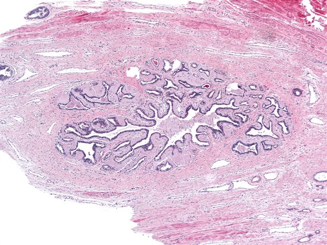
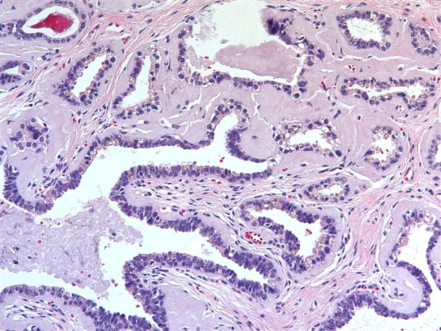
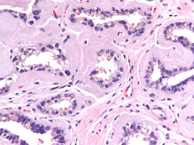
Microscopic images:
What is your diagnosis?
Diagnosis: Bilateral seminal vesicle amyloidosis
Discussion:
Amyloidosis commonly occurs in the seminal vesicles of men. The incidence increases with age, reaching 21% in men age 75 years and older (Histopathology 1993;22:173, Am J Pathol 1983;110:64). At this site, it is usually a localized finding and not part of a systemic process. Although immunohistochemistry often detects lactoferrin within the deposits, a recent study suggests that the amyloid derives from semenogelin I, the major secretory product of the seminal vesicles (Ann Pathol 2004;24:236, J Lab Clin Med 2005;145:187). Semenogelin I and II are mainly responsible for immediate gel formation of freshly ejaculated semen and are degraded by the proteolytic action of prostate specific antigen (PSA) (J Androl 1996;17:17).
The case contributor notes that trichrome is useful in detecting amyloid. It stains the amyloid a dusky gray, in sharp contrast to the strong blue staining of collagen. This is an alternate to Congo red staining and polarization, which often does not work well.
All cases are archived on our website. To view them sorted by case number, diagnosis or category, visit our main Case of the Month page. To subscribe or unsubscribe to Case of the Month or our other email lists, click here.
This case was contributed by Dr. Ankur Sangoi, Stanford University, Stanford, California (USA).
Vision BioSystems is advancing the science of histology. By uniting superior reagents with precision automation we improve quality, increase productivity and create diagnostic confidence.
The Vision BioSystems product range includes the revolutionary Bond system that delivers brilliant IHC and ISH staining; the Peloris rapid tissue processor that performs faster than any other instrument; and the Novocastra range of antibodies that are renowned for their sensitivity, specificity and consistency.
Vision BioSystems is a global business with over 20 years of histology experience. With expertise in histological science, laboratory automation and advanced applications support, Vision BioSystems is the ideal histology partner.
Website news:
(1) Sign up for our new Books email by subscribing to our Monthly Updates newsletter here. Every month, we will send you a list of new pathology related books with a short summary and link to Amazon.com and the publisher for further information. The email is free, and you can unsubscribe at any time.
Visit and follow our Blog to see recent updates to the website.
Case #85
Clinical history:
A 66 year old man had a prostate biopsy showing adenocarcinoma (Gleason 3+3) in 1 of 12 cores. He had a radical prostatectomy.
Microscopic images:
Trichrome
What is your diagnosis?
Click here for diagnosis and discussion:
Diagnosis: Bilateral seminal vesicle amyloidosis
Discussion:
Amyloidosis commonly occurs in the seminal vesicles of men. The incidence increases with age, reaching 21% in men age 75 years and older (Histopathology 1993;22:173, Am J Pathol 1983;110:64). At this site, it is usually a localized finding and not part of a systemic process. Although immunohistochemistry often detects lactoferrin within the deposits, a recent study suggests that the amyloid derives from semenogelin I, the major secretory product of the seminal vesicles (Ann Pathol 2004;24:236, J Lab Clin Med 2005;145:187). Semenogelin I and II are mainly responsible for immediate gel formation of freshly ejaculated semen and are degraded by the proteolytic action of prostate specific antigen (PSA) (J Androl 1996;17:17).
The case contributor notes that trichrome is useful in detecting amyloid. It stains the amyloid a dusky gray, in sharp contrast to the strong blue staining of collagen. This is an alternate to Congo red staining and polarization, which often does not work well.