23 March 2005 - Case of the Week #1
All cases are archived on our website. To view them sorted by case number, diagnosis or category, visit our main Case of the Month page. To subscribe or unsubscribe to Case of the Month or our other email lists, click here.
This case was contributed by Dr. N. Volkan Adsay, Wayne State University, Department of Pathology, Detroit, Michigan (USA). Due to some technical problems, additional images from other sources are also included.
Case of the Week #1
Clinical history:
The patient was a 46 year old man with a 4 cm mediastinal mass but no lesions in the lung or at other sites. The lesion was excised.
Gross:
Not available in this case but typically well circumscribed, light tan and solid. Hemorrhage and necrosis may be present.
Microscopic images:
Microscopic description:
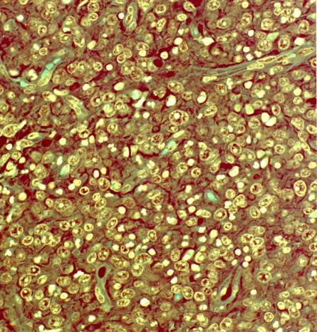
The tumor consists of fascicles, sheets, storiform or whorled patterns of a syncytium of oval and spindled cells, with pale to eosinophilic cytoplasm, oval nuclei with finely dispersed chromatin and small nucleoli; variable lymphocytes and plasma cells, often with perivascular cuffing. Multinucleated tumor giant cells are often present.
Immunohistochemistry:
The tumor cells are positive for CD21 and CD35, negative for CD1a and variable for S100, CD68 and EBV
Electron microscopy images:
Not available in this case but typically numerous interwoven long cytoplasmic processes joined focally by desmosomes.
What is your diagnosis?
Diagnosis: Follicular dendritic reticulum cell tumor / follicular dendritic cell sarcoma
Discussion:
Follicular dendritic cell sarcoma tumors affect lymph nodes and extranodal sites, including the liver, oral cavity, bowel and spleen. They typically have low grade malignant behavior, with local recurrence common and occasional distant metastases to liver or lung. Poor prognostic factors are intraabdominal location, size > 6 cm, 6 or more mitotic figures/10 HPF, atypia and coagulative necrosis. An inflammatory pseudotumor-like variant, localizing in the liver and spleen, has also been reported (AJSP 2001;25:721).
These tumors are often misdiagnosed. The differential diagnosis for these tumors includes other reticulum cell tumors (see below), as well as melanoma, thymoma, other sarcoma, some carcinomas and possibly inflammatory myofibroblastic tumors. Immunostains are usually required for definitive diagnosis.
Lymph nodes have several types of reticulum cells (reticulum means netlike formation or structure), which are nonlymphoid, nonphagocytic cells that capture and present antigens and immune complexes. Follicular dendritic reticulum cells are found in B cell zones, particularly in germinal centers. Interdigitating reticulum cells are found in T cell zones and are related to Langerhans cells. Fibroblastic reticulum cells are found in the parafollicular and deep cortex areas.
Immunohistochemical markers for these reticulum cells and related tumors are:
Interdigitating dendritic cells: positive for S100, vimentin, fascin, focal CD68; negative for CD1a, CD21, CD35, B and T cell markers, actin, desmin, keratin
Langerhans cells: positive for S100, CD1a, CD68, vimentin
Fibroblastic reticulum cells: positive for vimentin, smooth muscle actin, desmin, focal CD68; negative for CD21, CD35, S100 and EBV
Histiocytic tumors: positive for CD68 and lysozyme
Additional references: Am J Surg Pathol 1994;18:148, Am J Surg Pathol 1996;20:944, Mod Pathol 2001;14:354, Hum Pathol 2003;34:954, Am J Surg Pathol 2002;26:530, Am J Surg Pathol 1999;23:1141
All cases are archived on our website. To view them sorted by case number, diagnosis or category, visit our main Case of the Month page. To subscribe or unsubscribe to Case of the Month or our other email lists, click here.
This case was contributed by Dr. N. Volkan Adsay, Wayne State University, Department of Pathology, Detroit, Michigan (USA). Due to some technical problems, additional images from other sources are also included.
Case of the Week #1
Clinical history:
The patient was a 46 year old man with a 4 cm mediastinal mass but no lesions in the lung or at other sites. The lesion was excised.
Gross:
Not available in this case but typically well circumscribed, light tan and solid. Hemorrhage and necrosis may be present.
Microscopic images:
Microscopic description:
The tumor consists of fascicles, sheets, storiform or whorled patterns of a syncytium of oval and spindled cells, with pale to eosinophilic cytoplasm, oval nuclei with finely dispersed chromatin and small nucleoli; variable lymphocytes and plasma cells, often with perivascular cuffing. Multinucleated tumor giant cells are often present.
Immunohistochemistry:
The tumor cells are positive for CD21 and CD35, negative for CD1a and variable for S100, CD68 and EBV
Electron microscopy images:
Not available in this case but typically numerous interwoven long cytoplasmic processes joined focally by desmosomes.
What is your diagnosis?
Click here for diagnosis and discussion:
Diagnosis: Follicular dendritic reticulum cell tumor / follicular dendritic cell sarcoma
Discussion:
Follicular dendritic cell sarcoma tumors affect lymph nodes and extranodal sites, including the liver, oral cavity, bowel and spleen. They typically have low grade malignant behavior, with local recurrence common and occasional distant metastases to liver or lung. Poor prognostic factors are intraabdominal location, size > 6 cm, 6 or more mitotic figures/10 HPF, atypia and coagulative necrosis. An inflammatory pseudotumor-like variant, localizing in the liver and spleen, has also been reported (AJSP 2001;25:721).
These tumors are often misdiagnosed. The differential diagnosis for these tumors includes other reticulum cell tumors (see below), as well as melanoma, thymoma, other sarcoma, some carcinomas and possibly inflammatory myofibroblastic tumors. Immunostains are usually required for definitive diagnosis.
Lymph nodes have several types of reticulum cells (reticulum means netlike formation or structure), which are nonlymphoid, nonphagocytic cells that capture and present antigens and immune complexes. Follicular dendritic reticulum cells are found in B cell zones, particularly in germinal centers. Interdigitating reticulum cells are found in T cell zones and are related to Langerhans cells. Fibroblastic reticulum cells are found in the parafollicular and deep cortex areas.
Immunohistochemical markers for these reticulum cells and related tumors are:
- Follicular dendritic cells: positive for CD21
Additional references: Am J Surg Pathol 1994;18:148, Am J Surg Pathol 1996;20:944, Mod Pathol 2001;14:354, Hum Pathol 2003;34:954, Am J Surg Pathol 2002;26:530, Am J Surg Pathol 1999;23:1141


