9 June 2010 - Case #181
All cases are archived on our website. To view them sorted by case number, diagnosis or category, visit our main Case of the Month page. To subscribe or unsubscribe to Case of the Month or our other email lists, click here.
Thanks to Dr. Juan Jos Segura Fonseca, Laboratorio de Patologa Diagnstica, S.A., San Jos, Costa Rica, for contributing this case and much of the discussion. This case was reviewed in May 2020 by Dr. Jennifer Bennett, University of Chicago and Dr. Carlos Parra-Herran, University of Toronto.
Case #181
Clinical history:
A 28 year old woman was seen in the outpatient clinic because of a 4 month history of progressive enlargement and swelling of the right labium major, with obvious asymmetry.
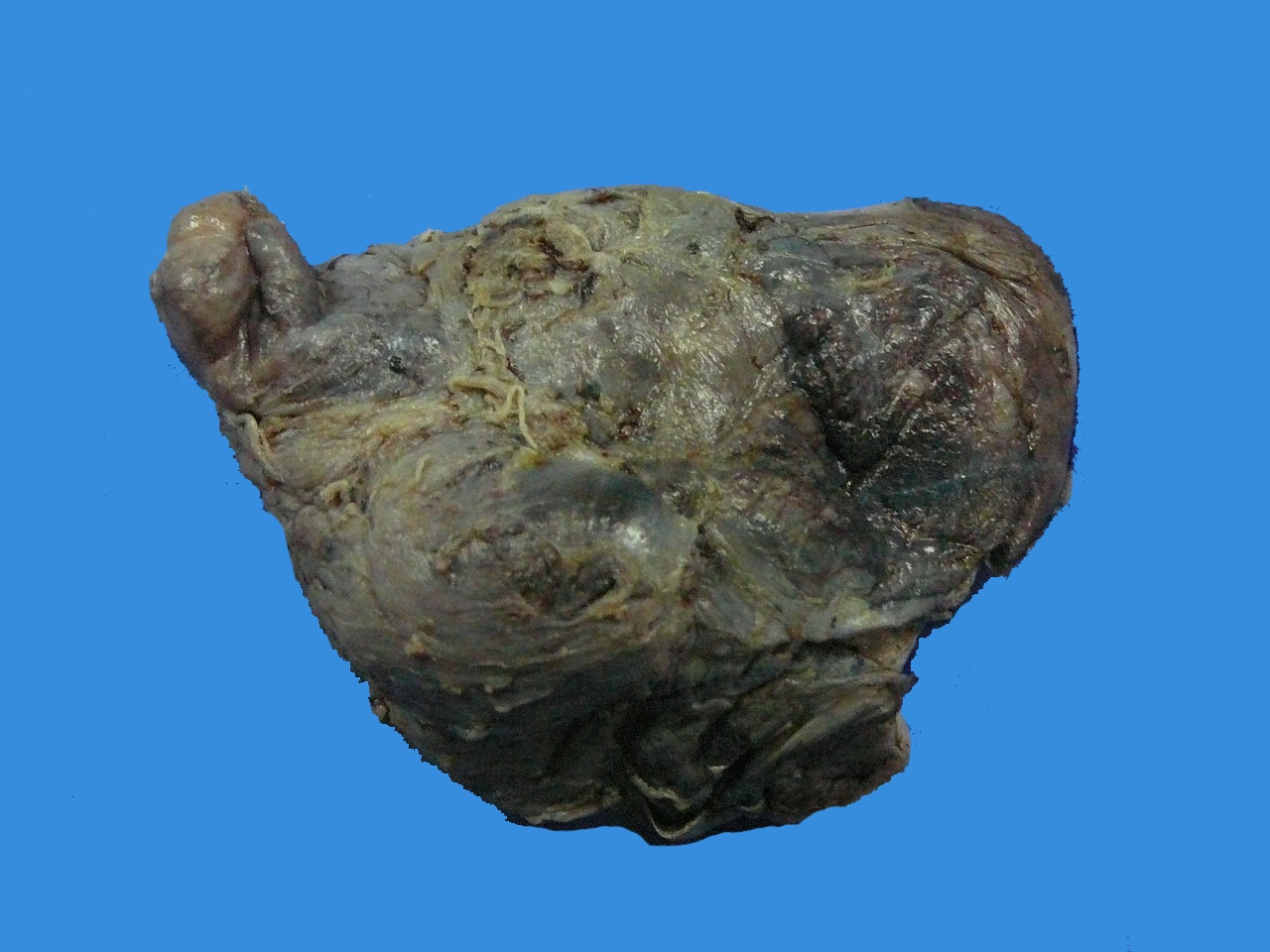
During surgery, a nonencapsulated tumor was found, which infiltrated down to the pelvic floor, making a complete resection difficult. The tumor was 9 x 6 x 5 cm with a white, gelatinous consistency.
Gross images:
Microscopic images:
What is your diagnosis?
Diagnosis: Aggressive angiomyxoma of the vulva
Immunohistochemistry images:
Discussion:
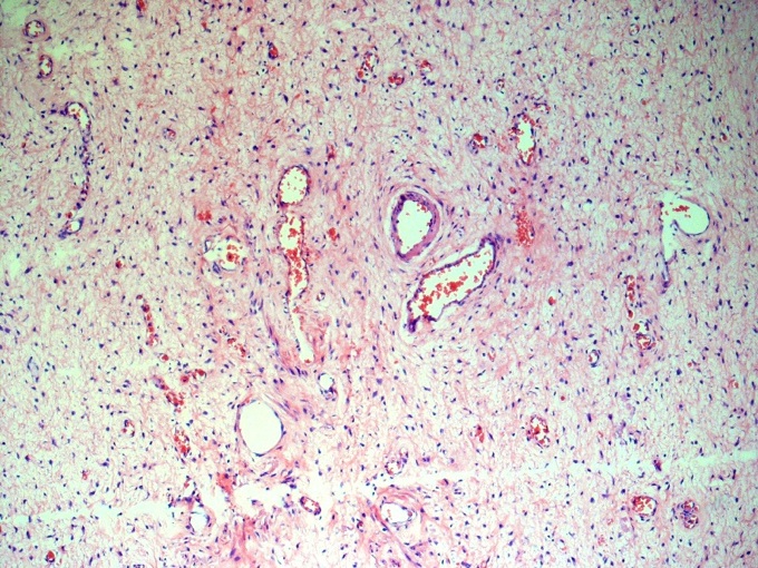
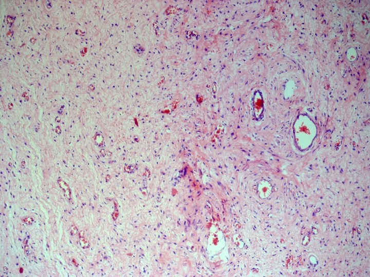
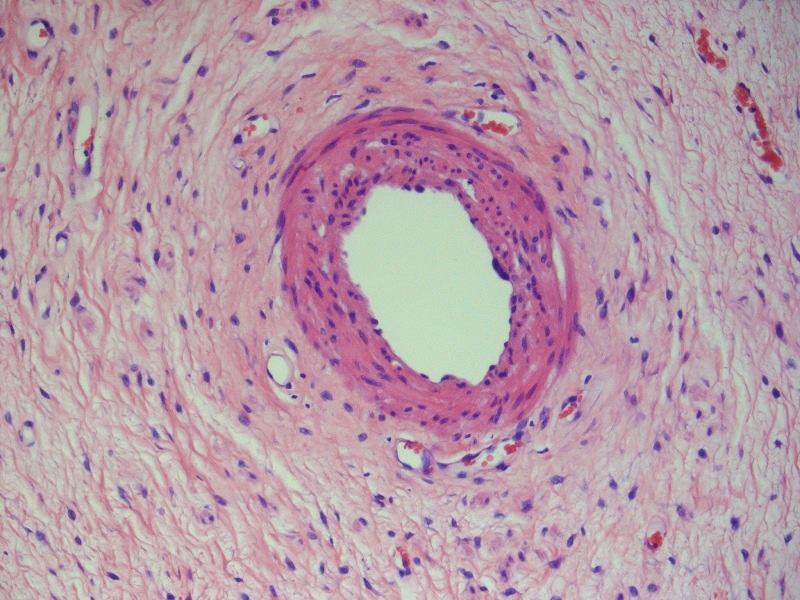
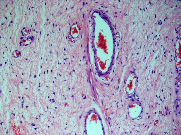
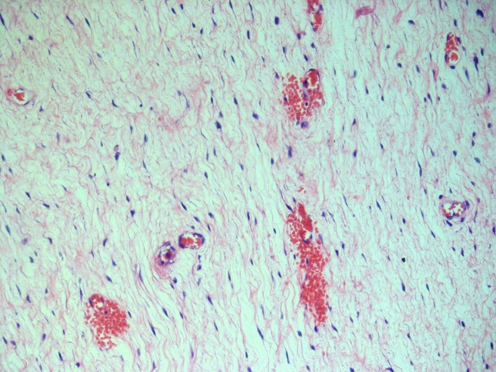
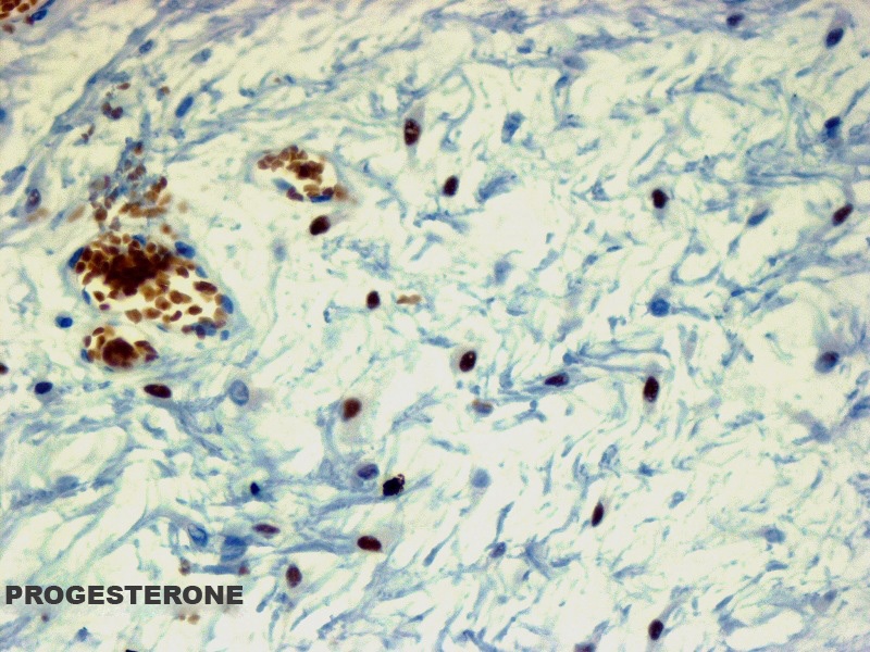
Many small and medium sized vessels in a myxomatous stroma were present (figures 3 and 4). The stromal cells resembled spindle shaped fibroblasts (figure 5). There was prominent condensation of collagen around vessels, with spinning off of muscle fibers (figures 6 and 7). Small hemorrhages were present around small capillaries (figure 8). The spindle cells were strongly positive for desmin (figure 9) and progesterone receptor (figure 10).
Aggressive angiomyxoma is a rare, distinctive, infiltrative mesenchymal tumor usually found in women of reproductive age, frequently in their third decade of life. It was first described by Steeper and Rosai as a slow growing, low grade neoplasm involving the pelvis and vulvoperineal region, with a high risk for local recurrence, which may occur after many years (Am J Surg Pathol 1983;7:463). In their original report of 9 cases, 6 were located in the vulva. Of 29 cases reported by Fetsch et al, 10 were vulvoperineal tumors (Cancer 1996;78:79). Although the tumor is locally aggressive with a propensity to infiltrate deep soft tissue in a diffuse manner, the metastatic potential is very low. Only 2 cases with metastasis have been reported (N Engl J Med 1999;341:1772, Hum Pathol 2003;34:1072).
Grossly, the tumor is rubbery, white and gelatinous. Most tumors are 6 to 9 cm but they are rarely huge and pedunculated (Kaohsiung J Med Sci 2006;22:301, Indian J Pathol Microbiol 2008;51:259). Histologically, there is a myxomatous stroma and a hypocellular pattern of mesenchymal stellate and spindle shaped cells with a myofibroblastic morphology, without nuclear atypia or mitoses. Numerous small capillaries, venules, veins and medium size arterioles are present. In some vessels, there is a peculiar perivascular eosinophilic condensation of collagen. Short bundles of smooth muscle fibers seem to spin off from the arterial walls into the stroma. Small hemorrhages around capillaries with fibrin thrombi are present. There are also entrapped nerves and adipocytes.
The stromal cells are strongly immunoreactive for desmin and are also positive for vimentin and actin. The tumor appears to be hormone dependent, based on immunostaining for estrogen and progesterone receptors (J Clin Pathol 2000;53:603). Tumor cells are variably positive for smooth muscle actin and negative for S100 (Int J Gynecol Cancer 2005;15:140).
The differential diagnosis includes other vulvar myxoid tumors:
Treatment is surgical excision, although the tumors are difficult to completely excise and there is a high recurrence rate. GnRH agonist therapy has also been used with success (Gynecol Oncol 2006;100:623).
All cases are archived on our website. To view them sorted by case number, diagnosis or category, visit our main Case of the Month page. To subscribe or unsubscribe to Case of the Month or our other email lists, click here.
Thanks to Dr. Juan Jos Segura Fonseca, Laboratorio de Patologa Diagnstica, S.A., San Jos, Costa Rica, for contributing this case and much of the discussion. This case was reviewed in May 2020 by Dr. Jennifer Bennett, University of Chicago and Dr. Carlos Parra-Herran, University of Toronto.
Website news:
(1) Thanks to the following image contributors: Dr. R.F. Chinoy, Prince Aly Khan Hospital, India: Plexiform Neurofibroma for Soft Tissue 3 chapter; Dr. Semir Vranic, University of Sarajevo, Bosnia & Herzegovina: Apocrine Carcinoma for Breast Malignant chapter.
(2) We posted a new article on our Management page, Pathology Practices: Are Your Payers Paying You Correctly? Are You Sure? Can You Prove It?, by Al H. Sirmon and Stephanie Denham, PSA, click here. This page now has 38 articles giving details of pathology practice billing, auditing, compliance and marketing.
(3) Welcome to new corporate advertisers Propath and McKesson. Propath is a medical practice and laboratory specializing in anatomic pathology and specialty clinical testing, and supporting hospitals and medical centers for full service inpatient pathology services. McKesson, a Fortune 15 corporation with 40 years of laboratory information solution experience and world-class support, is committed to helping your organization improve patient safety, productivity and profitability.
Visit and follow our Blog to see recent updates to the website.
Case #181
Clinical history:
A 28 year old woman was seen in the outpatient clinic because of a 4 month history of progressive enlargement and swelling of the right labium major, with obvious asymmetry.
During surgery, a nonencapsulated tumor was found, which infiltrated down to the pelvic floor, making a complete resection difficult. The tumor was 9 x 6 x 5 cm with a white, gelatinous consistency.
Gross images:
Microscopic images:
What is your diagnosis?
Click here for diagnosis and discussion:
Diagnosis: Aggressive angiomyxoma of the vulva
Immunohistochemistry images:
Discussion:
Many small and medium sized vessels in a myxomatous stroma were present (figures 3 and 4). The stromal cells resembled spindle shaped fibroblasts (figure 5). There was prominent condensation of collagen around vessels, with spinning off of muscle fibers (figures 6 and 7). Small hemorrhages were present around small capillaries (figure 8). The spindle cells were strongly positive for desmin (figure 9) and progesterone receptor (figure 10).
Aggressive angiomyxoma is a rare, distinctive, infiltrative mesenchymal tumor usually found in women of reproductive age, frequently in their third decade of life. It was first described by Steeper and Rosai as a slow growing, low grade neoplasm involving the pelvis and vulvoperineal region, with a high risk for local recurrence, which may occur after many years (Am J Surg Pathol 1983;7:463). In their original report of 9 cases, 6 were located in the vulva. Of 29 cases reported by Fetsch et al, 10 were vulvoperineal tumors (Cancer 1996;78:79). Although the tumor is locally aggressive with a propensity to infiltrate deep soft tissue in a diffuse manner, the metastatic potential is very low. Only 2 cases with metastasis have been reported (N Engl J Med 1999;341:1772, Hum Pathol 2003;34:1072).
Grossly, the tumor is rubbery, white and gelatinous. Most tumors are 6 to 9 cm but they are rarely huge and pedunculated (Kaohsiung J Med Sci 2006;22:301, Indian J Pathol Microbiol 2008;51:259). Histologically, there is a myxomatous stroma and a hypocellular pattern of mesenchymal stellate and spindle shaped cells with a myofibroblastic morphology, without nuclear atypia or mitoses. Numerous small capillaries, venules, veins and medium size arterioles are present. In some vessels, there is a peculiar perivascular eosinophilic condensation of collagen. Short bundles of smooth muscle fibers seem to spin off from the arterial walls into the stroma. Small hemorrhages around capillaries with fibrin thrombi are present. There are also entrapped nerves and adipocytes.
The stromal cells are strongly immunoreactive for desmin and are also positive for vimentin and actin. The tumor appears to be hormone dependent, based on immunostaining for estrogen and progesterone receptors (J Clin Pathol 2000;53:603). Tumor cells are variably positive for smooth muscle actin and negative for S100 (Int J Gynecol Cancer 2005;15:140).
The differential diagnosis includes other vulvar myxoid tumors:
- Plexiform neurofibroma: neural morphology, S100+
- Angiomyofibroblastoma: hypo and hypercellular with perivascular multinucleated giant cells
- Myxoid smooth muscle tumors
- Superficial angyomyxoma: tumor of superficial dermis
- Vulvar hypertrophy with lymphedema: bilateral, similar histologically but with prominent ectatic tortuous lymphatic vessels and no thick walled vessels (Arch Pathol Lab Med 2000;124:1697)
Treatment is surgical excision, although the tumors are difficult to completely excise and there is a high recurrence rate. GnRH agonist therapy has also been used with success (Gynecol Oncol 2006;100:623).


