31 October 2008 - Case #133
All cases are archived on our website. To view them sorted by case number, diagnosis or category, visit our main Case of the Month page. To subscribe or unsubscribe to Case of the Month or our other email lists, click here.
This case was contributed by Dr. Angela Bohlke, Tulane University Hospital, New Orleans, Louisiana (USA).

2nd Annual Winter Update in Clinical and Laboratory Medicine:
Clinical Chemistry, Immunology, Microbiology and Molecular Medicine
This 24 hour review and update in the areas of clinical chemistry, immunology, microbiology, and molecular medicine is intended to improve knowledge about the pathogenesis and clinical manifestations of a wide variety of metabolic, infectious, immunologic, and genetic disorders along with the selection, performance, and interpretation of clinical laboratory tests.

Approximately 60% of the diagnoses in medicine are based on the results of laboratory testing. The 2nd Annual Winter Update program will address this major gap in knowledge and inform the participant about developments in clinical laboratory testing and their relevance to clinical medicine. The conference is held at The Canyons in Park City, Utah.
Advertisement
Case #133
Clinical history:
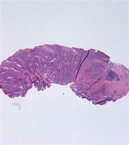
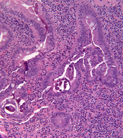
A 43 year old Honduran man presented with diarrhea and abdominal pain for one month. Physical findings and endoscopy were unremarkable. Duodenal biopsies were obtained.
Microscopic images:
What is your diagnosis?
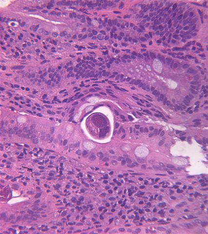
Diagnosis: Strongyloides stercoralis
Discussion:
Subsequent stool findings showed Strongyloides ova.
Strongyloides is a nematode whose larvae buries into the mucosa of the duodenum and jejunum, where they mature into adults. The females then lay eggs, which develop into larvae that pass into the stool, where they mature and become infective. The infective larvae penetrate intact skin, usually through the feet. The larvae enter the circulatory system, are transported to the lungs, and enter the alveolar spaces. They then are carried to the trachea and pharynx, are swallowed and enter the intestinal tract, where the process is repeated. If the larvae become infective before leaving the body, they may invade the intestinal mucosa or perianal skin, causing autoinfection.
Most patients suffer diarrhea, malabsorption or no symptoms. Immunocompromised individuals can acquire disseminated strongyloidiasis, a possibly fatal condition in which worms move into other organs (WormBook 2007:1).
Diagnosis is by stool exam, looking for larvae, or by biopsy of small intestinal mucosa, looking for the adult female or eggs. There is often granulomatous or eosinophilic inflammation. In female worms, the intestine or ovaries may be prominent (image). In gravid females, an egg (green arrow) may be identified within the uterus.
Treatment is with antihelminths, such as thiabendazole (Ann Pharmacother 2007;41:1992). Prevention is by wearing shoes in endemic areas.
All cases are archived on our website. To view them sorted by case number, diagnosis or category, visit our main Case of the Month page. To subscribe or unsubscribe to Case of the Month or our other email lists, click here.
This case was contributed by Dr. Angela Bohlke, Tulane University Hospital, New Orleans, Louisiana (USA).

Clinical Chemistry, Immunology, Microbiology and Molecular Medicine
This 24 hour review and update in the areas of clinical chemistry, immunology, microbiology, and molecular medicine is intended to improve knowledge about the pathogenesis and clinical manifestations of a wide variety of metabolic, infectious, immunologic, and genetic disorders along with the selection, performance, and interpretation of clinical laboratory tests.

Approximately 60% of the diagnoses in medicine are based on the results of laboratory testing. The 2nd Annual Winter Update program will address this major gap in knowledge and inform the participant about developments in clinical laboratory testing and their relevance to clinical medicine. The conference is held at The Canyons in Park City, Utah.
Website news:
(1) How can you learn about new fellowship openings? We have a new email list sent out every two weeks, listing the new fellowships posted at our website. To subscribe, click here.
(2) We have started a new section on Board Review Questions. If you have sample questions and answers, just email them to us.
(3) Tell your colleagues about our Other Laboratory Jobs page, which includes jobs for PAs, cytotechs, med techs, histotechs, managers, etc., and now includes related corporate jobs, such as pathology related sales.
Visit and follow our Blog to see recent updates to the website.
Case #133
Clinical history:
A 43 year old Honduran man presented with diarrhea and abdominal pain for one month. Physical findings and endoscopy were unremarkable. Duodenal biopsies were obtained.
Microscopic images:
What is your diagnosis?
Click here for diagnosis and discussion:
Diagnosis: Strongyloides stercoralis
Discussion:
Subsequent stool findings showed Strongyloides ova.
Strongyloides is a nematode whose larvae buries into the mucosa of the duodenum and jejunum, where they mature into adults. The females then lay eggs, which develop into larvae that pass into the stool, where they mature and become infective. The infective larvae penetrate intact skin, usually through the feet. The larvae enter the circulatory system, are transported to the lungs, and enter the alveolar spaces. They then are carried to the trachea and pharynx, are swallowed and enter the intestinal tract, where the process is repeated. If the larvae become infective before leaving the body, they may invade the intestinal mucosa or perianal skin, causing autoinfection.
Most patients suffer diarrhea, malabsorption or no symptoms. Immunocompromised individuals can acquire disseminated strongyloidiasis, a possibly fatal condition in which worms move into other organs (WormBook 2007:1).
Diagnosis is by stool exam, looking for larvae, or by biopsy of small intestinal mucosa, looking for the adult female or eggs. There is often granulomatous or eosinophilic inflammation. In female worms, the intestine or ovaries may be prominent (image). In gravid females, an egg (green arrow) may be identified within the uterus.
Treatment is with antihelminths, such as thiabendazole (Ann Pharmacother 2007;41:1992). Prevention is by wearing shoes in endemic areas.


