11 September 2008 - Case #129
All cases are archived on our website. To view them sorted by case number, diagnosis or category, visit our main Case of the Month page. To subscribe or unsubscribe to Case of the Month or our other email lists, click here.
This case was contributed by Dr. Sharon Bihlmeyer, University of Vermont (USA).
Case #129
Clinical history:
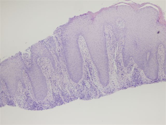
A 52 year old man had a clinical history of Condyloma acuminata peri-rectal wart lesions present for 2 months. They were excised.
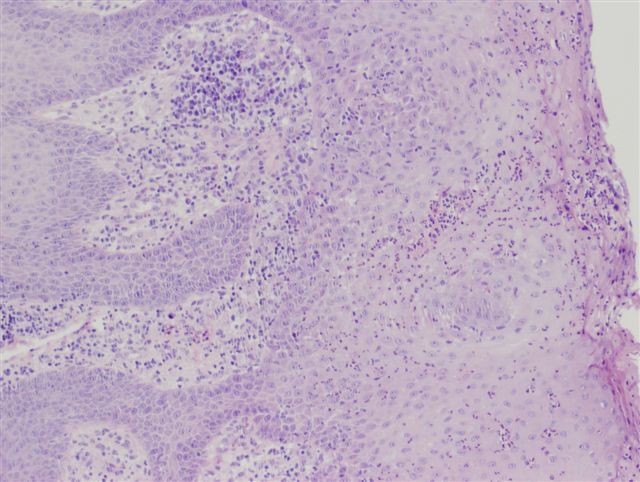
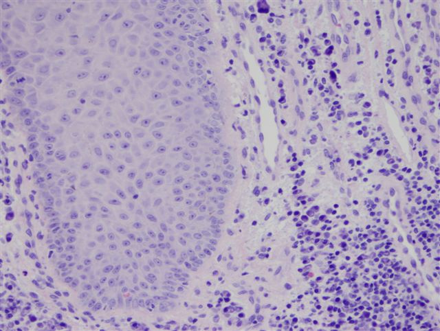
Microscopic images:
What is your diagnosis?
Diagnosis: Anal syphilis
Immunostains:
Discussion:
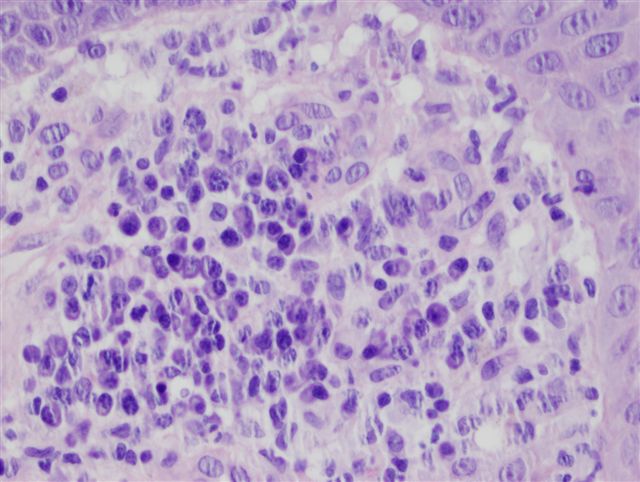
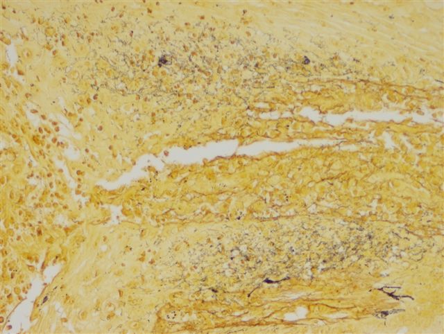
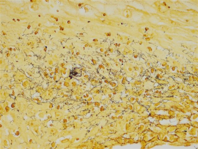
At high power, numerous plasma cells were evident (figure 4). The Steiner stain demonstrated the spirochetes.
Syphilis is a genital ulcerative disease caused by the spirochete Treponema pallidum, that causes significant complications if untreated. It also facilitates the transmission of HIV. Although the rate of primary and secondary syphilis in the United States declined 90% between 1990 and 2000, it increased 25% between 2001 and 2006 (CDC: STD Surveillance Network (SSuN) [Accessed 3 May 2024]). Most of the increase appears due to MSM (men having sex with men), with the estimated proportion of primary syphilis cases from this group increasing from 4% in 2000 to 62% in 2004 (Am J Public Health 2007;97:1076).
Microscopy shows capillary proliferation, obliterative endarteritis and heavy plasma cell infiltration. Lymphocytes and macrophages may also be present.
In this case, the diagnosis of syphilis was missed initially but was caught at a secondary review. Thus, this case reminds us that multiple diagnoses may be present, particularly for sexually transmitted diseases and that explanations for histologic findings extraneous to a diagnosis (such as plasma cells with condyloma) should be sought.
References: eMedicine: Syphilis [Accessed 3 May 2024]
All cases are archived on our website. To view them sorted by case number, diagnosis or category, visit our main Case of the Month page. To subscribe or unsubscribe to Case of the Month or our other email lists, click here.
This case was contributed by Dr. Sharon Bihlmeyer, University of Vermont (USA).
Website news:
(1) We have updated the Breast-Malignant chapter, now with 1300+ image links and new references and text. Please visit this chapter to assist with signout, answer proficiency testing questions or for teaching.
Visit and follow our Blog to see recent updates to the website.
(1) We have updated the Breast-Malignant chapter, now with 1300+ image links and new references and text. Please visit this chapter to assist with signout, answer proficiency testing questions or for teaching.
Visit and follow our Blog to see recent updates to the website.
Case #129
Clinical history:
A 52 year old man had a clinical history of Condyloma acuminata peri-rectal wart lesions present for 2 months. They were excised.
Microscopic images:
What is your diagnosis?
Click here for diagnosis and discussion:
Diagnosis: Anal syphilis
Immunostains:
Steiner stain
Discussion:
At high power, numerous plasma cells were evident (figure 4). The Steiner stain demonstrated the spirochetes.
Syphilis is a genital ulcerative disease caused by the spirochete Treponema pallidum, that causes significant complications if untreated. It also facilitates the transmission of HIV. Although the rate of primary and secondary syphilis in the United States declined 90% between 1990 and 2000, it increased 25% between 2001 and 2006 (CDC: STD Surveillance Network (SSuN) [Accessed 3 May 2024]). Most of the increase appears due to MSM (men having sex with men), with the estimated proportion of primary syphilis cases from this group increasing from 4% in 2000 to 62% in 2004 (Am J Public Health 2007;97:1076).
Microscopy shows capillary proliferation, obliterative endarteritis and heavy plasma cell infiltration. Lymphocytes and macrophages may also be present.
In this case, the diagnosis of syphilis was missed initially but was caught at a secondary review. Thus, this case reminds us that multiple diagnoses may be present, particularly for sexually transmitted diseases and that explanations for histologic findings extraneous to a diagnosis (such as plasma cells with condyloma) should be sought.
References: eMedicine: Syphilis [Accessed 3 May 2024]