20 June 2008 - Case #123
All cases are archived on our website. To view them sorted by case number, diagnosis or category, visit our main Case of the Month page. To subscribe or unsubscribe to Case of the Month or our other email lists, click here.
This case was contributed by Dr. Oscar Sanabria Monney, National Children's Hospital, San Jose, Costa Rica.
Case #123
Clinical history:
A 1 year old boy had a slow growing cervical mass for 8 months, with recent laryngeal stridor.
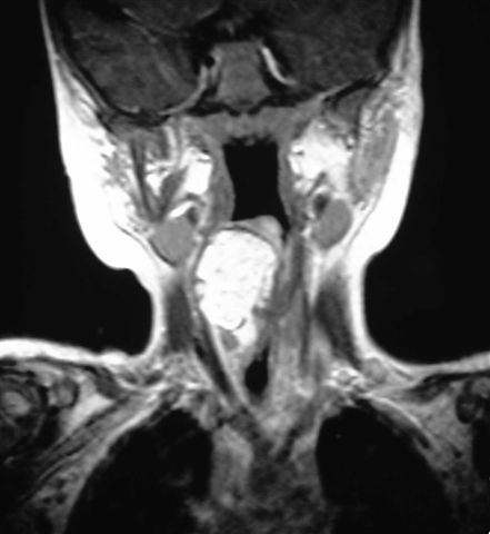
A neck ultrasound showed a solid homogenous retropharyngeal mass that pushed against the trachea. MRI identified a 3 x 2 cm mass with the density of fatty tissue. A fine needle aspiration and excision were performed.
Radiology images:
Cytology images:
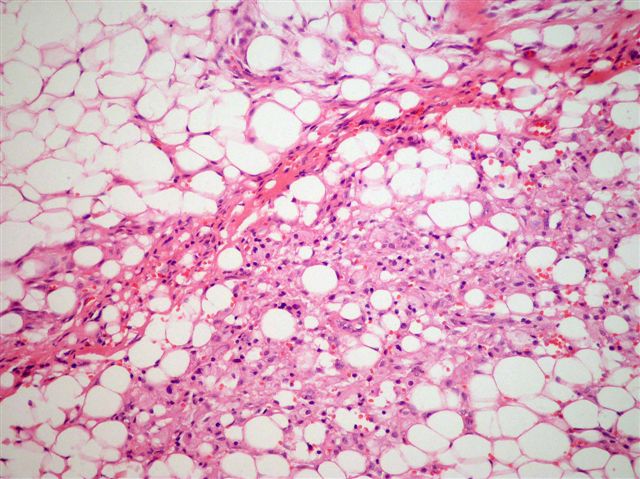
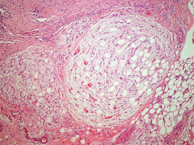
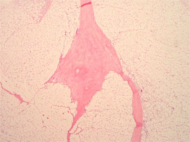
Microscopic images:
What is your diagnosis?
Diagnosis: Lipoblastoma
Discussion:
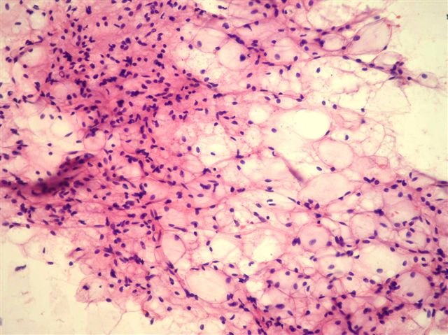
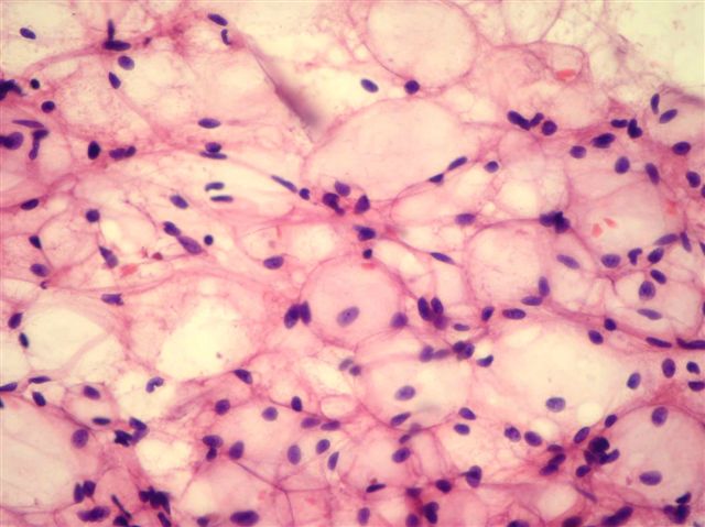
Cytologic smears showed fragments of mature adipose tissue with occasional vacuolated adipocytes (lipoblasts), in a myxoid stroma. The cells have centrically located small nuclei, without indentations. No necrosis, atypia or mitotic figures were present. Cytologic features are suggestive of lipoblastoma, although the differential diagnosis also includes lipoma with regressive changes, well-differentiated liposarcoma and myxoid liposarcoma. In addition to the cytologic features, the patient's age is very useful (Diagn Cytopathol 2005;33:195)
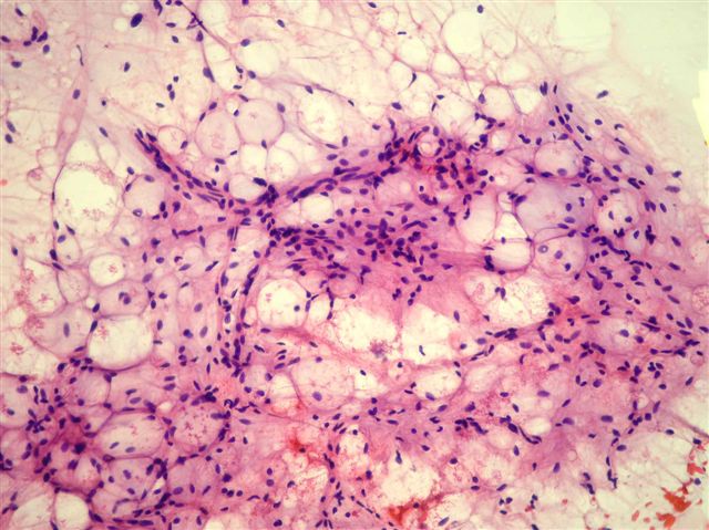
Lipoblastoma is a benign and uncommon tumor of adipose tissue that occurs primarily in young children, usually age 5 years or younger (Am J Surg Pathol 1997;21:1131). It resembles fetal adipose tissue, with lobules of immature fat cells that are separated by fibrous septa and myxoid matrix. Chromosome 8q11-13 is rearranged in 82%, which includes the PLAG1 gene (Cancer Genet Cytogenet 2008;183:60, Am J Pathol 2001;159:955). FISH may be helpful to confirm the diagnosis (Histopathology 2008;52:294).
The differential diagnosis includes myxoid liposarcoma and well differentiated liposarcoma. Myxoid liposarcoma is rare before age 10 years and is commonly associated with t(12;16)(q13;p11). It has no distinct lobulation and often has nuclear atypia, mitotic figures, hyperchromasia and hypercellularity. Well differentiated liposarcoma is also rare in children. It has mature fat but no lipoblasts. It contains spindle cells with large, deep staining nuclei and marked nuclear enlargement or pleomorphism; cellularity is low. MDM2 and CDK4 immunostains are sensitive and specific markers (Am J Surg Pathol 2005;29:1340).
Complete excision of lipoblastoma is the treatment of choice. The diffuse form (lipomatosis) can be difficult to excise completely, which may lead to recurrence. Follow up for 3 years or more is recommended (Pediatr Surg Int 2005;21:809).
All cases are archived on our website. To view them sorted by case number, diagnosis or category, visit our main Case of the Month page. To subscribe or unsubscribe to Case of the Month or our other email lists, click here.
This case was contributed by Dr. Oscar Sanabria Monney, National Children's Hospital, San Jose, Costa Rica.
Case #123
Clinical history:
A 1 year old boy had a slow growing cervical mass for 8 months, with recent laryngeal stridor.
A neck ultrasound showed a solid homogenous retropharyngeal mass that pushed against the trachea. MRI identified a 3 x 2 cm mass with the density of fatty tissue. A fine needle aspiration and excision were performed.
Radiology images:
Cytology images:
Microscopic images:
What is your diagnosis?
Click here for diagnosis and discussion:
Diagnosis: Lipoblastoma
Discussion:
Cytologic smears showed fragments of mature adipose tissue with occasional vacuolated adipocytes (lipoblasts), in a myxoid stroma. The cells have centrically located small nuclei, without indentations. No necrosis, atypia or mitotic figures were present. Cytologic features are suggestive of lipoblastoma, although the differential diagnosis also includes lipoma with regressive changes, well-differentiated liposarcoma and myxoid liposarcoma. In addition to the cytologic features, the patient's age is very useful (Diagn Cytopathol 2005;33:195)
Lipoblastoma is a benign and uncommon tumor of adipose tissue that occurs primarily in young children, usually age 5 years or younger (Am J Surg Pathol 1997;21:1131). It resembles fetal adipose tissue, with lobules of immature fat cells that are separated by fibrous septa and myxoid matrix. Chromosome 8q11-13 is rearranged in 82%, which includes the PLAG1 gene (Cancer Genet Cytogenet 2008;183:60, Am J Pathol 2001;159:955). FISH may be helpful to confirm the diagnosis (Histopathology 2008;52:294).
The differential diagnosis includes myxoid liposarcoma and well differentiated liposarcoma. Myxoid liposarcoma is rare before age 10 years and is commonly associated with t(12;16)(q13;p11). It has no distinct lobulation and often has nuclear atypia, mitotic figures, hyperchromasia and hypercellularity. Well differentiated liposarcoma is also rare in children. It has mature fat but no lipoblasts. It contains spindle cells with large, deep staining nuclei and marked nuclear enlargement or pleomorphism; cellularity is low. MDM2 and CDK4 immunostains are sensitive and specific markers (Am J Surg Pathol 2005;29:1340).
Complete excision of lipoblastoma is the treatment of choice. The diffuse form (lipomatosis) can be difficult to excise completely, which may lead to recurrence. Follow up for 3 years or more is recommended (Pediatr Surg Int 2005;21:809).