Table of Contents
Definition / general | Clinical features | Radiology images | Case reports | Treatment | Gross description | Gross images | Microscopic (histologic) description | Microscopic (histologic) images | Differential diagnosisCite this page: Williamson S. Vasculitis. PathologyOutlines.com website. https://www.pathologyoutlines.com/topic/testisvasculitis.html. Accessed December 2nd, 2024.
Definition / general
- Polyarteritis nodosa (PAN) is the most common form of testicular vasculitis, predominantly seen in the fourth to sixth decades of life; often associated with hepatitis B virus and human immunodeficiency virus; although 60% to 86% of patients with PAN show testicular involvement at autopsy (ScientificWorldJournal 2010;10:1915), testicular symptoms are present in less than 20% of patients (Hum Pathol 1988;19:186)
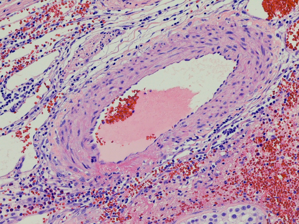
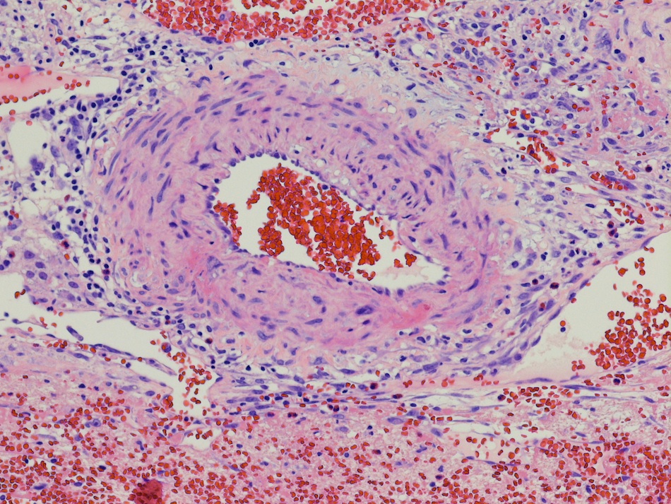
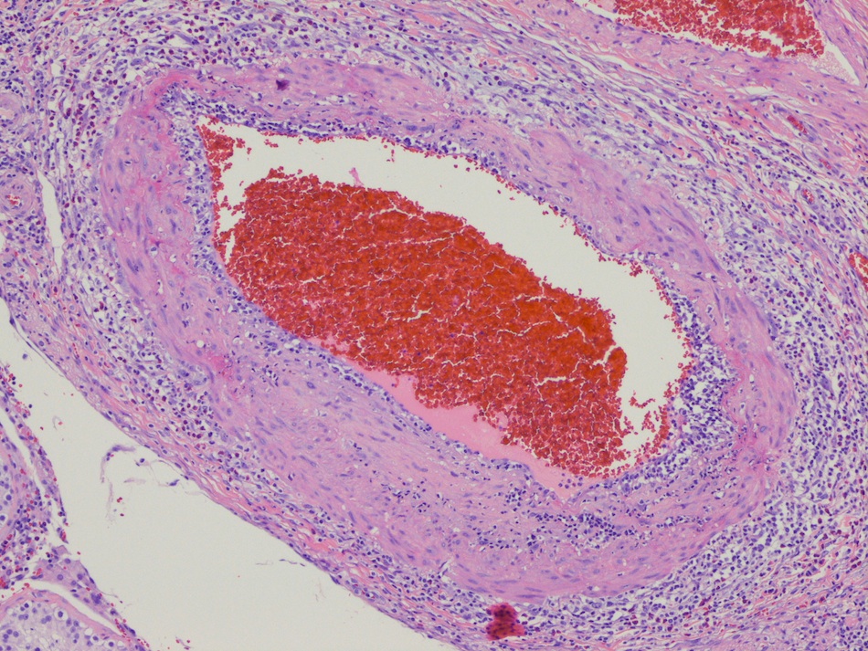
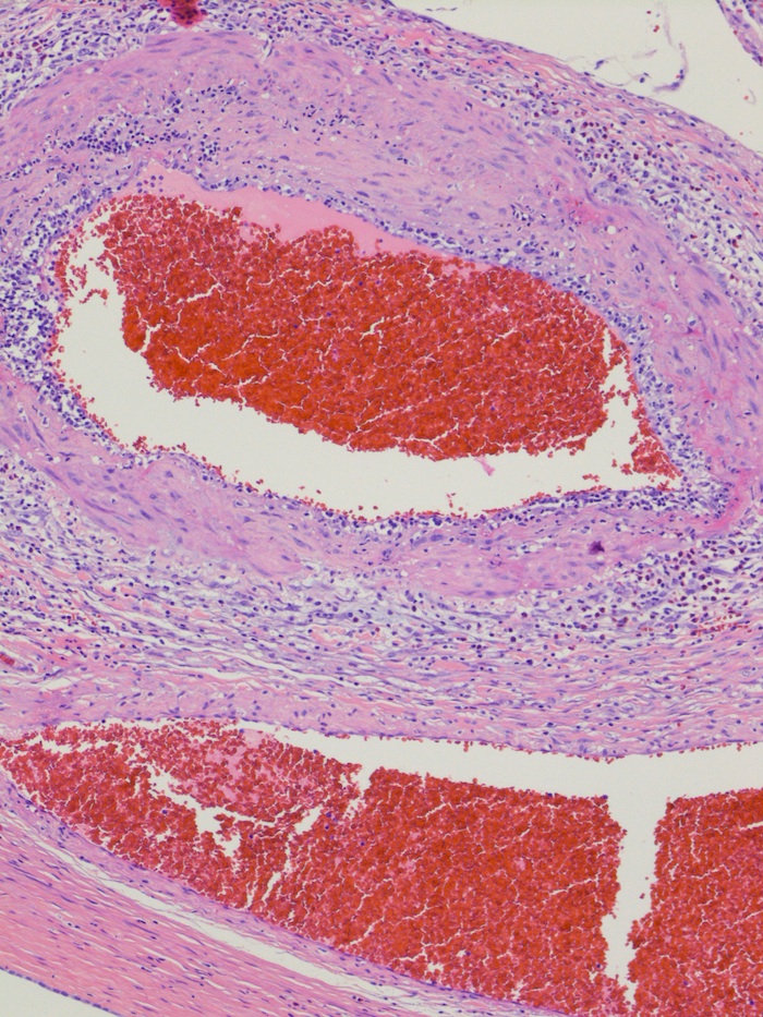
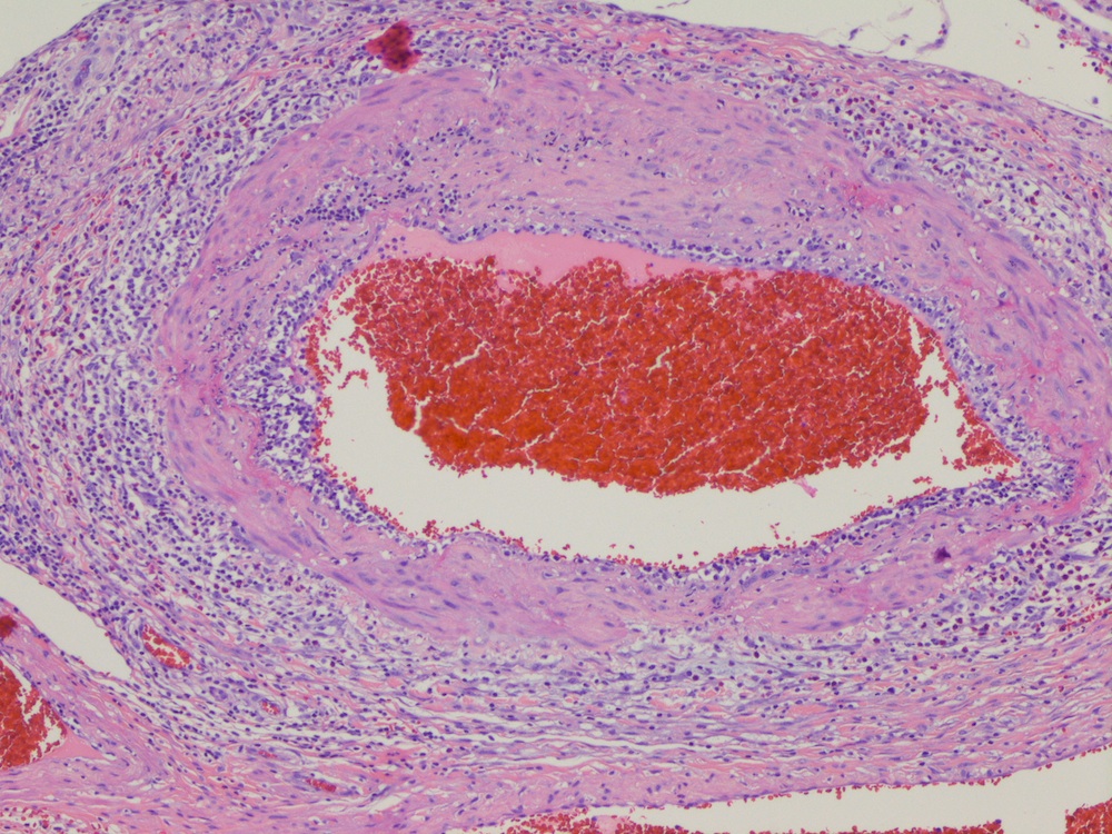
- Necrotizing vasclulitis: systemic disease which manifests as inflammation of arteries with fibrinoid necrosis and fibrosis of the vascular wall
- May be associated with polyarteritis nodosa or an isolated finding; can be associated with a variety of other entities or may be idiopathic (Cardiovascular Pathology 1994;3:197)
- While necrotizing vasculitis can involve any organ, isolated testicular involvement is rare and can pose diagnostic difficulties, especially in the absence of clinical and laboratory evidence of systemic involvement (World J Surg Oncol 2011;9:63)
- Other vasculitides that may involve the testes include Granulomatosis with polyangiitis (Wegener's), Henoch-Schönlein purpura, giant cell arteritis and various autoimmune connective tissue disorders such as systemic lupus erythematosis (J Urol 2006;176:2682)
Clinical features
- Can present with acute onset of pain and swelling, presumably related to intratesticular hemorrhage (Ir Med J 2006;99:27)
- Systemic disease is frequently already present at the time of initial presentation of testicular vasculitis or develops shortly thereafter
- Some cases, however, remain isolated to the testes with no evidence of progression to systemic disease (J Clin Pathol 1994;47:1121)
- It is, therefore, unclear whether isolated testicular vasculitis represents an early site of involvement by systemic disease or a separate entity (World J Surg Oncol 2011;9:63)
- Only several cases of isolated testicular vasculitis were reported in the literature and a presentation suggestive of a testicular neoplasm is even less common (J Clin Pathol 1994;47:1121)
- Since testicular biopsy is contraindicated when malignancy is suspected, the diagnosis is usually reported postorchiectomy in these cases (ScientificWorldJournal 2010;10:1915, Urology 2003;61:1035)
- Biopsy most likely diagnostic in patients with testicular symptoms (pain, enlargement or shrinking)
- Wedge biopsy should contain capsule, tunica vasculosa and testicular parenchyma
Case reports
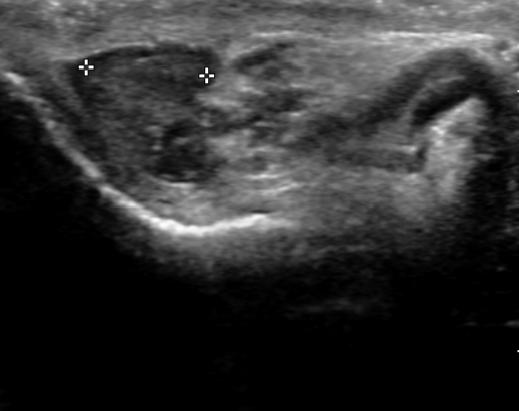
- 34 year old man with hypoechoic testicular lesion (Case #328)
Treatment
- Treatment options are still debated and frequently hinge on individual factors which need to be carefully considered in each patient
- Immunosuppressive therapy has been administered to some of the patients
- In other cases, careful follow up to exclude progression to systemic disease was preferred (J Clin Pathol 1994;47:1121)
- Some authors have even postulated that the excision of the affected organ might be curative, obviating the need for pharmacologic therapy (ScientificWorldJournal 2010;10:1915)
- Close follow up, including serology, is recommended since the risk of progression to systemic disease is unknown (J Clin Pathol 1994;47:1121)
Gross description
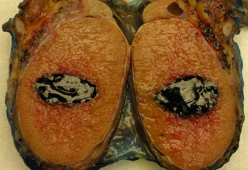
- Testis was bivalved to show a hemorrhagic, well defined, soft, dark red lesion (Case of the Week #328)
Microscopic (histologic) description
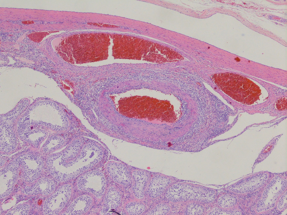
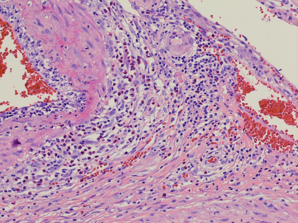
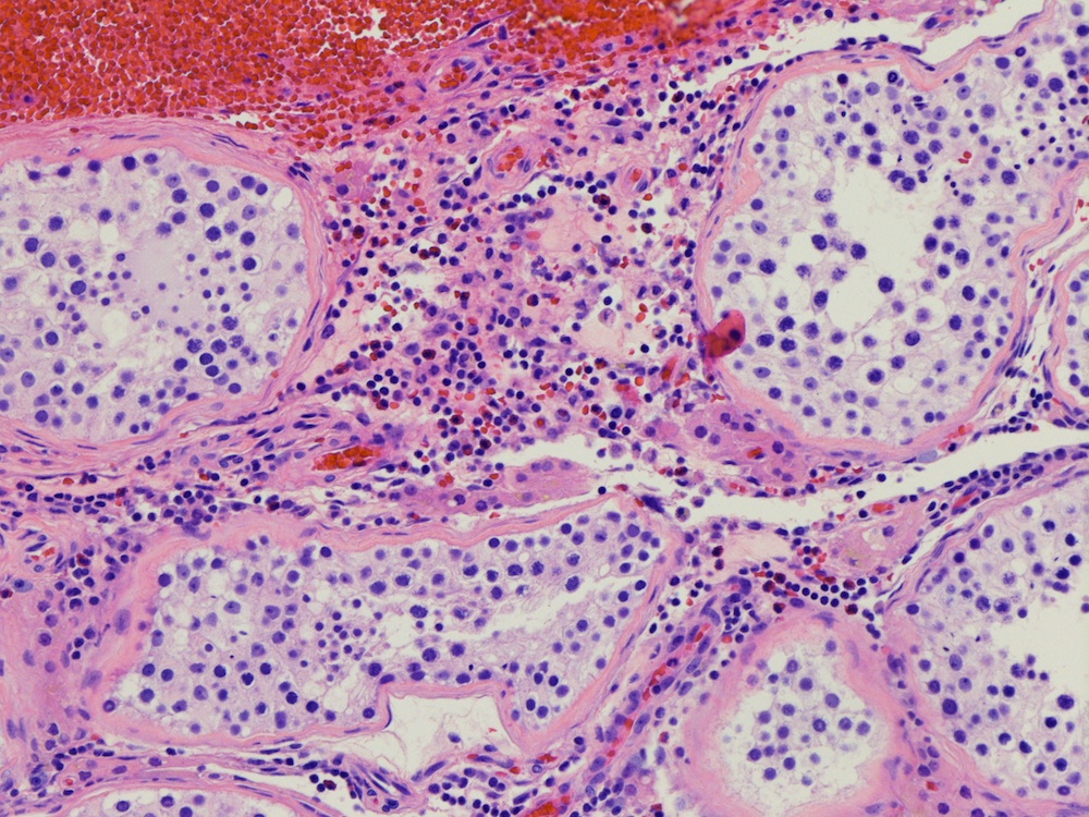
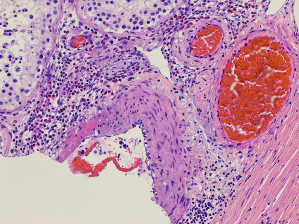
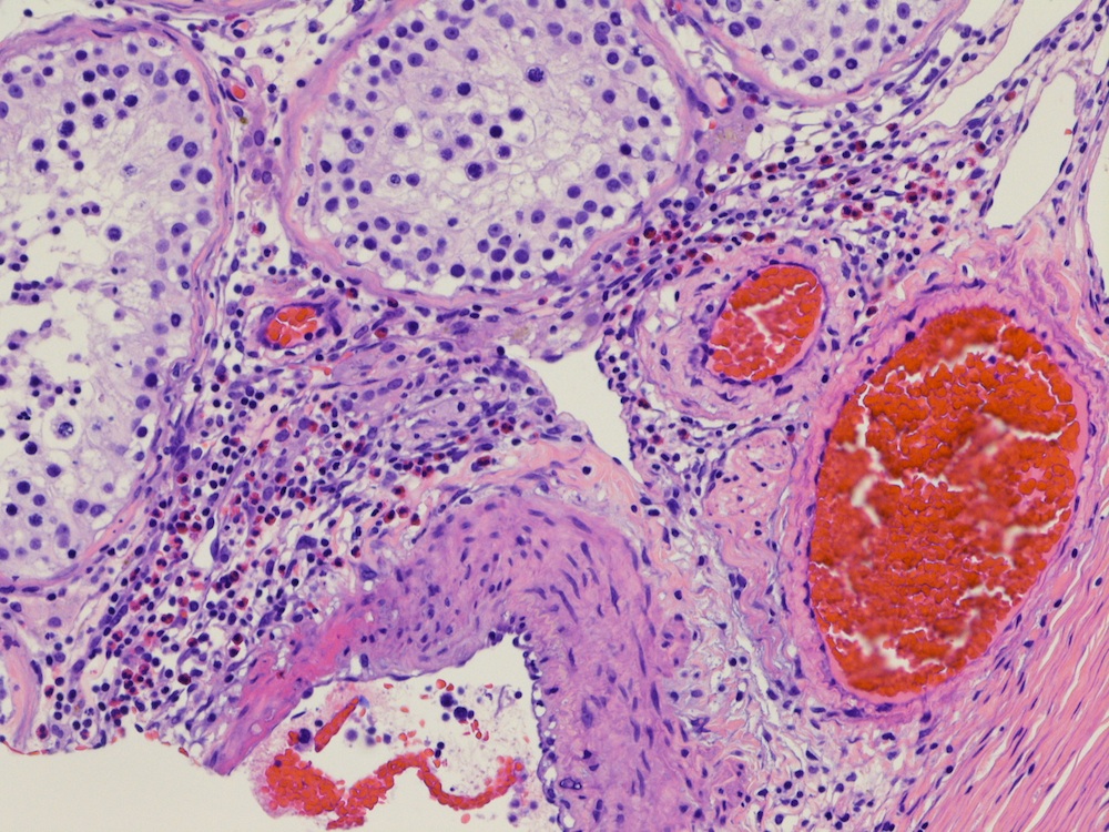
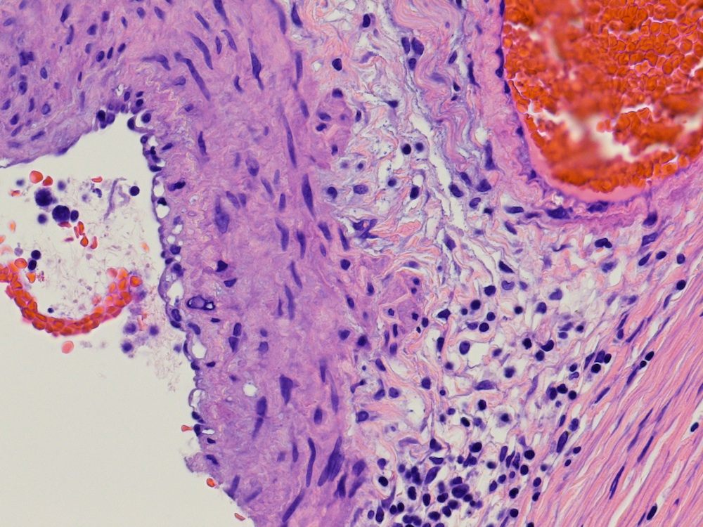
- Focal hemorrhage with early organization and small arteries around the area of hemorrhage (Case of the Week #328)
Differential diagnosis
- Although uncommon, isolated necrotizing testicular vasculitis remains in the differential diagnosis of hypovascular testicular lesions; long term follow up and additional research is needed in order to further elucidate the pathogenesis of isolated organ vasculitis and the subsequent risk of disease progression


