
Testis & paratestis
Nonneoplastic lesions
Epidermoid cyst
Last author update: 1 December 2012
Last staff update: 19 March 2024
Copyright: 2002-2025, PathologyOutlines.com, Inc.
PubMed Search: Teratoma-epidermoid cyst testis


Table of Contents
Definition / general | Case reports | Gross description | Microscopic (histologic) description | Microscopic (histologic) images | Molecular / cytogenetics description | Molecular / cytogenetics images | Differential diagnosisCite this page: Gordetsky J, Williamson S. Epidermoid cyst. PathologyOutlines.com website. https://www.pathologyoutlines.com/topic/testisepidermoidcyst.html. Accessed March 30th, 2025.
Definition / general
- Rare benign cyst, < 1% of testicular tumors
- Usually ages 10 - 39
- Unclear if neoplastic - occasionally has allelic losses, favoring neoplasia (Arch Pathol Lab Med 2003;127:858)
- Pilomatricoma / pilomatrixoma (histologic changes including shadow cells, which resemble cutaneous piiomatrixoma), is considered a subtype (Am J Surg Pathol 2001;25:788, Arch Pathol Lab Med 1995;119:96)
Case reports
- 21 and 27 year old men (Cesk Patol 2005;41:102)
Gross description
- Intraparenchymal lesion, usually adjacent to tunica albuginea, mean 2 cm, contains white grumous keratin debris
Microscopic (histologic) description
- Cyst with keratinized squamous epithelial lining containing a granular cell layer, cyst filled with laminated keratin
- Cyst rupture may cause granulomatous reaction
- No adnexal structures, no other tissue types
- Pilomatrixoma-like variant:
- Adjacent testis does not show intratubular germ cell neoplasia, unclassified type (IGCNU)
- Eosinophilic islands of "shadow" squamous epithelium with focal, peripheral calcification and ossification
- The ghost-like cells are similar to the shadow cells of cutaneous pilomatrixoma (Am J Surg Pathol 2001;25:788)
Microscopic (histologic) images
Contributed by Debra L. Zynger, M.D. and @ThatGlassTho on Twitter
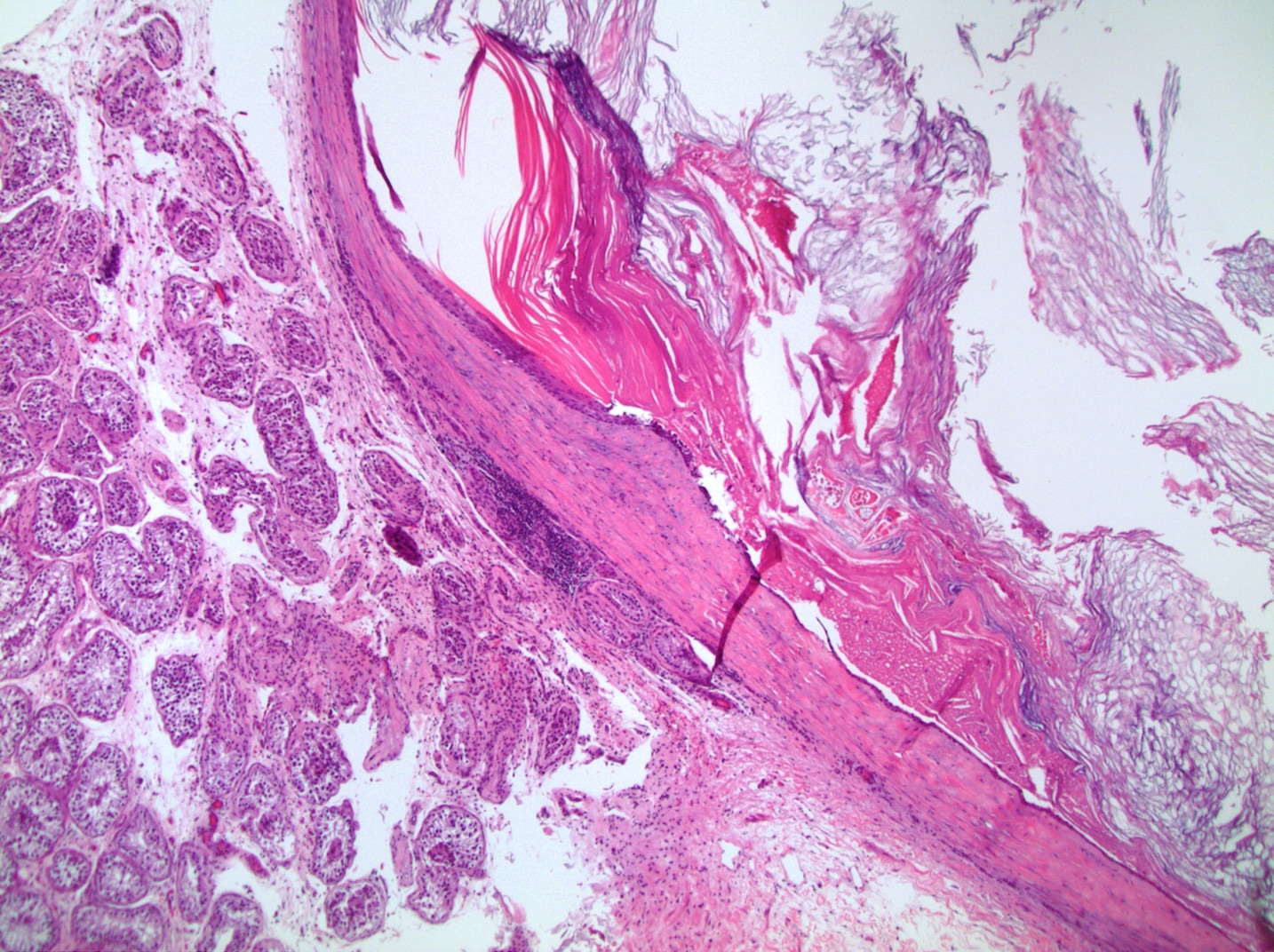
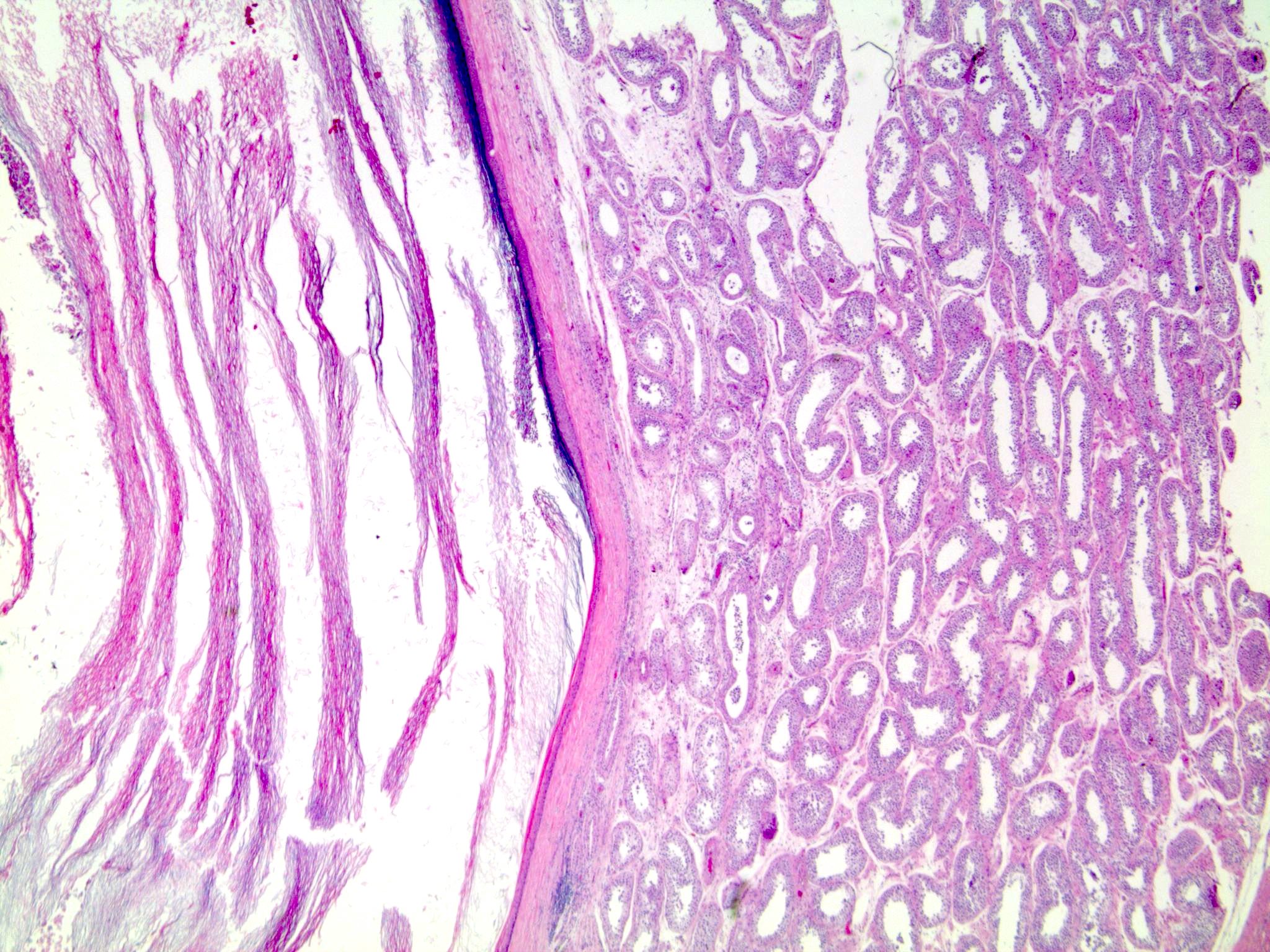
Testicular epidermoid cyst
Cyst wall with adjacent normal seminiferous tubule
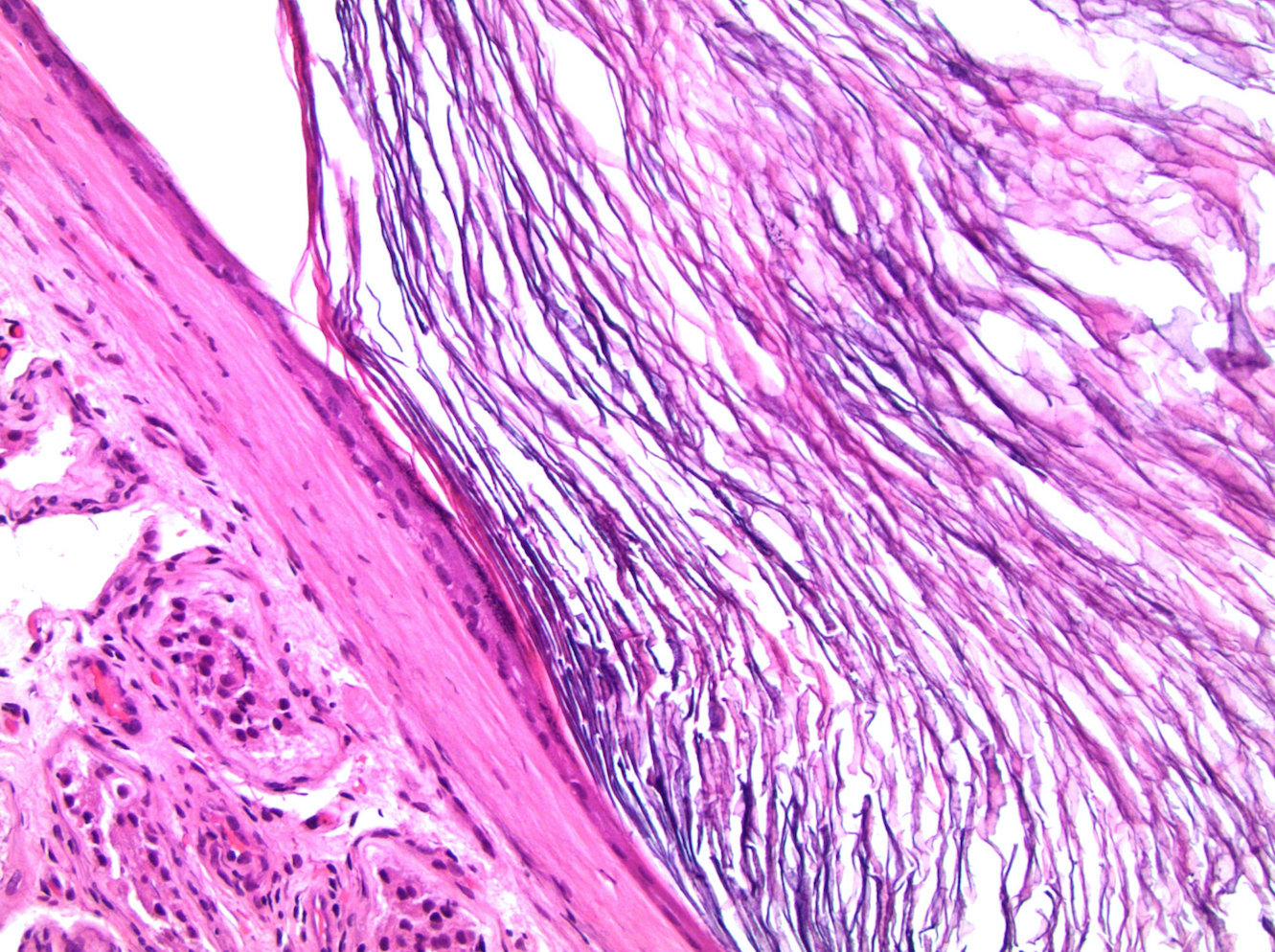
Lamellar keratin within the cyst
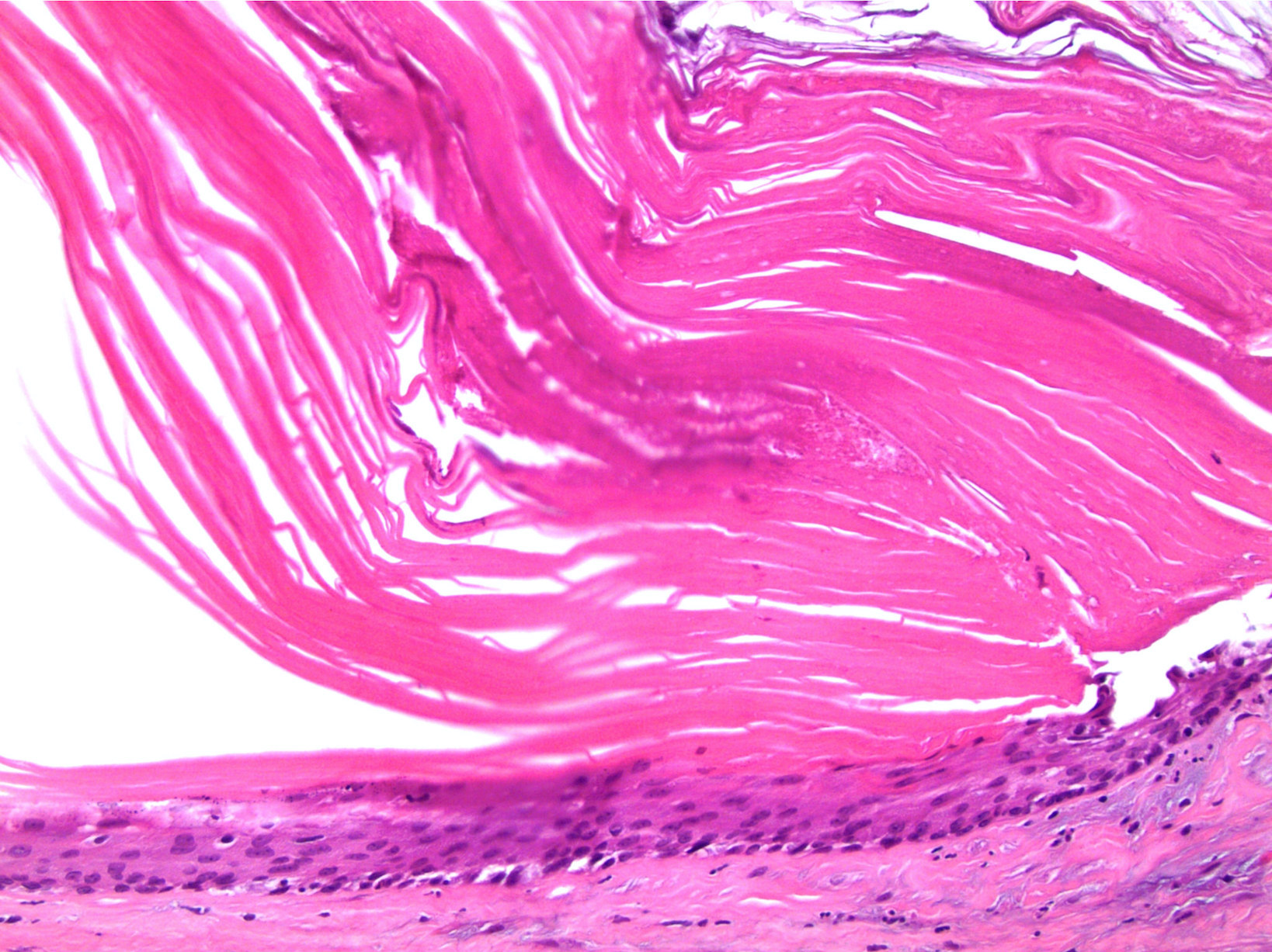
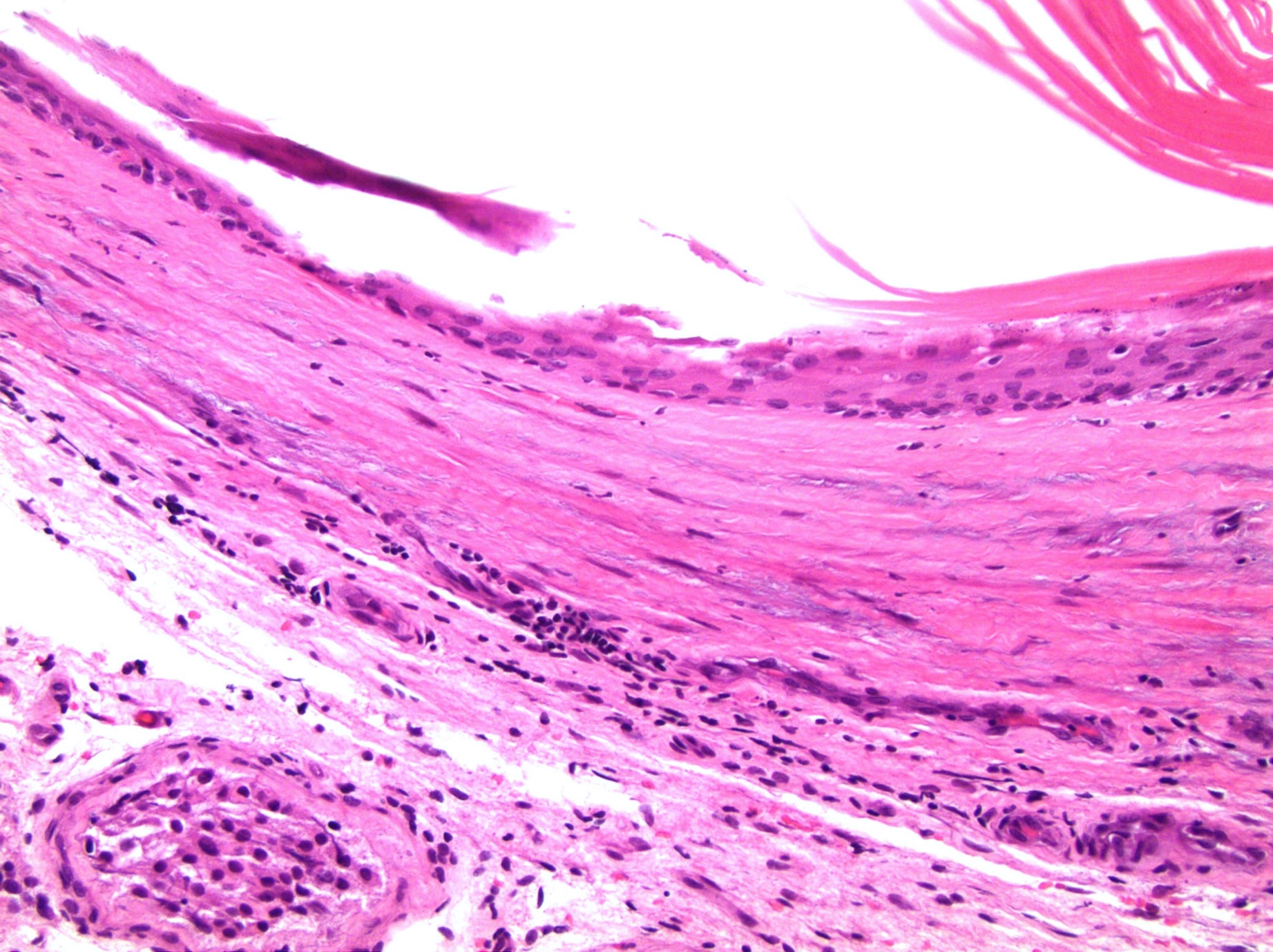
Stratified squamous epithelium with adjacent keratin

Teratoma - epidermoid cyst
Molecular / cytogenetics description
- Isochromosome 12p and other overrepresentation of 12p are present in usual teratoma but not in epidermoid cyst (Clin Cancer Res 2006;12:5668)
Molecular / cytogenetics images
Images hosted on other servers:

Normal FISH for 12p
Differential diagnosis
- Teratoma: usually postpubertal testis, IGCNU present, often atypia in teratomatous elements
