Stains & molecular markers
p53

Copyright: 2002-2025, PathologyOutlines.com, Inc.
PubMed Search: p53 pathology
p53
Author: Till Braunschweig, M.D.
Editorial Board Member: Christian M. Schürch, M.D., Ph.D.
Deputy Editor-in-Chief: Jennifer A. Bennett, M.D.
Last author update: 9 December 2021
Last staff update: 24 August 2023
Copyright: 2002-2025, PathologyOutlines.com, Inc.
PubMed Search: p53 pathology
Table of Contents
Definition / general | Essential features | Pathophysiology | Clinical features | Interpretation | Uses by pathologists | Prognostic factors | Microscopic (histologic) description | Microscopic (histologic) images | Wildtype (normal) staining - normal tissues | Wildtype (normal) staining - disease | Aberrant (mutational type) staining | Molecular / cytogenetics description | Sample pathology report | Additional references | Board review style question #1 | Board review style answer #1Cite this page: Braunschweig T. p53. PathologyOutlines.com website. https://www.pathologyoutlines.com/topic/stainsp53.html. Accessed March 27th, 2025.
Definition / general
- Tumor suppressor gene at 17p13, 53 kDa
- Mutations are among most commonly detected genetic abnormalities in human neoplasia; however, presence of TP53 mutation is usually not, by itself, specific enough for a diagnosis for malignancy and its absence does not rule out malignancy
Essential features
- Strong nuclear positivity or complete absence by immunohistochemistry has a strong correlation with the presence of a mutation
- Low to intermediate expression suggests a nonmutated gene
Pathophysiology
- Tumor suppressor gene at 17p13, 53 kDa
- p53 ensures that cells repair any damaged DNA before cell division by inducing cell cycle arrest to allow time for DNA repair or to force the cell to undergo apoptosis via activation of BAX gene (J Biomed Biotechnol 2011;2011:603925, Cancers (Basel) 2018;10:135)
- Produces nuclear phosphoprotein involved in transcriptional regulation (Nat Rev Cancer 2006;6:909)
- N terminus amino acids bind (a) TAFs (TATA binding protein associated factors), which attract other proteins needed to initiate gene expression, as well as (b) MDM2, which inhibits p53 and has the opposite effect as TAFs (Cell 1997;88:323)
- p53 induces p21 WAF1, which inhibits cyclin dependent kinases (Oncogene 2004;23:303)
- p53 has half life of only 5 - 20 minutes, which increases when a mutation is present (Genes Dev 1998;12:2973)
- In unstressed cells, p53 is inactivated by MDM2 (OMIM: Tumor Protein p53 [Accessed 12 February 2021])
- Also inactivated by SV40 T antigen and E1B adenovirus product (Cell 1997;88:323)
- Sequestered by HPV E6 protein
Clinical features
- Li Fraumeni syndrome: hereditary TP53 mutation causes multiple tumors (J Natl Cancer Inst 1969;43:1365)
- According to several review publications, incidences of different tumor types are different for women and men (Nat Rev Clin Oncol 2014;11:260, Eur J Hum Genet 2020;28:1379, Cold Spring Harb Perspect Med 2017;7:a026187)
- Women: almost 100% develop TP53 mutation associated cancer with a risk of 49% by the age of 30
- Men: 79% develop TP53 mutation associated cancer with a risk of 21% by the age of 30
- Distribution: breast 28%, soft tissues 14%, brain 13%, adrenocortical 11%, bones 8%, hematological 4%, colorectal 3%, other 19% (gastric, melanoma, lung, pancreas, ovary, bladder, prostate)
- For children, the spectrum of cancers includes mostly brain tumors as choroid plexus carcinoma, glioma, medulloblastoma and rhabdomyosarcoma (Nat Rev Clin Oncol 2014;11:260)
Interpretation
- Wildtype (normal) - scattered nuclear staining, mid epithelial (basal sparing)
- Aberrant (mutational type) → 80% strong and diffuse nuclear staining, complete absence of nuclear staining in all cells, moderate to strong cytoplasmic staining (Int J Gynecol Pathol 2021;40:32)
Uses by pathologists
- Differentiate reactive changes or low grade intraepithelial neoplasms occurring in mucosa or adenoma from high grade intraepithelial neoplasms (p53+) in the esophagus (Barrett metaplasia), stomach, small intestine, large bowel (Gut 2020;69:811)
- Differentiate reactive epithelial changes from high grade, in situ urothelial carcinoma (p53+) (Hum Pathol 2020;98:81)
- Detect high grade intraepithelial neoplasms or carcinoma cells in fine needle aspiration of the pancreas (p53+) (Am J Surg Pathol 2018;42:1556)
- Differentiate low grade from high grade serous ovarian carcinoma (aberrant p53+) (Mod Pathol 2011;24:1248)
- Differentiate subtypes of glioma and medulloblastoma (J Neuroimaging 2021;31:306)
- Determine if children with choroid plexus carcinoma should be screened for Li Fraumeni syndrome (Pediatr Blood Cancer 2012;58:905)
- Differentiate prognostic subgroups in vulvar squamous cell carcinoma (Mod Pathol 2020;33:1595)
- Surrogate marker for classifying endometrial carcinomas as TCGA copy number high (Int J Gynecol Pathol 2019;38 Suppl 1:S123, Br J Cancer 2015;113:299)
- Detect HPV independent, differentiated vulvar intraepithelial neoplasia (Pharmaceuticals (Basel) 2021;14:324, Histopathology 2021;78:424)
- Differentiate serous tubal intraepithelial carcinoma (STIC) from reactive changes in epithelium of the fallopian tube or fimbriae, although serous tubal intraepithelial lesions may also have aberrant p53 phenotype and histologic differentiation is important (Int J Gynecol Pathol 2011;30:417, Int J Gynecol Pathol 2021;40:419)
Prognostic factors
- Poor outcome in p53 aberrant hematopoetic malignancies (Mol Clin Oncol 2017;6:876, Pediatr Hematol Oncol 2014;31:327)
- p53 aberrant basal cell carcinomas respond better to photodynamic therapy (PLoS One 2019;14:e0215537)
- Stratify TP53 mutated medulloblastoma, SHH (sonic hedgehog) subgroup as high risk patients (Acta Neuropathol 2016;131:821)
- Worse outcome in p53+ aberrant vulva squamous cell carcinoma (Am J Obstet Gynecol 2021;224:595.e1, Mod Pathol 2020;33:1595)
- Worse outcome in p53 aberrant morphologically ambiguous endometroid carcinoma (Mod Pathol 2010;23:80)
- Worse outcome in TP53 mutated salivary carcinoma (Cancer Immunol Immunother 2020;69:1363)
- Worse outcome in p53+ / p16- anal squamous cell carcinoma (Mod Pathol 2021;34:1017)
Microscopic (histologic) description
- Low to intermediate inhomogeneous nuclear expression in up to 50% of wildtype (WT) cases
- Cutoff values vary in publications, some suggest > 5%, > 10% or > 50% strong positive nuclear stain as positive
- Cutoff value of > 10% in glioblastoma has low predictive value (Oncotarget 2019;10:6204)
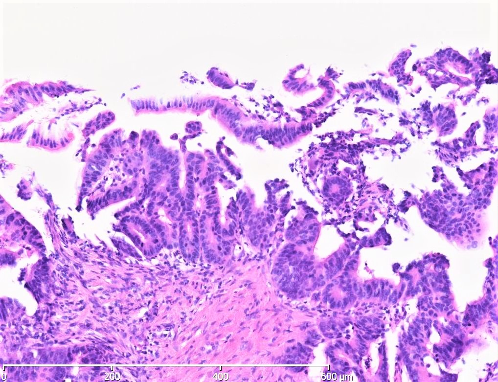
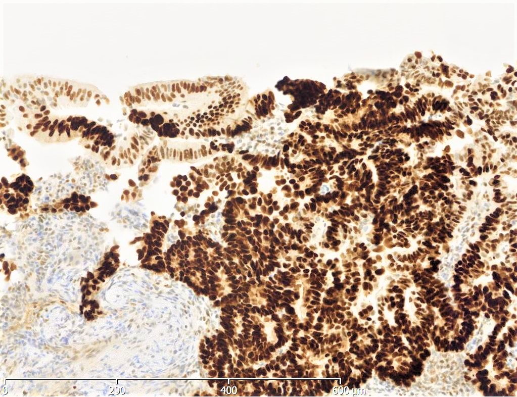
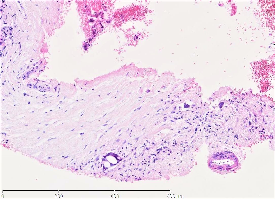
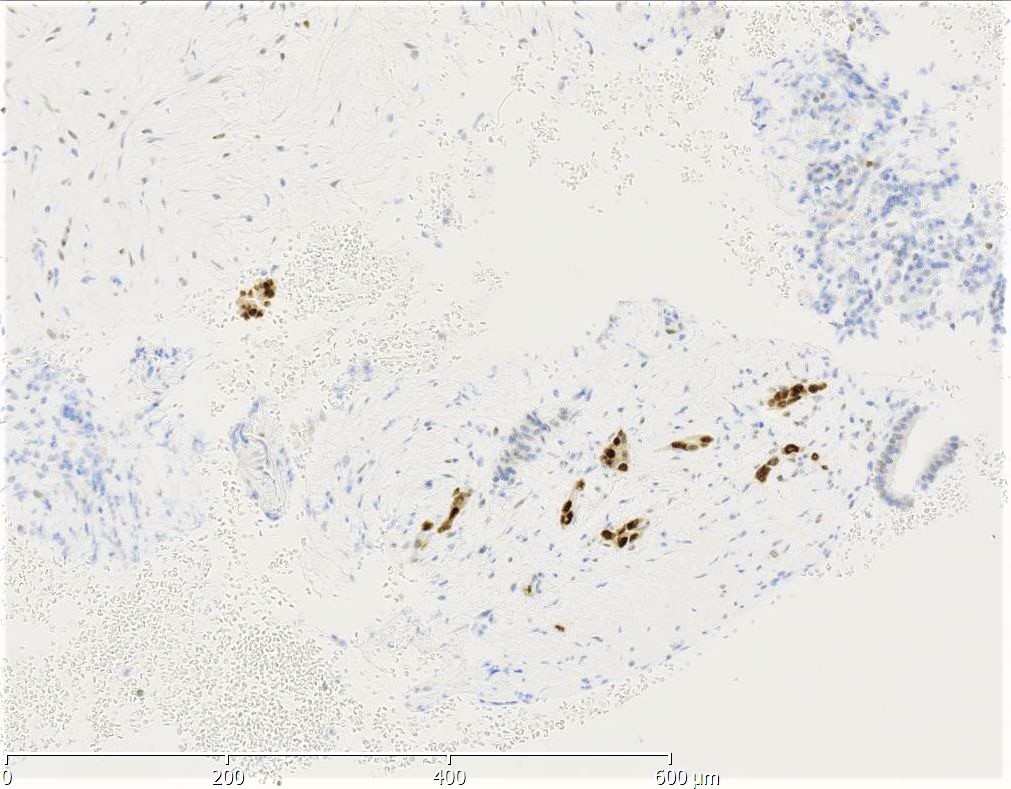
Microscopic (histologic) images
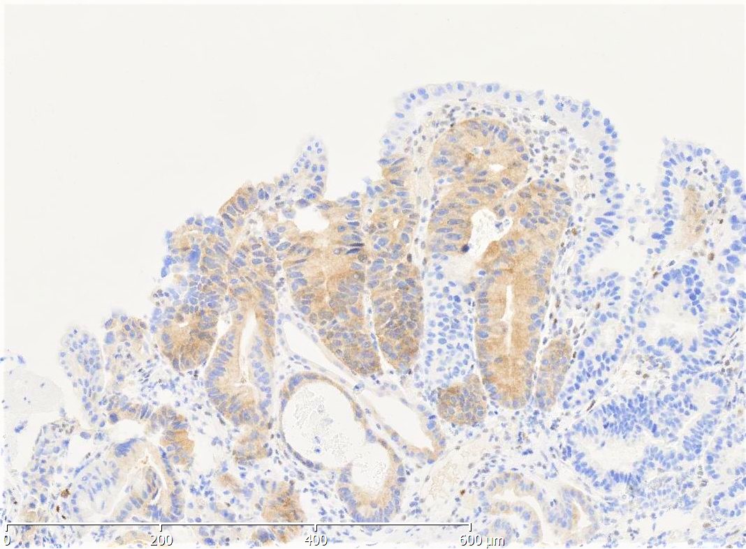
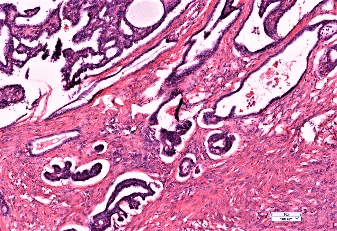
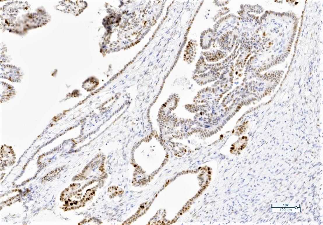
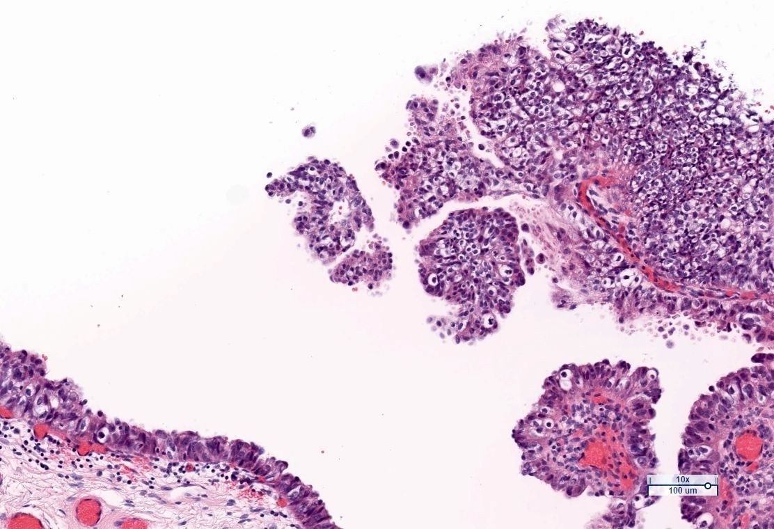
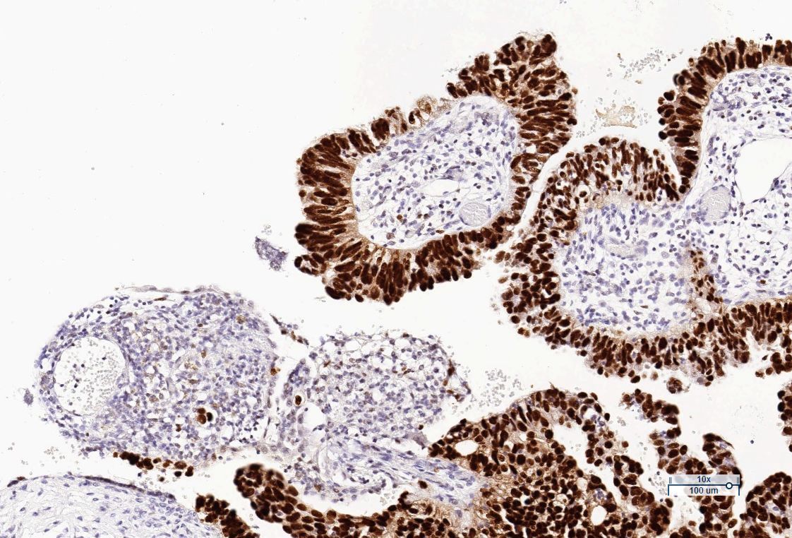
Contributed by Till Braunschweig, M.D.
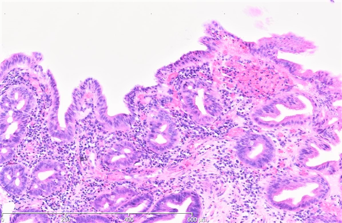

Barrett esophagus, high grade intraepithelial neoplasia, p53+

Barrett esophagus, high grade intraepithelial neoplasia, p53 absent
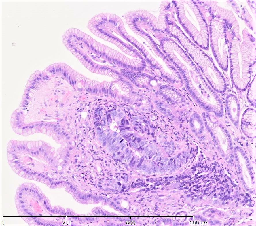
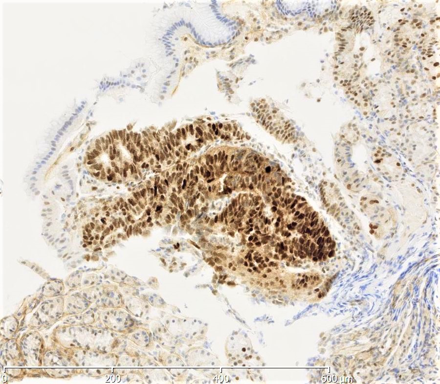
Stomach mucosa, high grade intraepithelial neoplasia, p53+
Tubular adenoma of the duodenum, high grade intraepithelial neoplasia, p53+
Adenocarcinoma in core biopsy of the pancreas, p53+
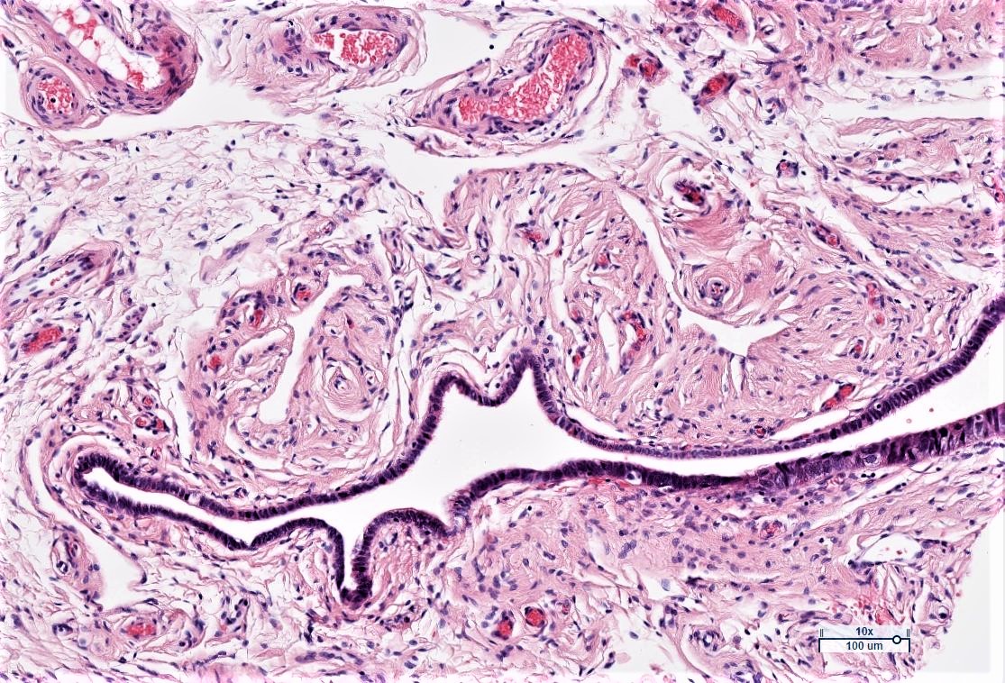
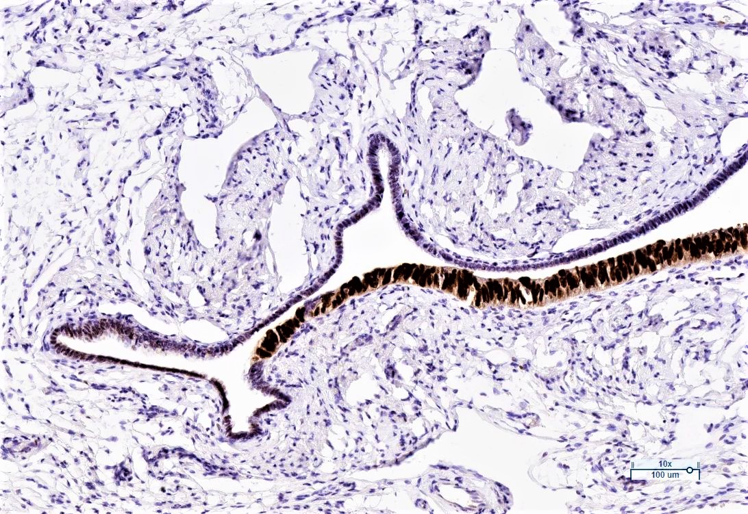
Fallopian tube, intraepithelial carcinoma, p53 overexpressed
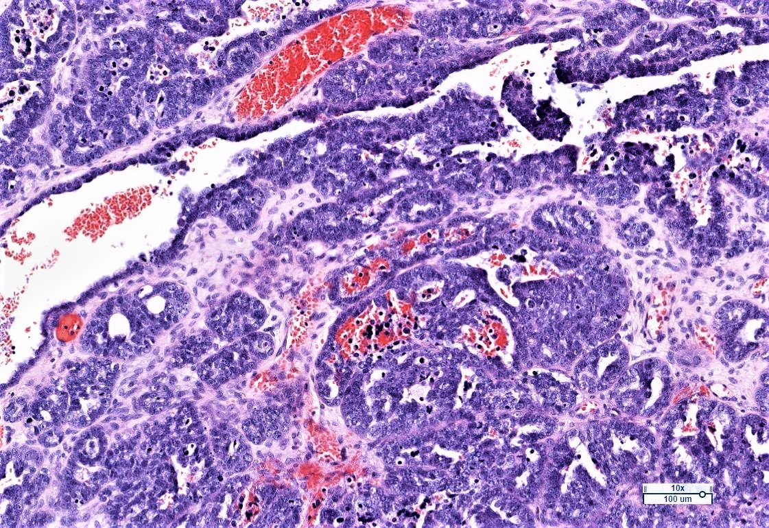
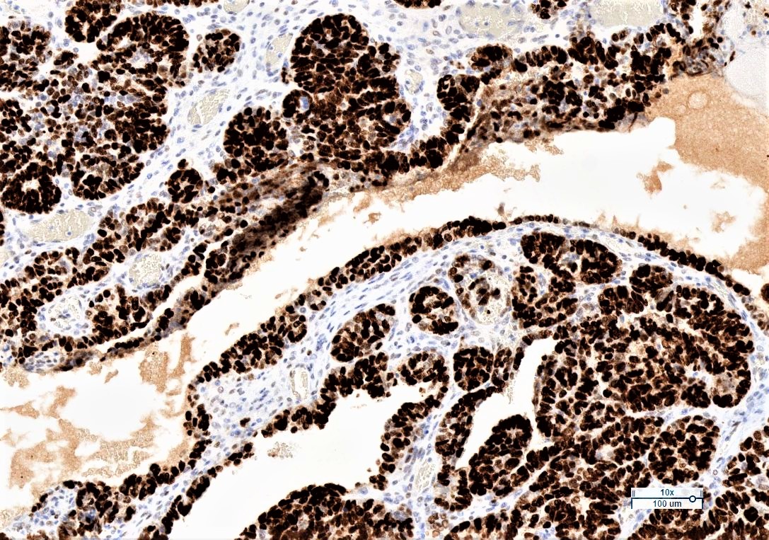
Serous ovarian carcinoma, high grade, p53 overexpressed
Serous ovarian carcinoma, low grade, p53 wildtype
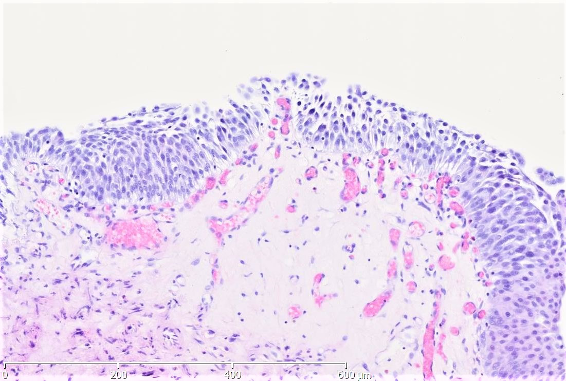
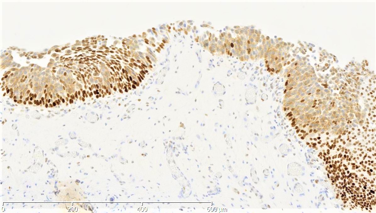
Urothelial carcinoma in situ, p53 absent
Wildtype (normal) staining - normal tissues
- Virtually all normal tissues should show wildtype p53 expression
Wildtype (normal) staining - disease
- Low grade intraepithelial neoplasia in Barrett esophagus (Gut 2020;69:811)
- Low grade serous ovarian carcinoma (Mod Pathol 2011;24:1248, J Pathol Clin Res 2016;2:247)
- Differentiate reactive changes in flat urothelial lesions with nuclear atypia (Hum Pathol 2020;98:81)
- Differentiate reactive changes in ductal structures in pancreas tissue (biopsies, cytological specimen) (Pathobiology 2017;84:192)
Aberrant (mutational type) staining
- High grade intraepithelial neoplasia and carcinoma of Barrett esophagus, stomach, small intestine, colon, exocrine pancreas, biliary system, urinary tract (Histopathology 2020 Sep;77:481, Gut 2020;69:811, Gastric Cancer 2003;6:71, Am J Surg Pathol 2018;42:1556, Hum Pathol 2016;57:61, Hum Pathol 2020;98:81)
- High grade serous ovarian carcinoma (Mod Pathol 2011;24:1248, J Pathol Clin Res 2016;2:247)
- High grade endometrial carcinoma (Int J Gynecol Pathol 2021;40:32, Int J Gynecol Pathol 2019;38 Suppl 1:S123)
- Subtypes of glioma (Brain Pathol 2015;25:256)
- Subtypes of medulloblastoma (J Neuroimaging 2021;31:306)
- Subset of HPV independent vulvar squamous cell carcinoma (Mod Pathol 2020;33:1595)
Molecular / cytogenetics description
- Mostly missense or nonsense mutations, loss of function or splicing mutations uncommon (Cold Spring Harb Perspect Med 2016;6:a026179)
- Mutations in several hotspots and similarly distributed over several cancer types, mostly in the DNA binding domain (Hum Mutat 2007;28:622)
Sample pathology report
- Right ovary, oophorectomy:
- High grade serous carcinoma (see comment)
- Comment: Immunohistochemistry for p53 shows strong nuclear positivity in over 80% of tumor cells, consistent with aberrant (mutational type) expression. Controls show appropriate reactivity.
Additional references
Board review style question #1
Immunohistochemistry of p53 can serve as a distinct marker for the confirmation of high grade neoplastic changes of different tissues. What kind of staining pattern is most common?
- Circumferential membranous staining
- Cytoplasmic and nuclear staining
- Partial membranous staining
- Strong cytoplasmic staining
- Strong nuclear staining
Board review style answer #1
