
Stains & CD markers
Napsin A

Copyright: 2002-2024, PathologyOutlines.com, Inc.
PubMed Search: Napsin A
Napsin A
Author: Nick Baniak, M.D.
Editorial Board Members: Christian M. Schürch, M.D., Ph.D., Brandon Umphress, M.D.
Last author update: 23 March 2023
Last staff update: 23 March 2023
Copyright: 2002-2024, PathologyOutlines.com, Inc.
PubMed Search: Napsin A

Table of Contents
Definition / general | Essential features | Interpretation | Uses by pathologists | Microscopic (histologic) images | Positive staining - normal | Positive staining - disease | Negative staining | Sample pathology report | Board review style question #1 | Board review style answer #1Cite this page: Baniak N. Napsin A. PathologyOutlines.com website. https://www.pathologyoutlines.com/topic/stainsnapsina.html. Accessed April 19th, 2024.
Definition / general
- Functional aspartic protease of the pepsin family A encoded by the NAPSA gene located at chromosome 19q13.3, essential for maturation of prosurfactant protein B (Semin Diagn Pathol 2018;35:143, Pathol Oncol Res 2021;27:613099, Am J Surg Pathol 2015;39:1742, Appl Immunohistochem Mol Morphol 2020;28:593)
- Potentially involved in phagocytosis by macrophages and lysosomal protein catabolism in renal cells (Pathol Oncol Res 2021;27:613099, Adv Anat Pathol 2012;19:66, Arch Histol Cytol 2002;65:359)
- Predominantly expressed in the cytoplasm of type II pneumocytes, intra-alveolar macrophages, proximal and convoluted renal tubules, and pancreatic acini and ducts (Pathol Oncol Res 2021;27:613099, Am J Surg Pathol 2015;39:1742, Jpn J Cancer Res 2000;91:1015)
- Regulated by TTF1 (Pathol Oncol Res 2021;27:613099)
- On slide external positive controls:
- Kidney: almost all epithelial cells of the proximal tubules should show at least moderate, granular cytoplasmic staining
- Lung: type 2 pneumocytes and alveolar macrophages show strong staining
- Some diagnostically validated clones include IP64, MRQ-60, TMU-Ad02, polyclonal (Pathology 2019;51:240, Am J Surg Pathol 2014;38:189, Turk Patoloji Derg 2021;37:7, Appl Immunohistochem Mol Morphol 2016;24:648, Pathology 2015;47:105, APMIS 2018;126:45, Am J Surg Pathol 2018;42:989)
Essential features
- Usually positive (cytoplasmic) in lung adenocarcinomas and clear cell carcinomas of the gynecologic tract while negative in the primary differentials of these entities
- Clear cell carcinomas often have focal or rare staining (Am J Surg Pathol 2018;42:989, Hum Pathol 2015;46:957)
Interpretation
- Cytoplasmic (Histopathology 2017;70:375)
- Staining can be subtle; often focal and granular cytoplasmic (Int J Gynecol Pathol 2020;39:344)
Uses by pathologists
- Aids in histotype classification of primary lung tumors
- Aids in histotype classification of gynecologic tumors when clear cell carcinoma is in the differential
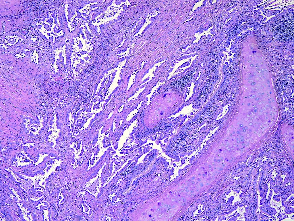
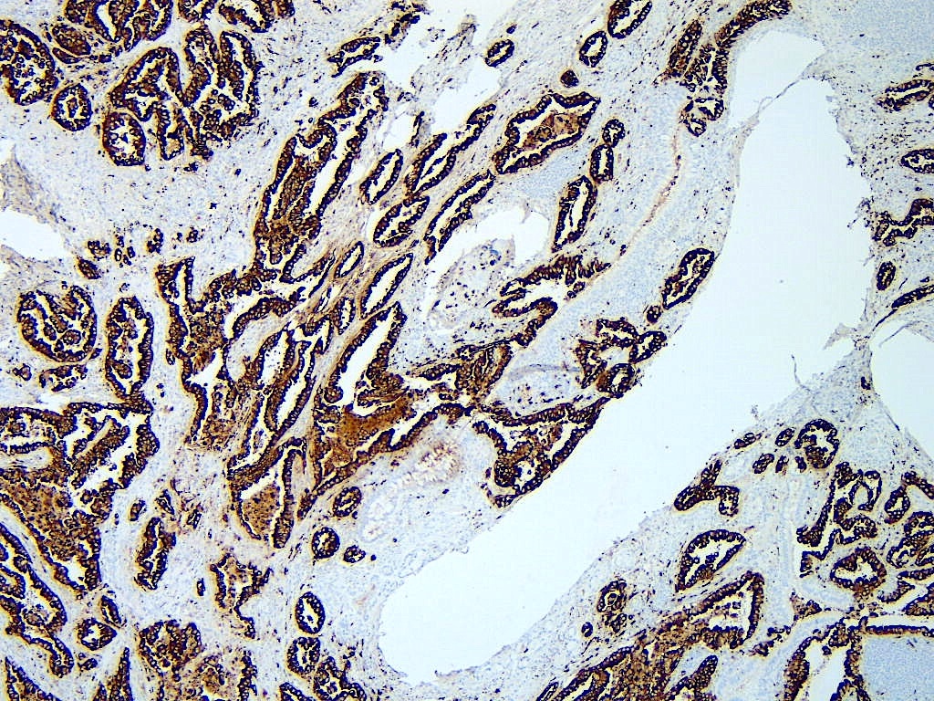
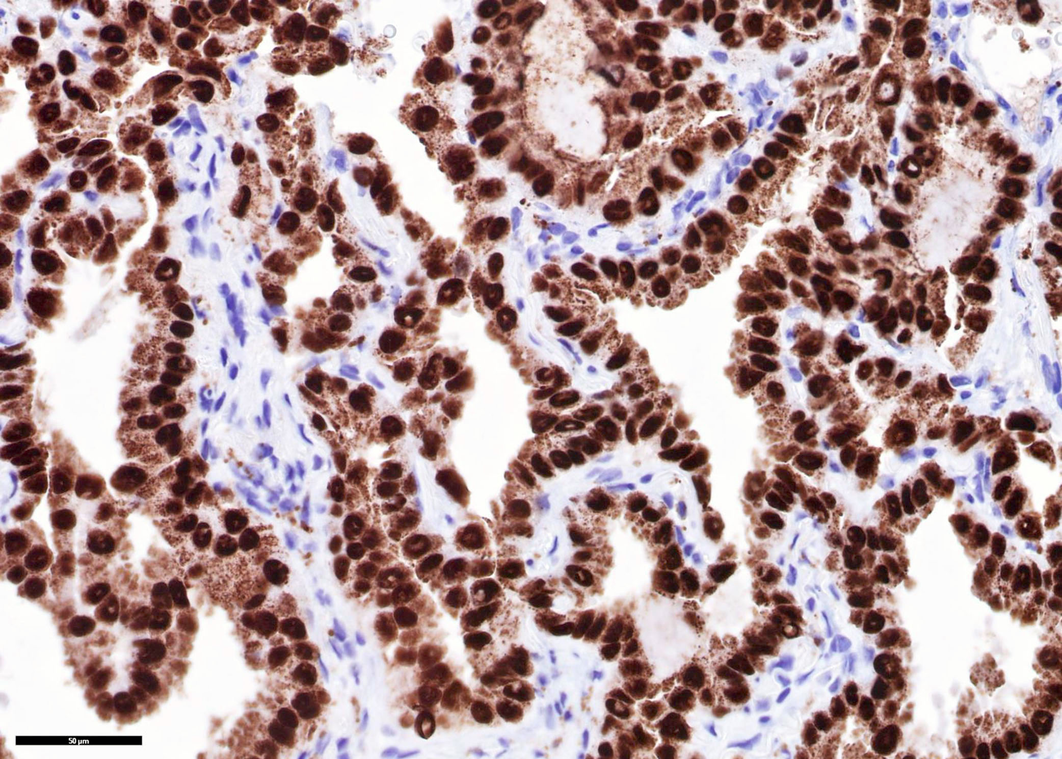
Microscopic (histologic) images
Contributed by Nick Baniak, M.D. and Andrey Bychkov, M.D., Ph.D.
Lung resection, adenocarcinoma
Lung resection, adenocarcinoma staining
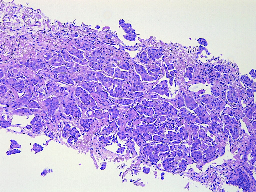
Lung biopsy, adenocarcinoma
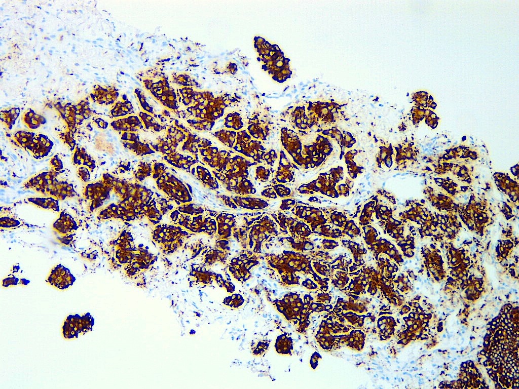
Lung biopsy, adenocarcinoma staining
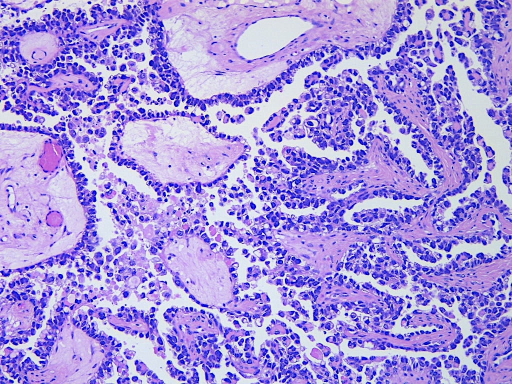
Ovarian clear cell carcinoma
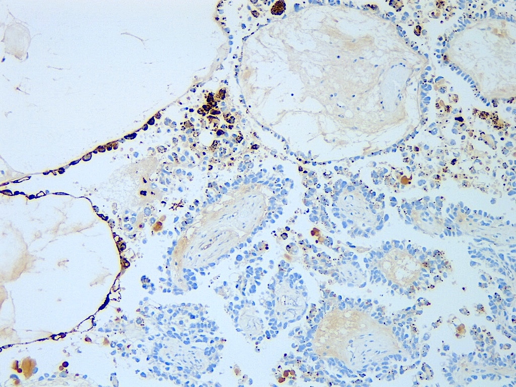
Ovarian clear cell carcinoma staining
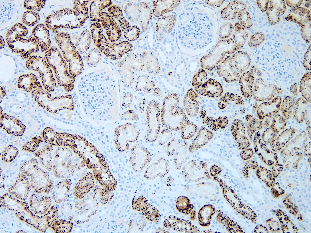
External on slide control kidney
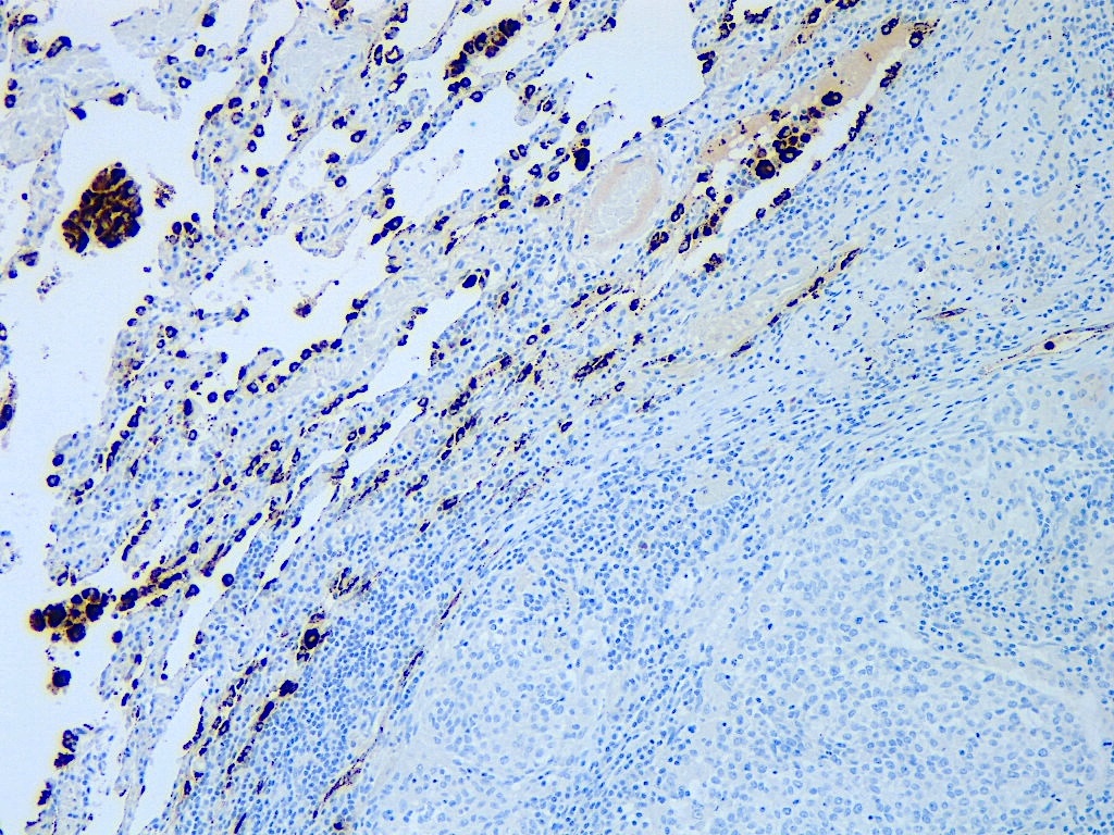
External on slide control lung
Lung adenocarcinoma
Positive staining - normal
- Pneumocytes and alveolar macrophages (Pathol Oncol Res 2021;27:613099)
- Epithelial cells of the renal tubules (Pathol Oncol Res 2021;27:613099)
- Endometrial glands in decidualized stroma (Pathol Oncol Res 2021;27:613099)
- Tubules of the epididymis (1+ / 2+) (Pathol Oncol Res 2021;27:613099)
- Nephrogenic adenoma (100%) (Hum Pathol 2020;102:23)
- Arias-Stella reaction (100%) (Int J Gynecol Pathol 2020;39:344)
Positive staining - disease
- Lung adenocarcinoma (74 - 92%) (Pathology 2019;51:240, Pathol Oncol Res 2021;27:613099, Cancer Cytopathol 2016;124:472, Turk Patoloji Derg 2021;37:7, Asian Pac J Cancer Prev 2020;21:3345, Appl Immunohistochem Mol Morphol 2016;24:648)
- Plus or minus 3% if using 1% versus 10% tumor cell staining as positive cutoff (Appl Immunohistochem Mol Morphol 2016;24:648)
- Sensitivity 75 - 87%, specificity 95 - 98% (Appl Immunohistochem Mol Morphol 2016;24:648, Biotech Histochem 2018;93:364)
- Mucinous lung adenocarcinoma (50%) (Arch Pathol Lab Med 2014;138:1067)
- Ovarian clear cell carcinoma (72 - 100%) (Pathol Oncol Res 2021;27:613099, Hum Pathol 2015;46:957, Am J Surg Pathol 2021;45:1452, Pathology 2015;47:105, APMIS 2018;126:45)
- Endometrial clear cell carcinoma (43 - 93%) (Pathol Oncol Res 2021;27:613099, Appl Immunohistochem Mol Morphol 2017;25:632, Am J Surg Pathol 2014;38:189, Am J Surg Pathol 2015;39:1061, Hum Pathol 2015;46:957, Histopathology 2015;66:664)
- Endocervical clear cell carcinoma (43 - 57%) (Am J Surg Pathol 2018;42:989)
- Kidney:
- Papillary renal cell carcinoma (40 - 84%) (Pathol Oncol Res 2021;27:613099, Diagn Pathol 2015;10:4)
- Acquired cystic disease renal cell carcinoma (100%) (Diagn Pathol 2015;10:4)
- Metanephric adenoma (100%) (Diagn Pathol 2015;10:4)
- Gastrointestinal mucinous adenocarcinoma (83%) (Arch Pathol Lab Med 2014;138:1067)
Negative staining
- Lung
- Squamous cell carcinoma (0 - 8%) (Pathology 2019;51:240, Histopathology 2017;70:375, Pathol Oncol Res 2021;27:613099, Cancer Cytopathol 2016;124:472, Appl Immunohistochem Mol Morphol 2016;24:648)
- Plus or minus 2% if using 1% versus 10% tumor cell staining as positive cutoff (Appl Immunohistochem Mol Morphol 2016;24:648)
- Poorly differentiated neuroendocrine (small cell) carcinoma (0 - 20%) (Pathol Oncol Res 2021;27:613099, Am J Clin Pathol 2014;142:320, Biotech Histochem 2018;93:364)
- Carcinoid tumors (0%) (Am J Clin Pathol 2014;142:320)
- Squamous cell carcinoma (0 - 8%) (Pathology 2019;51:240, Histopathology 2017;70:375, Pathol Oncol Res 2021;27:613099, Cancer Cytopathol 2016;124:472, Appl Immunohistochem Mol Morphol 2016;24:648)
- Epithelioid mesothelioma (0%) (Histopathology 2017;70:375, Pathol Oncol Res 2021;27:613099)
- Head and neck tumors (laryngeal squamous cell carcinoma, oral squamous cell carcinoma, pleomorphic adenoma, Warthin tumor, basal cell adenoma, adenoid cystic carcinoma) (0%) (Pathol Oncol Res 2021;27:613099, J Histotechnol 2021;44:139, Hum Pathol 2019;84:221)
- Thymus
- Thymic paragangliomas and neuroendocrine tumors (0%) (Hum Pathol 2014;45:2463)
- Thymoma (0%) (Pathol Oncol Res 2021;27:613099, Hum Pathol 2019;84:221)
- Thyroid carcinomas
- Papillary (9 - 30%) (Pathol Oncol Res 2021;27:613099, Endocr Pathol 2020;31:39)
- Follicular (1%) (Pathol Oncol Res 2021;27:613099)
- Medullary (0%) (Pathol Oncol Res 2021;27:613099)
- Poorly differentiated (13%) (Am J Surg Pathol 2013;37:1215)
- Anaplastic (0 - 15%) (Pathol Oncol Res 2021;27:613099, Endocr Pathol 2020;31:39, Am J Surg Pathol 2013;37:1215)
- Adrenal
- Cortical carcinoma (0%) (Pathol Oncol Res 2021;27:613099)
- Paraganglioma (pheochromocytoma) (0%) (Pathol Oncol Res 2021;27:613099)
- Gynecologic tract
- Squamous cell carcinoma (0%) (Pathol Oncol Res 2021;27:613099)
- Endocervix:
- Adenocarcinoma, usual type (0 - 23%) (Pathol Oncol Res 2021;27:613099, Am J Surg Pathol 2018;42:989)
- Adenocarcinoma, gastric type (21 - 25%) (Int J Gynecol Pathol 2019;38:276, Am J Surg Pathol 2018;42:989)
- Endometrial
- Endometrioid (0 - 12%) (Pathol Oncol Res 2021;27:613099, Appl Immunohistochem Mol Morphol 2017;25:632, Am J Surg Pathol 2014;38:189, Am J Surg Pathol 2015;39:1061, Hum Pathol 2015;46:957, Am J Surg Pathol 2021;45:1452)
- Serous (7 - 22%) (Pathol Oncol Res 2021;27:613099, Appl Immunohistochem Mol Morphol 2017;25:632, Am J Surg Pathol 2014;38:189)
- Carcinosarcoma (0%) (Pathol Oncol Res 2021;27:613099)
- Stromal sarcoma (0%) (Pathol Oncol Res 2021;27:613099)
- Mesonephric carcinoma (22%; often focal, < 5%) (Appl Immunohistochem Mol Morphol 2020;28:593)
- Ovarian
- Endometrioid (0 - 8%) (Pathol Oncol Res 2021;27:613099, Hum Pathol 2015;46:957, Am J Surg Pathol 2020;44:982)
- High grade serous (0 - 1%) (Pathol Oncol Res 2021;27:613099, Pathology 2015;47:105, APMIS 2018;126:45)
- Low grade serous (0%) (Pathol Oncol Res 2021;27:613099, Pathology 2015;47:105)
- Mucinous (1%) (Pathol Oncol Res 2021;27:613099)
- Yolk sac tumor (0 - 17%) (Pathology 2015;47:105, APMIS 2018;126:45)
- Dysgerminoma (0%) (Pathology 2015;47:105)
- Sertoli-Leydig cell tumors (0%) (Pathology 2015;47:105)
- Vaginal adenocarcinoma, gastric type (0%) (Am J Surg Pathol 2018;42:958)
- Breast (lobular, no special type, medullary, tubular, mucinous, phyllodes) (0%) (Pathol Oncol Res 2021;27:613099)
- Skin tumors (pilomatrixoma, basal cell carcinoma, squamous cell carcinoma, melanoma, Merkel cell carcinoma) (0%) (Pathol Oncol Res 2021;27:613099)
- Gastrointestinal
- 0%: colonic adenocarcinoma, small intestinal adenocarcinoma, gastric adenocarcinoma, esophageal squamous cell carcinoma, anal squamous cell carcinoma, hepatocellular carcinoma, pancreatic adenocarcinoma, gallbladder adenocarcinoma neuroendocrine tumors, gastrointestinal stromal tumor (Pathol Oncol Res 2021;27:613099, Am J Surg Pathol 2015;39:1742, Hum Pathol 2019;84:221)
- Esophageal adenocarcinoma (0 - 2%) (Pathol Oncol Res 2021;27:613099, Hum Pathol 2019;84:221)
- One study: 79% (only 17% 3+) (clone: polyclonal) (Arch Pathol Lab Med 2013;137:1094)
- Extrahepatic biliary duct carcinoma (13%) (Int J Surg Pathol 2016;24:24)
- Cholangiocarcinoma (0 - 9%) (Pathol Oncol Res 2021;27:613099, Am J Surg Pathol 2014;38:224)
- Genitourinary
- Urothelial (0%) (Pathol Oncol Res 2021;27:613099, Hum Pathol 2020;102:23, Diagn Pathol 2015;10:4)
- Focal (< 5%) in areas of glandular differentiation (Hum Pathol 2020;102:23)
- Prostate (0%) (Pathol Oncol Res 2021;27:613099, Hum Pathol 2019;84:221)
- Testicular germ cell tumor (0%, though rare teratomatous elements can be positive) (Pathol Oncol Res 2021;27:613099)
- Clear cell renal cell carcinoma (8 - 44%) (Pathol Oncol Res 2021;27:613099, Diagn Pathol 2015;10:4)
- Chromophobe renal cell carcinoma (0 - 11%) (Pathol Oncol Res 2021;27:613099, Diagn Pathol 2015;10:4)
- Oncocytoma (2 - 57%) (Pathol Oncol Res 2021;27:613099)
- Clear cell tubulopapillary renal cell tumor (17 - 48%) (Pathol Oncol Res 2021;27:613099, Diagn Pathol 2015;10:4)
- Urothelial (0%) (Pathol Oncol Res 2021;27:613099, Hum Pathol 2020;102:23, Diagn Pathol 2015;10:4)
- Soft tissue
- Uniformly negative, including granular cell tumor, leiomyoma, acute myeloid leukemia, liposarcoma, leiomyosarcoma, dermatofibrosarcoma protuberans, angiosarcoma, ganglioneuroma, Kaposi sarcoma, malignant peripheral nerve sheath tumor, neurofibroma, schwannoma, paraganglioma, rhabdomyosarcoma, synovial sarcoma, osteosarcoma, chondrosarcoma (Pathol Oncol Res 2021;27:613099)
Sample pathology report
- Right ovary and fallopian tube, salpingo-oophorectomy:
- Ovarian clear cell carcinoma
- Fallopian tube, negative for tumor
- Immunohistochemistry performed shows the following staining profile in the lesional cells:
- Positive: Napsin A, AMACR
- Negative: ER
- Wild type staining: p53
Board review style question #1
A 64 year old woman presented with an ovarian mass shown in the Napsin A stained image above. What diagnosis does this staining reaction support?
- Clear cell carcinoma
- Endometrioid carcinoma
- High grade serous carcinoma
- Mucinous carcinoma
Board review style answer #1

