
Stains & CD markers
Myeloperoxidase (MPO)

Copyright: 2002-2024, PathologyOutlines.com, Inc.
PubMed Search: Myeloperoxidase
Myeloperoxidase (MPO)
Author: Nat Pernick, M.D.
Editorial Board Members: Christian M. Schürch, M.D., Ph.D., Anamarija M. Perry, M.D.
Last author update: 26 February 2024
Last staff update: 26 February 2024
Copyright: 2002-2024, PathologyOutlines.com, Inc.
PubMed Search: Myeloperoxidase

Table of Contents
Definition / general | Essential features | Terminology | Pathophysiology | Clinical features | Interpretation | Uses by pathologists | Microscopic (histologic) description | Microscopic (histologic) images | Positive staining - normal | Positive staining - disease | Variable staining | Negative staining - normal | Negative staining - disease | Electron microscopy description | Sample pathology report | Board review style question #1 | Board review style answer #1 | Board review style question #2 | Board review style answer #2Cite this page: Pernick N. Myeloperoxidase (MPO). PathologyOutlines.com website. https://www.pathologyoutlines.com/topic/stainsmyeloperoxidase.html. Accessed April 20th, 2024.
Definition / general
- Myeloid marker that stains neutrophils strongly, other granulocytes variably
- Staining is either via immunohistochemistry (IHC), enzyme cytochemistry or flow cytometry (see Interpretation for details) (Br J Haematol 2021;193:922)
Essential features
- Myeloid marker that stains neutrophils strongly, other granulocytes variably
- Myeloid marker useful for initial screening of primary marrow disorders and acute myeloid leukemia (AML) subtyping
Terminology
- Note that myeloperoxidase (MPO) antibodies known as antineutrophil cytoplasmic antibodies (ANCAs) are associated with and useful for the diagnosis of vasculitis
- However, MPO ANCA antibodies are not traditional IHC and are not discussed in this topic
Pathophysiology
- Lysosomal enzyme encoded by MPO gene on chromosome 17 (Wikipedia: Myeloperoxidase [Accessed 24 January 2024])
- This heme containing peroxidase is found in the primary granules of most granulocytes from promyelocytes to neutrophils and to a lesser degree, in primary lysosomes of monocytes (Arch Biochem Biophys 2018;640:47)
- Within neutrophils, catalyzes the conversion of hydrogen peroxide and chloride ions into hypochlorous acid, which plays an important role in microbial killing by neutrophils via reactive oxygen species (Wikipedia: Myeloperoxidase [Accessed 24 January 2024], Int J Mol Sci 2022;23:12250)
- Can prime and activate platelets, contributing to chronic inflammatory processes in vessels (Free Radic Biol Med 2013;61:357)
Clinical features
- Detected in > 80% of AML blasts
- Isolated MPO positivity is associated with unfavorable prognostic factors in B cell acute lymphoblastic leukemia (B ALL) in adults, including lower overall survival (Asian Pac J Cancer Prev 2021;22:2143)
- Isolated MPO expression can be detected, most commonly by IHC but also by flow cytometry, contributing to misdiagnosis in B ALL in adults (Asian Pac J Cancer Prev 2021;22:2143)
- In extensive, longstanding ulcerative colitis, MPO expression is associated with ulcerative colitis - colorectal cancer (Inflamm Bowel Dis 2012;18:275)
Interpretation
- IHC: most sensitive technique in discrepant cases (Indian J Hematol Blood Transfus 2018;34:233)
- Enzyme cytochemistry: sensitive and specific for myeloid leukemias and myeloid (granulocytic) sarcoma
- Useful to distinguish acute promyelocytic leukemia (APL) and myelomonocytic / monocytic leukemia
- Useful to diagnose mixed phenotype acute leukemias (Am J Hematol 2016;91:856)
- Flow cytometry: useful for diagnosis and classification of acute leukemias and for diagnosis of myelodysplastic syndrome in peripheral blood (Blood 2018;132:5194, Br J Haematol 2021;193:922, Haematologica 2019;104:2382)
- Diffuse granular staining pattern of MPO in neutrophil cytoplasm is due to staining of primary granules
Uses by pathologists
- For primary marrow disorder initial screening, along with CD34, CD61, CD71 and CD117
- For AML subtyping
- To distinguish chronic myelomonocytic leukemia from acute myeloid leukemia with monocytic differentiation as part of a comprehensive panel (Histopathology 2012;60:933)
- Note: Sudan Black B can be used as a substitute in patients with myeloperoxidase deficiency
- For enzyme cytochemistry, fresh smears are preferred; if not possible, store unstained slides away from light
Microscopic (histologic) description
- Diffuse granular staining pattern of MPO is present in neutrophils and other myeloid cells due to staining of primary granules
Microscopic (histologic) images
Contributed by Carolina Martinez Ciarpaglini, M.D., Ph.D. (Case #429), Yen-Chun Liu, M.D., Ph.D., Angel Fernandez-Flores, M.D., Ph.D. (Case #151), K.V. Vinu Balraam, M.D. and S. Venkatesan, M.D. (Case #463) and AFIP
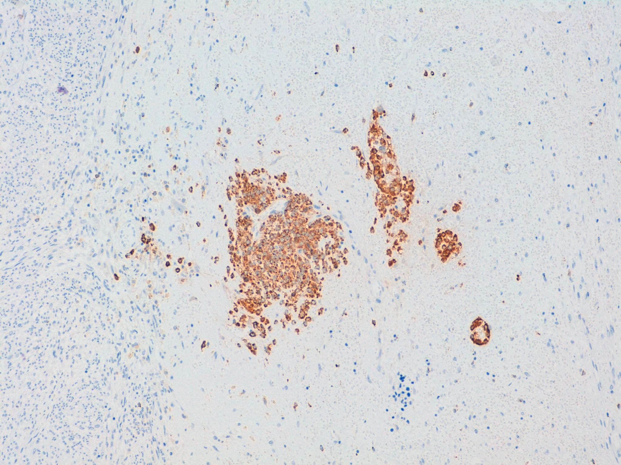
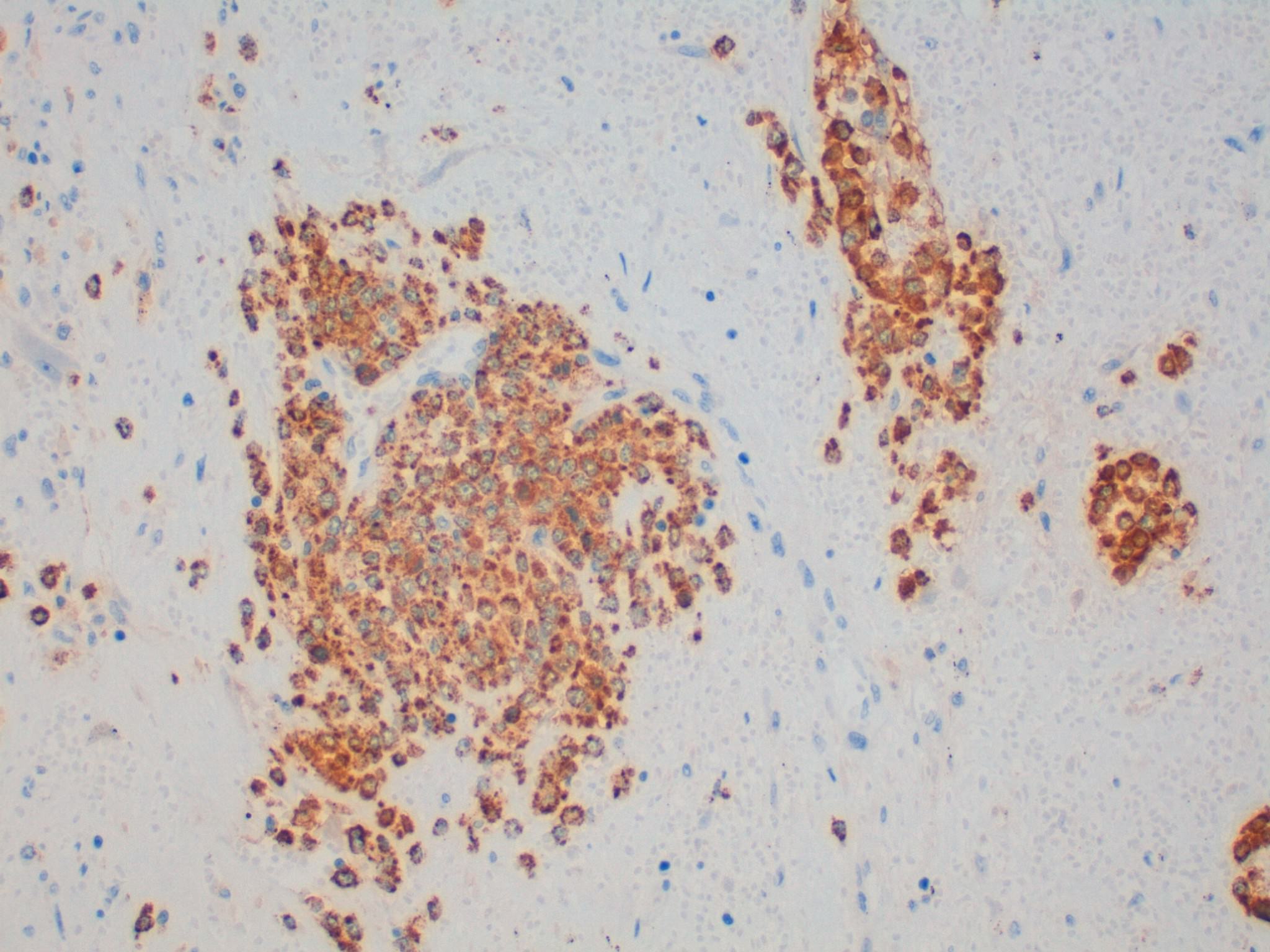
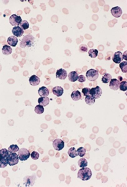
Myelocytes and promyelocytes in extramedullary hematopoiesis (EMH)
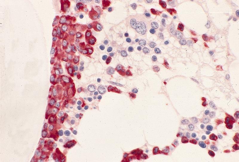
Normal bone trabeculae
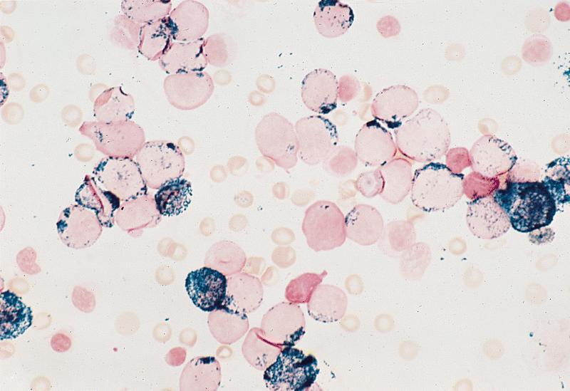
AML M0 is MPO negative
AML M1
AML M2
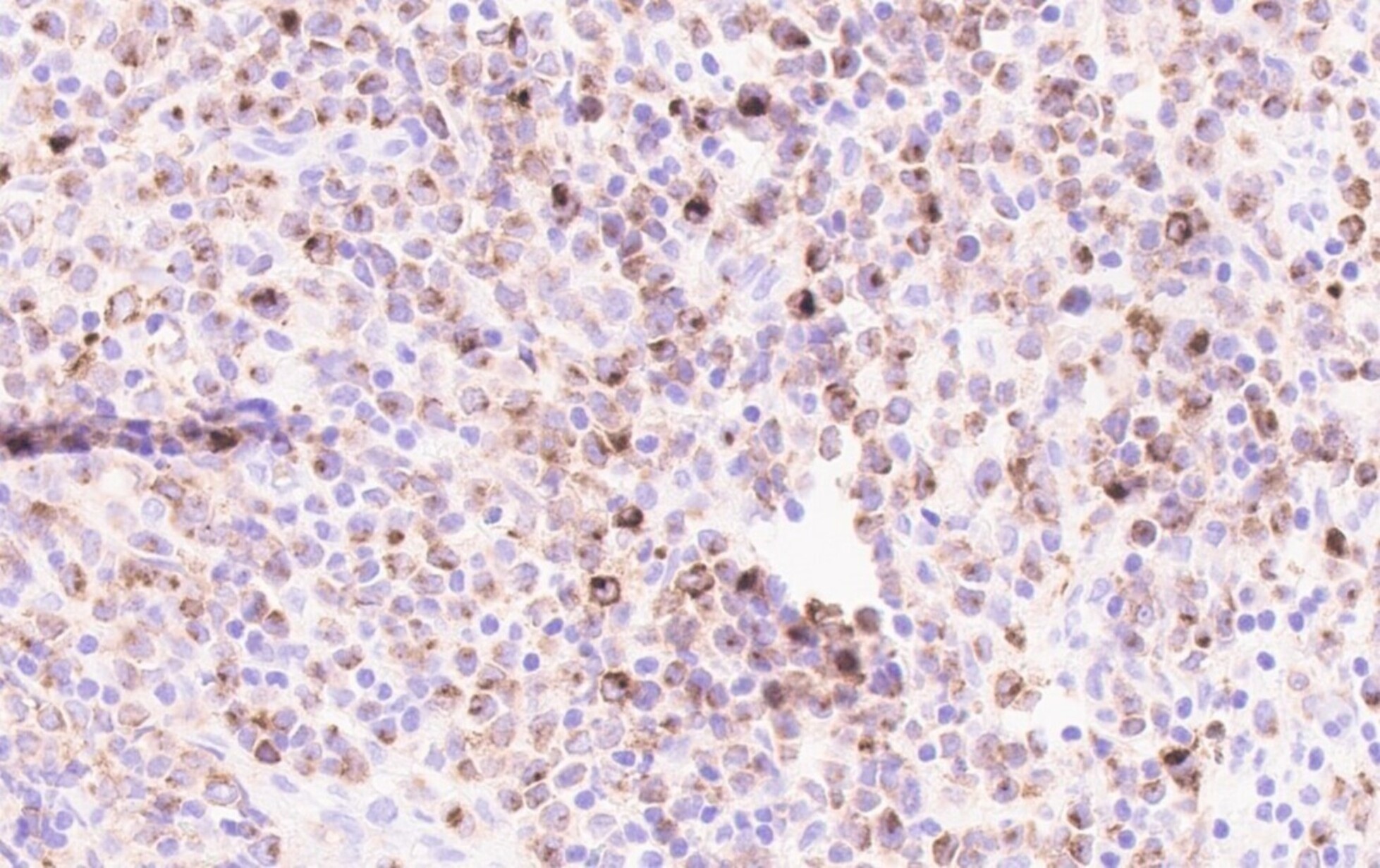
Myeloid sarcoma: myeloblasts are MPO+
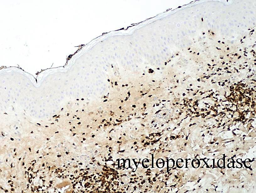
Leukemia cutis

CML: MPO negative
Positive staining - normal
- Myeloid cells: neutrophils (primary granules contain myeloperoxidase), eosinophils, mast cells, myeloblasts
- Reactive neutrophilia
- Monocytes and histiocytes (variable but no granules are present in contrast to myeloid cells)
- Platelets (Free Radic Biol Med 2013;61:357)
Positive staining - disease
- Chédiak-Higashi syndrome (Indian J Hematol Blood Transfus 2014;30:195)
- AML without maturation (FAB AML M1) (may be reduced) (Hematol Pathol 1989;3:153, HemepathReview.com: Acute Myeloid Leukemias (AML) [Accessed 8 February 2024])
- AML with maturation (FAB AML M2) (Blood 2010;115:453)
- APL with PML::RARA (formerly FAB AML M3)
- Acute myelomonocytic leukemia (AMML, FAB AML M4) (in cells of both lineages but may be weaker in monocytic cells) (Orphanet: Acute Myelomonocytic Leukemia [Accessed 26 January 2024])
- AML hypocellular (J Med Case Rep 2009;3:27)
- AML with mutated NPM1 (Clin Case Rep 2015;3:955)
- AML with KAT6A::CREBBP (Case Rep Hematol 2019;2019:4198415)
- AML with inv(3)(q21.3;q26.2) or t(3;3)(q21.3;q26.2); GATA2, MECOM
- AML with inv(16)(p13.1;q22) or t(16;16)(p13.1;q22); CBFB::MYH11 (Mol Cytogenet 2020;13:4)
- Myeloid (granulocytic) sarcoma (Proc (Bayl Univ Med Cent) 2014;27:246)
- Leukemia cutis (Dermatol Online J 2011;17:11)
- Note: myeloid cells, including myeloblasts, are stained in these disorders
Variable staining
- AML with BCR::ABL1 (depends on type of blast present)
- AML with mutated RUNX1 or AML with t(8;21)(q22;q22); RUNX1::RUNX1T1
- Acute panmyelosis with myelofibrosis (Mod Pathol 2005;18:603, Mediterr J Hematol Infect Dis 2013;5:e2013042)
Negative staining - normal
- Lymphocytes (low levels may be present), basophils (may be present in immature forms), megakaryocytes (Cell Immunol 2016;300:18, ScienceDirect: Myeloperoxidase [Accessed 25 January 2024], Free Radic Biol Med 2013;61:357)
- Chronic myeloid leukemia, medullary / bone marrow, chronic myeloid leukemia, extramedullary (negative or weak) (Ann Hematol 2005;84:767)
Negative staining - disease
- Acute lymphoblastic leukemia but isolated MPO expression can be detected, most commonly by IHC but also sometimes by flow cytometry, in a subset of typical B ALL in adults, contributing to misdiagnosis (Asian Pac J Cancer Prev 2021;22:2143)
- Acute basophilic leukemia
- Acute monoblastic and acute monocytic leukemia (AML M5)
- Acute monoblastic leukemia (AML M5a)
- Acute monoblastic / monocytic leukemia (AML M5b) (Front Pediatr 2022;10:911093)
- Acute erythroid leukemia / AML M6
- Acute megakaryoblastic leukemia (AML M7), AML (megakaryoblastic) with t(1;22)(p13.3;q13.1); RBM15::MRTF1
- AML with t(9;11)(p22;q23); KMT2A::MLLT3
- Myeloid leukemia associated with Down syndrome
- Chronic myelomonocytic leukemia (usually)
- Histiocytic sarcoma
- Systemic mastocytosis
- Blastic plasmacytoid dendritic cell neoplasm
- MGUS-IgM
Electron microscopy description
- Blasts in AML with minimal differentiation (FAB AML M0) may show focal myeloperoxidase+ granules
Sample pathology report
- Bone marrow, biopsy:
- Acute myeloid leukemia with mutated NPM1 (see comment)
- Comment: The bone marrow is massively infiltrated by blasts with a myelomonocytic appearance. By immunohistochemistry, they express cytoplasmic NPM1 and coexpress myeloperoxidase and macrophage restricted CD68 but CD34 is negative.
Board review style question #1
Which of the following entities are typically myeloperoxidase positive by immunocytochemistry or enzyme cytochemistry?
- Acute erythroid leukemia (AML M6)
- Acute lymphoblastic leukemia
- AML with minimal differentiation (FAB AML M0)
- AML without maturation (FAB AML M1)
Board review style answer #1
D. AML without maturation (FAB AML M1). In AML without maturation (FAB AML M1), the myeloblasts are positive for myeloperoxidase. Answers A, B and C are incorrect because these entities are typically negative for myeloperoxidase, although acute lymphoblastic leukemia can show isolated myeloperoxidase positivity.
Comment Here
Reference: Myeloperoxidase
Comment Here
Reference: Myeloperoxidase
Board review style question #2
Which normal cells are typically strongly myeloperoxidase positive by immunocytochemistry or enzyme cytochemistry?
- Basophils
- Lymphocytes
- Megakaryocytes
- Neutrophils
Board review style answer #2
D. Neutrophils. Myeloperoxidase strongly stains the primary granules in neutrophils. Answers A, B and C are incorrect because myeloperoxidase does not strongly stain basophils (immature forms are MPO+), lymphocytes (some subsets are weakly positive) or megakaryocytes.
Comment Here
Reference: Myeloperoxidase
Comment Here
Reference: Myeloperoxidase

