
Stains & CD markers
Chromogranin

Copyright: 2003-2024, PathologyOutlines.com, Inc.
PubMed Search: Chromogranin stain
Chromogranin
Author: Nat Pernick, M.D.
Last author update: 1 February 2012
Last staff update: 13 June 2022
Copyright: 2003-2024, PathologyOutlines.com, Inc.
PubMed Search: Chromogranin stain

Table of Contents
Definition / general | Terminology | Uses by pathologists | Microscopic (histologic) images | Positive staining - normal | Positive staining - disease | Negative stainingCite this page: Pernick N. Chromogranin. PathologyOutlines.com website. https://www.pathologyoutlines.com/topic/stainschromogranin.html. Accessed April 25th, 2024.
Definition / general
- Granin protein located in secretory vesicles of neurons and endocrine cells (Clin Invest Med 1995;18:47, Endocr Rev 2011;32:755)
- Immunostain is specific but not sensitive for neuroendocrine cells; more sensitive in well differentiated versus poorly differentiated tumors
- Antibody binds acidic glycoproteins in the soluble fraction of neurosecretory granules
- Serum levels may not be useful for diagnosis but changes in levels may reflect response to therapy or recurrence (Biomarkers 2012;17:186, Folia Biol (Praha) 2011;57:173)
Terminology
- Typically refers to chromogranin A or parathyroid secretory protein 1 (gene name CHGA)
- Chromogranin B is not commonly used and has a different distribution (Mod Pathol 2000;13:140)
Uses by pathologists
- Commonly used neuroendocrine marker (also synaptophysin and CD56) for normal cells and neuroendocrine tumors
- Helps differentiate pheochromocytoma (almost always positive) from adrenocortical carcinoma (almost always negative, Am J Surg Pathol 2010;34:423)
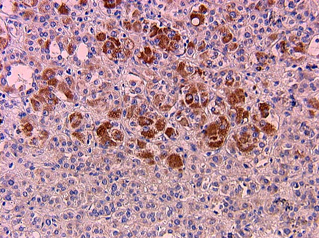
Microscopic (histologic) images
Cases #195, #204, #108 and AFIP images

Bladder: paraganglioma
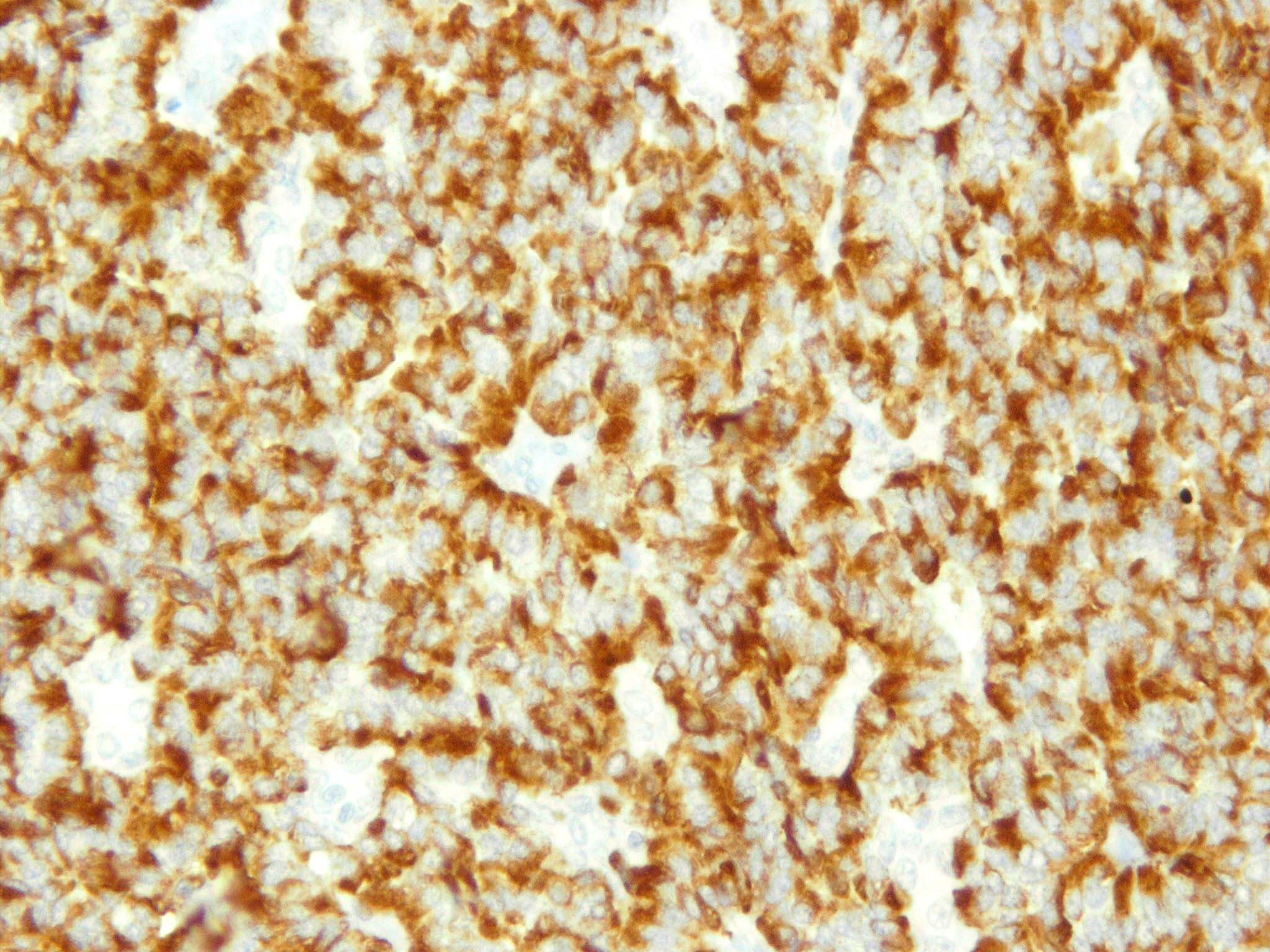
Kidney: carcinoid tumor

Parathyroid gland: ectopic tissue
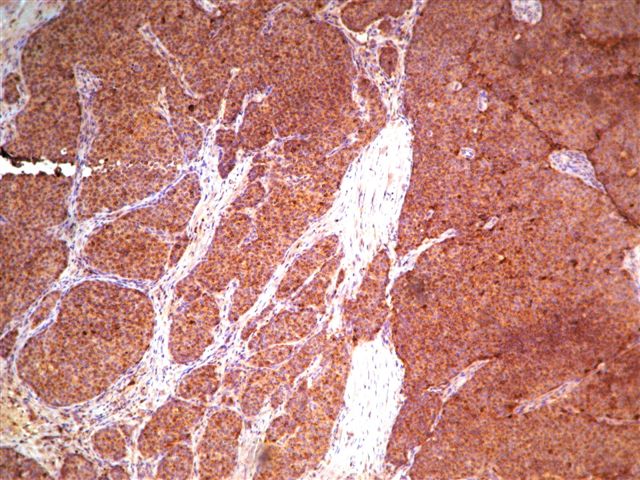
Breast: neuroendocrine carcinomas
Thyroid: medullary carcinoma
Contributed by Jijgee Munkhdelger, M.D., Ph.D. and Andrey Bychkov, M.D., Ph.D.
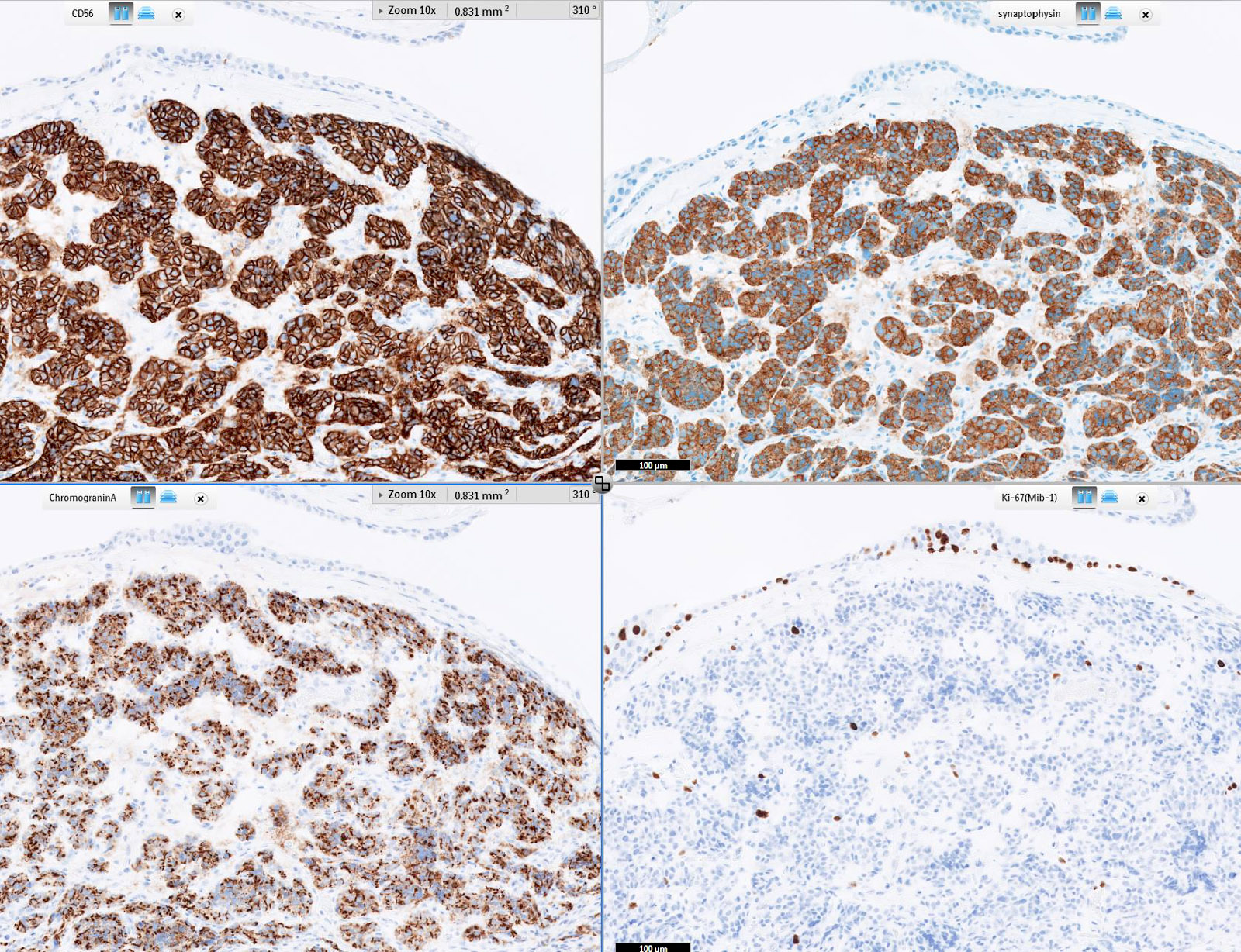
Typical carcinoid immunoprofile
Images hosted on other servers:
Adrenal medulla:
pheochromocytoma

Breast: small cell carcinoma

Heart: metastatic
pheochromocytoma
(right side)
Positive staining - normal
- Neuroendocrine and ganglion cells in adrenal medulla (chromaffin cells), heart AV node, pancreas (islets), parathyroid (chief cells), thyroid (C cells), other tissues
- Also bile ductules-reactive and pancreatic acinar cells (occasional, Am J Surg Pathol 2009;33:66)
Positive staining - disease
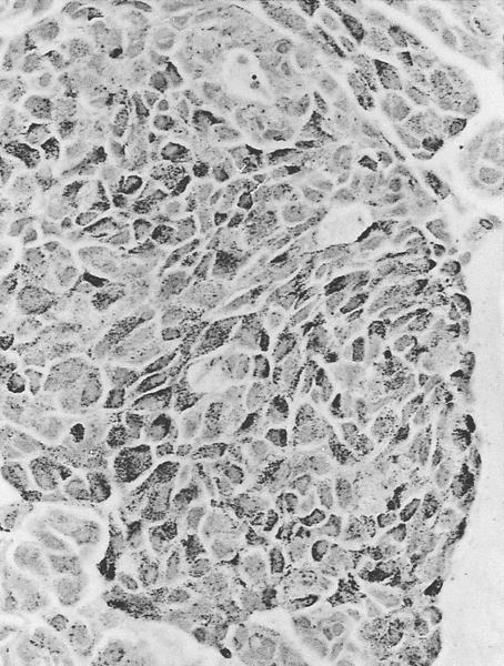
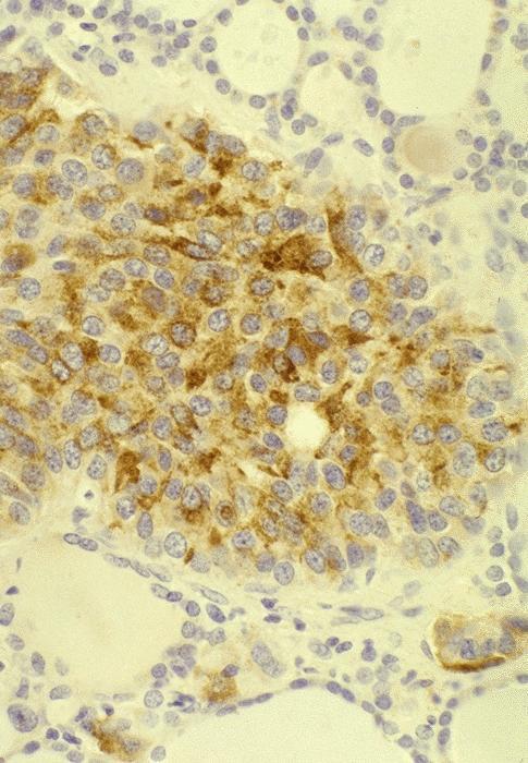
- Neuroendocrine and ganglion cell tumors (carcinoid, Merkel cell carcinoma-lung, neuroblastoma, neuroendocrine carcinoma, paraganglioma, small cell carcinoma), neuroendocrine hyperplasia
- Desmoplastic small cell tumor, middle ear adenoma, parathyroid cyst, pituitary adenoma (Mod Pathol 2002;15:543)
- Many fetal-type tumors (hepatoblastoma, lung adenocarcinoma-fetal type)
- Note: granular cytoplasmic pattern in small cell carcinoma reflects neurosecretory granules
Negative staining
- Tumors without neuroendocrine components including adrenocortical tumors, chordoma, Ewing sarcoma / PNET
- Alveolar rhabdomyosarcoma (may have rare positive cells), pancreatic solid pseudopapillary neoplasm (occasionally focal staining) (Mod Pathol 2008;21:795)

