
Stains & CD markers
H3.3 G34W (H3F3A)

Copyright: 2023-2024, PathologyOutlines.com, Inc.
PubMed Search: H3.3 G34W
H3.3 G34W (H3F3A)
Author: Jose G. Mantilla, M.D.
Editorial Board Member: Christian M. Schürch, M.D., Ph.D.
Deputy Editor-in-Chief: Borislav A. Alexiev, M.D.
Last author update: 20 February 2024
Last staff update: 29 April 2024
Copyright: 2023-2024, PathologyOutlines.com, Inc.
PubMed Search: H3.3 G34W

Table of Contents
Definition / general | Essential features | Terminology | Pathophysiology | Interpretation | Uses by pathologists | Microscopic (histologic) images | Positive staining - disease | Negative staining | Molecular / cytogenetics description | Sample pathology report | Board review style question #1 | Board review style answer #1 | Board review style question #2 | Board review style answer #2Cite this page: Mantilla JG. H3.3 G34W (H3F3A). PathologyOutlines.com website. https://www.pathologyoutlines.com/topic/stainsH3F3AG34W.html. Accessed May 11th, 2024.
Definition / general
- Mutation specific immunohistochemical stain directed against the G34W mutation in histone 3F3A
- Useful to support the diagnosis of giant cell tumor of bone
Essential features
- Mutations in H3F3A in position G34 are present in up to 96% of giant cell tumors of bone
- Most common mutation is G34W
- Antibody directed to the G34W mutant protein is a robust immunohistochemical marker to support this diagnosis (Cancer Cytopathol 2018;126:552, Am J Surg Pathol 2017;41:1059)
- Strong nuclear stain in neoplastic mononuclear cells
- No expression of G34W has been reported in other giant cell rich histologic mimics
Terminology
- H3F3A G34W
Pathophysiology
- Point mutations in the glycine 34 position in H3F3A have been reported in up to 96% of giant cell tumors of bone
- The most common mutation in these tumors, by far, is G34W (J Pathol Clin Res 2015;1:113, Nat Genet 2013;45:1479)
- These mutations affect histone methylation
Interpretation
- Strong and diffuse nuclear staining in neoplastic mononuclear cells
- Giant cells are usually negative
Uses by pathologists
- Nuclear immunoreactivity helps support the diagnosis of giant cell tumor of bone, including lesions formerly designated as benign fibrous histiocytoma of bone
- Sensitivity: 92 - 96%; specificity: 100% (Nat Genet 2013;45:1479, J Pathol Clin Res 2015;1:113)
- Not typically expressed in other giant cell rich lesions of bone such as chondroblastoma, aneurysmal bone cyst or others
- Not expressed in central giant cell granuloma (Oral Surg Oral Med Oral Pathol Oral Radiol 2014;118:583)
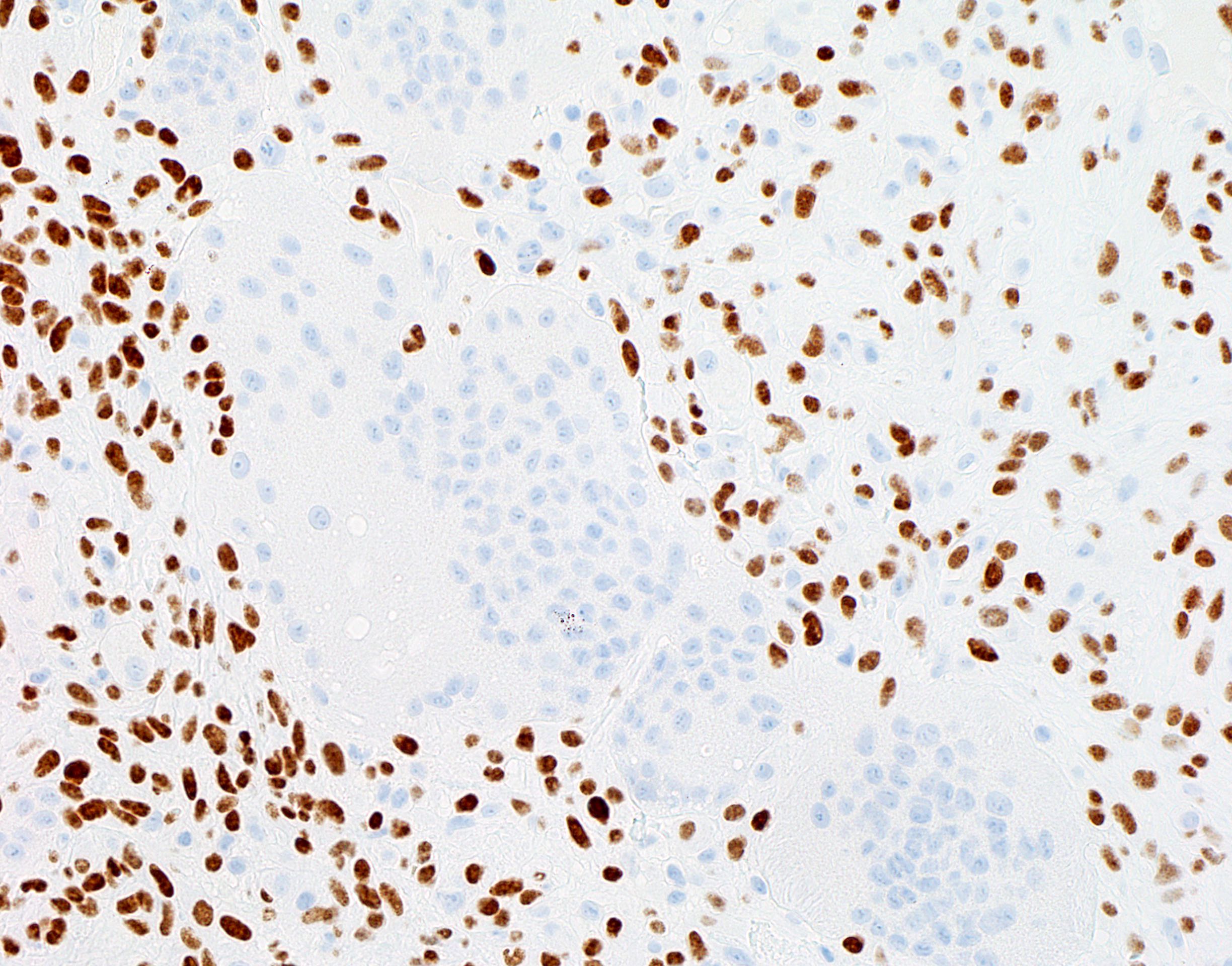
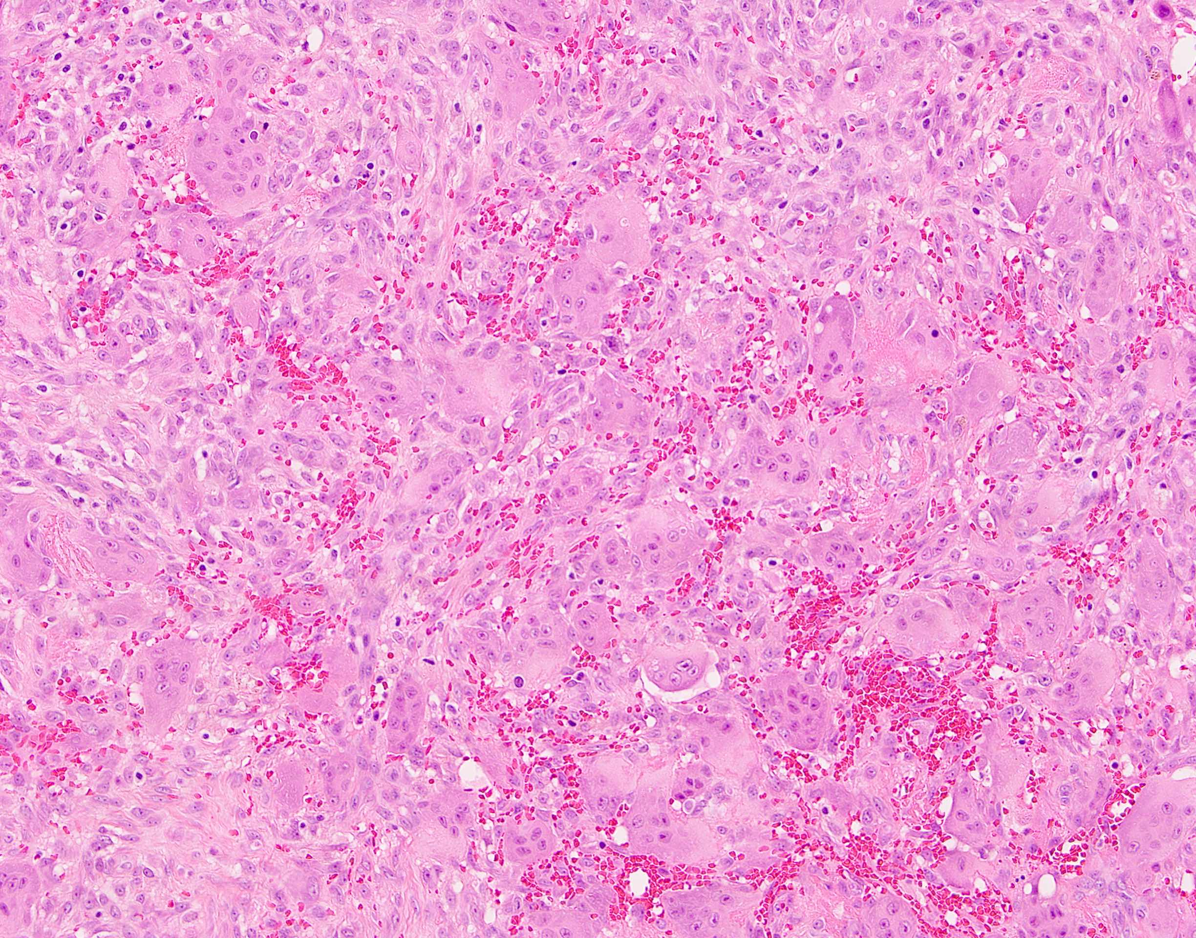
Microscopic (histologic) images
Contributed by Jose G. Mantilla, M.D.


Giant cell tumor of bone
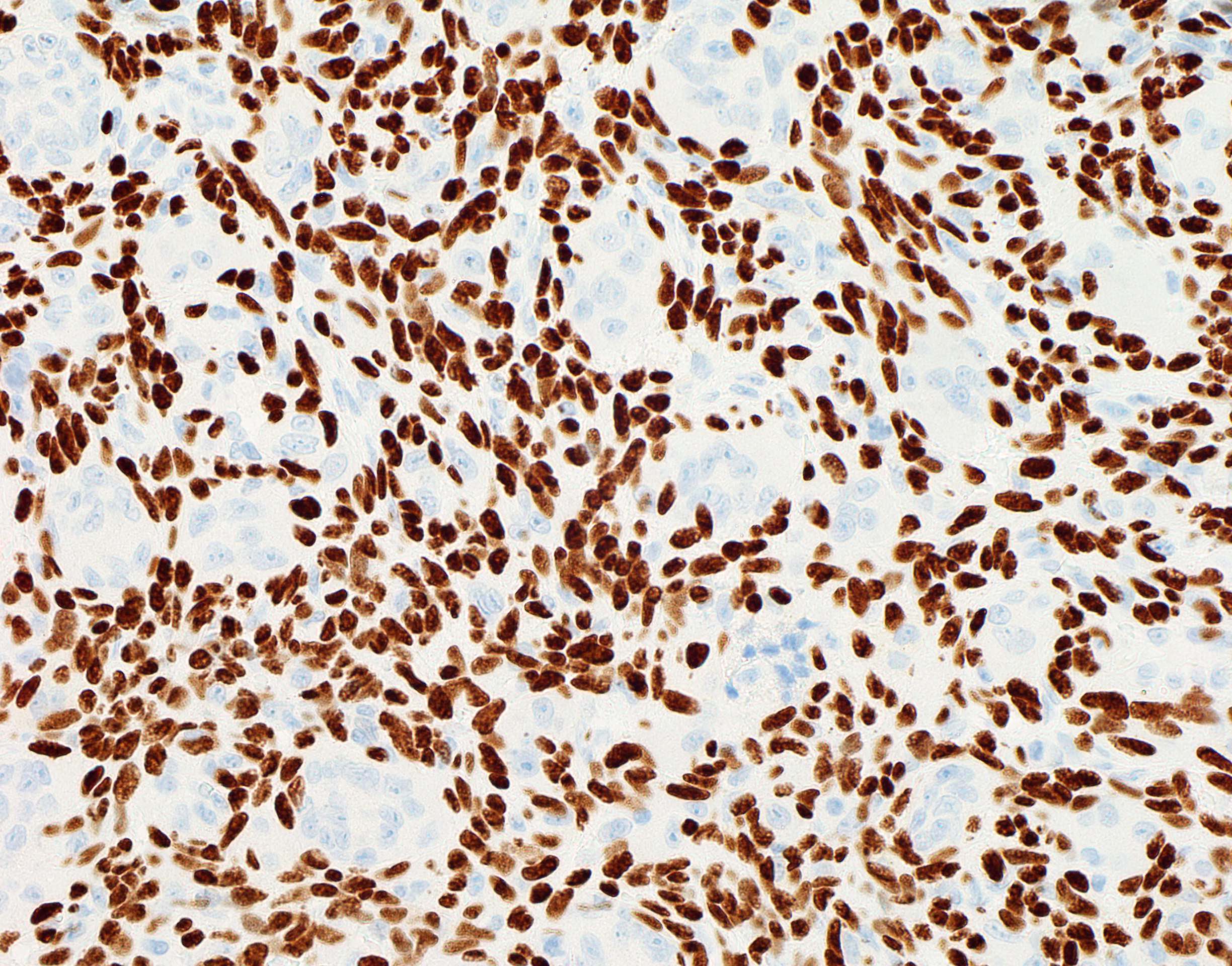
G34W reactivity in giant cell tumor

Giant cell poor giant cell tumor of bone
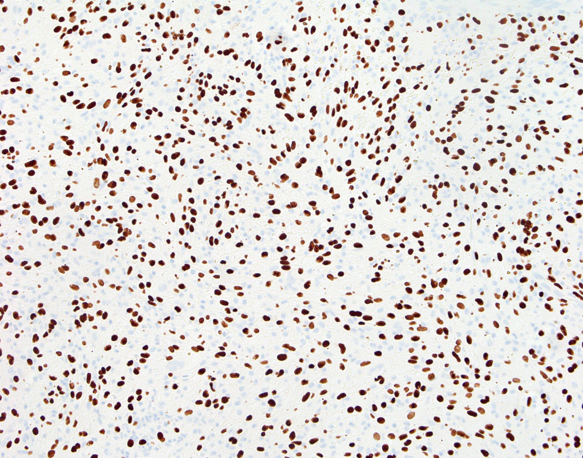
G34W reactivity in giant cell poor giant cell tumor
Positive staining - disease
- Giant cell tumor of bone (92 - 96% of cases)
Negative staining
- Primary aneurysmal bone cyst (0%) (J Pathol Clin Res 2015;1:113)
- Chondroblastoma (0%) (J Pathol Clin Res 2015;1:113, Nat Genet 2013;45:1479)
- Central giant cell granuloma (0%) (Oral Surg Oral Med Oral Pathol Oral Radiol 2014;118:583, J Pathol Clin Res 2015;1:113)
Molecular / cytogenetics description
- See Pathophysiology
Sample pathology report
- Distal femur lesion, biopsy:
- Giant cell tumor of bone (see comment)
- Comment: Histologic sections contain a neoplasm composed of cytologically bland mononuclear cells in a syncytial growth pattern accompanied by abundant multinucleated giant cells, which share similar cytologic features with the mononuclear cell population. No overt features of malignancy are identified. A G34W immunohistochemical stain is positive. These findings support the diagnosis of giant cell tumor of bone.
Board review style question #1
A radiolucent lesion in the mandible of a 38 year old man was biopsied and is shown in the image above. An H3.3 G34W immunohistochemical stain is negative. What is the correct diagnosis?
- Central giant cell granuloma
- Chondroblastoma
- Conventional type osteosarcoma
- Giant cell tumor of bone
- Solid aneurysmal bone cyst
Board review style answer #1
A. Central giant cell granuloma. Although this lesion has similar morphologic features to those of giant cell tumor of bone, it is an indolent lesion with different biological features. Its characteristic location and absence of G34W reactivity help support this diagnosis (Oral Surg Oral Med Oral Pathol Oral Radiol 2014;118:583).
Answer B is incorrect because the histologic features in the image are not representative of chondroblastoma, which is composed of cytologically bland, clear to epithelioid cells and may have a characteristic pattern of pericellular calcification. Giant cells are common. Answer C is incorrect because no features of malignancy are present in the image. Answer D is incorrect because giant cell tumor of bone has similar histologic features; however, most cases express G34W and typically arise in the epiphysis of long bones. Answer E is incorrect because although aneurysmal bone cysts may have prominent giant cells, the lesion lacks histologic features diagnostic of an aneurysmal bone cyst (e.g., aneurysmal spaces, characteristic myofibroblastic population).
Comment Here
Reference: H3.3 G34W (H3F3A)
Comment Here
Reference: H3.3 G34W (H3F3A)
Board review style question #2
Which of the following diseases has been reported to harbor point mutations in the histone 3.3 genes?
- Conventional type osteosarcoma
- Epithelioid sarcoma, proximal type
- Giant cell tumor of bone
- Malignant peripheral nerve sheath tumor
- Undifferentiated SMARCA4 deficient thoracic malignant neoplasm
Board review style answer #2
C. Giant cell tumor of bone. Point mutations in H3.3 have been described in the majority of cases of giant cell tumor of bone and chondroblastoma (Nat Genet 2013;45:1479). Answers D, B and E are incorrect because although alterations in the polycomb repressive complex 2 (PRC2) complex genes (as seen in malignant peripheral nerve sheath tumor) and switch / sucrose nonfermenting complex (SWI / SNF) (seen in both epithelioid sarcoma and undifferentiated SMARCA4 deficient thoracic neoplasm) lead to alterations in histone methylation, these are not due to intrinsic mutations in the histone genes. Answer A is incorrect because conventional type osteosarcoma lacks recurrent genetic alterations.
Comment Here
Reference: H3.3 G34W (H3F3A)
Comment Here
Reference: H3.3 G34W (H3F3A)

