

Soft tissue
Fibroblastic / myofibroblastic
Ischemic fasciitis
Author: Jerad M. Gardner, M.D.

Last author update: 1 June 2013
Last staff update: 26 July 2023
Copyright: 2002-2024, PathologyOutlines.com, Inc.
PubMed Search: Ischemic fasciitis
Table of Contents
Definition / general | Epidemiology | Sites | Etiology | Case reports | Treatment | Clinical images | Gross description | Microscopic (histologic) description | Microscopic (histologic) images | Cytology description | Positive stains | Negative stains | Differential diagnosis | Additional referencesCite this page: Chaudhri, A. Ischemic fasciitis. PathologyOutlines.com website. https://www.pathologyoutlines.com/topic/softtissueischemicfasciitis.html. Accessed April 27th, 2024.
Definition / general
- Painless pseudosarcomatous fibroblastic proliferation in soft tissue overlying bony prominences subject to intermittent pressure-induced ischemia
- Also spelled "ischaemic"
- First described in 1992 as "atypical decubital fibroplasia" (Am J Surg Pathol 1992;16:708)
Epidemiology
- Occurs primarily in immobilized and physically impaired patients (i.e., wheelchair bound, bedridden), with mean age of presentation 70 - 90 years
- Also occurs in younger patients not debilitated (Am J Surg Pathol 2008;32:1546) or patients with physical disabilities (Path Int 1998;48:160, Int J Gynecol Pathol 2004;23:65)
- Patients present with painless mass slowly growing over 3 weeks to 6 months
Sites
- Shoulder, chest wall, sacrococcygeal region, greater trochanter
Etiology
- Similar to decubitus ulcers; pressure-induced ischemia causes mass-producing reactive process, but is insufficient to cause skin ulceration
Case reports
- 10 year old girl with firm palpable abdominal mass associated with a torso brace for scoliosis (Dermatol Online J 2012;18:9)
- 45 year old woman with post-mastectomy axillary mass (The Internet Journal of Pathology 2008;7:1)
- 55 year old man with hip mass (Case of the Week #64)
- 76 year old woman with thigh mass (Arch Pathol Lab Med 2004;128:e139)
Treatment
- Local excision is curative, although may recur due to continuation of underlying ischemia and injury
Clinical images
Contributed by Mark R. Wick, M.D.
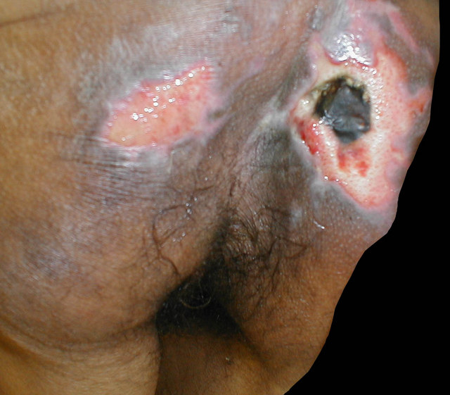
Decubital
Gross description
- Usually 1 - 8 cm, poorly circumscribed, often myxoid
- Usually involves deep subcutis, may involve adjacent skeletal muscle and fascia
- Ulceration is uncommon (i.e. overlying skin is intact)
Microscopic (histologic) description
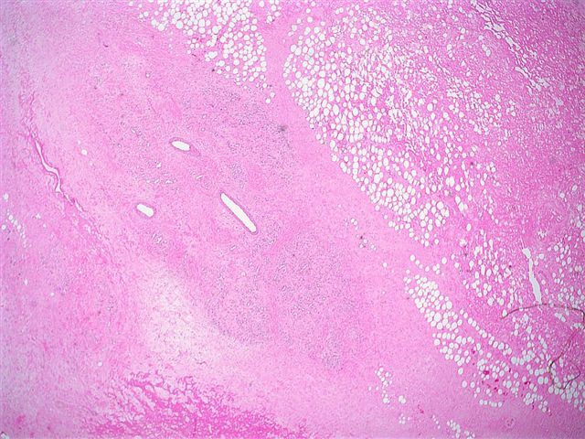
- Zonal pattern of central fibrinoid necrosis with uneven borders staining deep red / violet and prominent myxoid areas surrounded by ectatic, thin walled vessels and proliferating fibroblasts
- Endothelial cells may be atypical
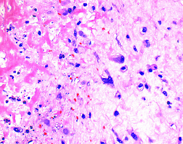
- Fibroblasts have degenerative features with abundant, eosinophilic to amphophilic cytoplasm, enlarged nuclei with smudged chromatin and prominent nucleoli (resembling ganglion-like cells in proliferative fasciitis)
- Variable mitotic activity, but no atypical mitotic figures
- Fibrin thrombi are common within peripheral vessels, which may show fibrinoid necrosis and recanalization but no true vasculitis
- May have multivacuolated macrophages in myxoid zones mimicking lipoblasts
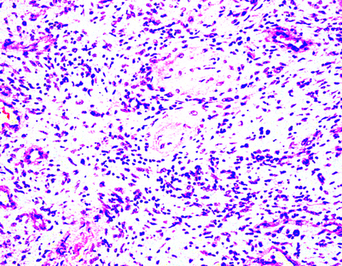
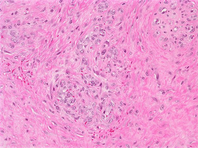
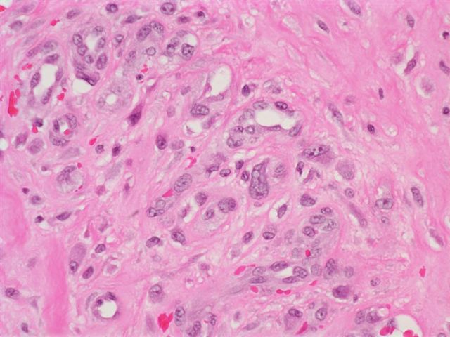
Microscopic (histologic) images
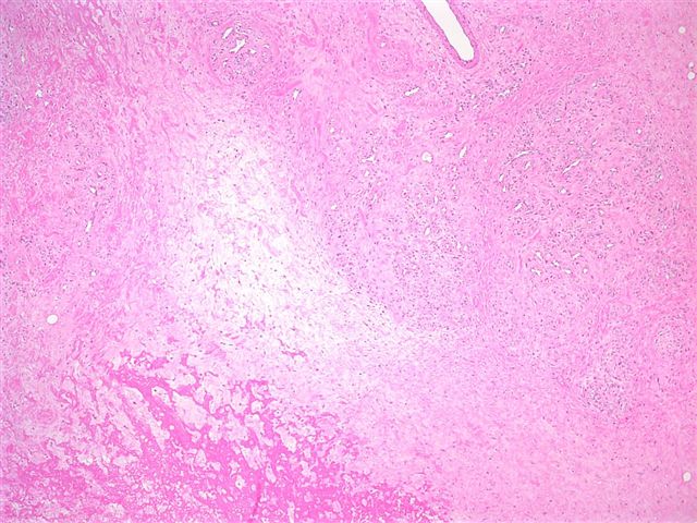
Contributed by Mark R. Wick, M.D., AFIP and Ronald Angeles, M.D. (Case #64)
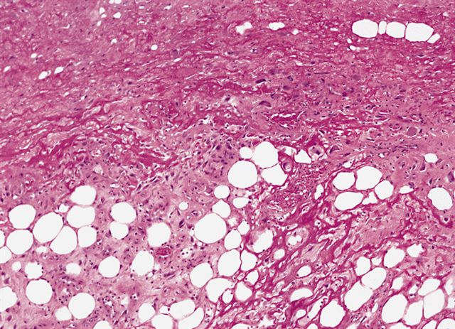
Cellular, fibrin rich proliferation
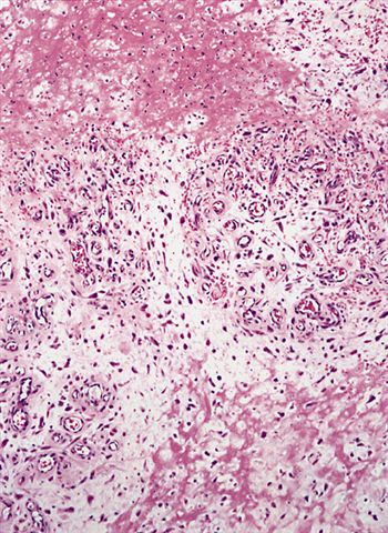
Hyalinized focus

Foci of ganglion-like cells
Decubital
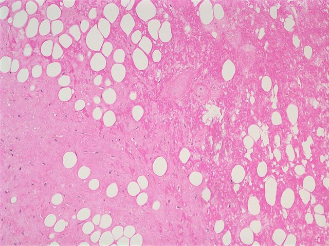

Hip mass in 55 year old bedridden man
Cytology description
- Spindled and ovoid cells with ample cytoplasm and occasional nuclear atypia (Acta Cytol 1997;41:598)
Differential diagnosis
- Epithelioid sarcoma:
- Young adults on distal extremities
- More cellular with central tumor cell necrosis
- Cells have eosinophilic cytoplasm
- Atypical mitotic figures
- Keratin+
- Myxofibrosarcoma:
- Marked atypia but no smudgy chromatin or fibrin thrombi
- Lacks zonation and degenerative features
- Myxoid liposarcoma:
- Prominent plexiform vasculature and lipoblasts
- Small monotonous cells rather than larger myofibroblasts
- Proliferative fasciitis:
- Younger patients
- Lesions not associated with pressure; zonation, myofibroblasts and fibroblasts with tissue culture type growth, also large ganglion cells
