

Soft tissue
Fibroblastic / myofibroblastic
Fibromatosis
Inclusion body fibromatosis
Author: Komal Arora, M.D.

Last author update: 1 July 2012
Last staff update: 26 July 2023
Copyright: 2002-2024, PathologyOutlines.com, Inc.
PubMed Search: Inclusion body fibromatosis
Table of Contents
Definition / general | Sites | Clinical features | Case reports | Treatment | Clinical images | Gross description | Microscopic (histologic) description | Microscopic (histologic) images | Cytology description | Positive stains | Negative stains | Electron microscopy description | Differential diagnosis | Additional referencesCite this page: Arora K. Inclusion body fibromatosis. PathologyOutlines.com website. https://www.pathologyoutlines.com/topic/softtissueinfantiledigitalfibromatosis.html. Accessed April 25th, 2024.
Definition / general
- Dermal fibroblastic and myofibroblastic lesion with cytoplasmic eosinophilic inclusions, usually in digits of infants
- Also called infantile digital fibromatosis, infantile digital fibroma (J Hand Surg Am 1995;20:1014)
- Distinct lesion from classic fibromatosis (Am J Surg Pathol 2009;33:1)
Sites
- Usually exterior surface of distal phalanges of fingers and toes, but not thumb or great toe, also oral cavity and breast
- 50% recur, do not metastasize
- Similar inclusions reported in breast fibroadenoma (Arch Pathol Lab Med 2007;131:1126), breast phyllodes tumor (Am J Surg Pathol 1994;18:506, Breast J 2008;14:198), cervical polyp (Pathology 1998;30:215), GI leiomyomas (Cesk Patol 2006;42:139)
Clinical features
- Rare; lesions usually present at birth or in first 2 years
- Similar lesions in adults
- Often are multiple
Case reports
- 6 month old girl (Indian J Pathol Microbiol 2010;53:827)
- 1 year old boy with spontaneous regression (J Dermatol 1998;25:523)
- 2 year old with post-surgical involvement of all 4 extremities (Ann Plast Surg 2008;61:472)
Treatment
- Conservative excision (preserve function because recurrences are not destructive and tumors do not metastasize) or watchful waiting (Am J Surg Pathol 2009;33:1, Pediatr Dermatol 2008;25:72)
Clinical images
Contributed by Mark R. Wick, M.D.
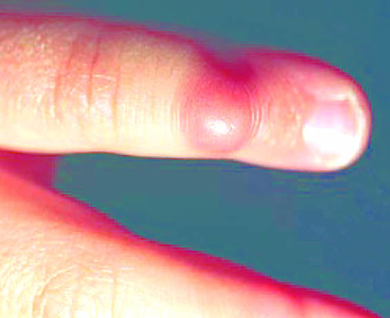
Images hosted on other servers:

Various images
Gross description
- Nodules with stretched overlying skin, lesions are ill defined, white-tan, usually 2 cm or less
- No hemorrhage or necrosis
Microscopic (histologic) description
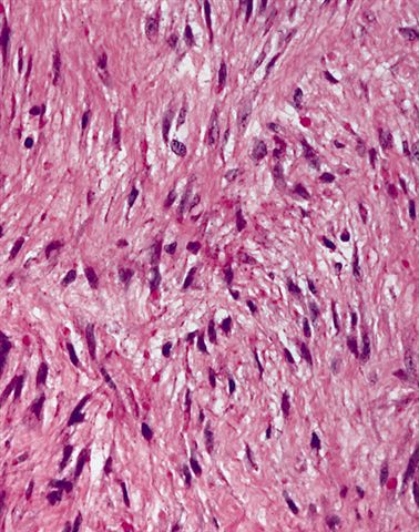
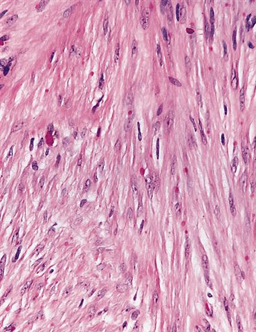
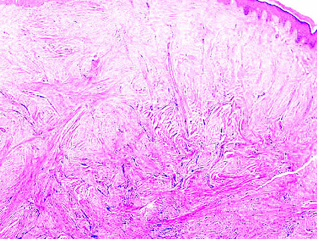
- Nonencapsulated, dermal proliferation of hypocellular sheets or fascicles of fibroblasts and myofibroblasts with variable collagen
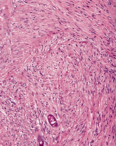
- Some spindle cells have peculiar eosinophilic (hyaline) cytoplasmic inclusions the size of a lymphocyte nucleus
- Usually mitotic figures
- May infiltrate into adjacent tissue
- No atypia
Microscopic (histologic) images
Contributed by Mark R. Wick, M.D. and AFIP
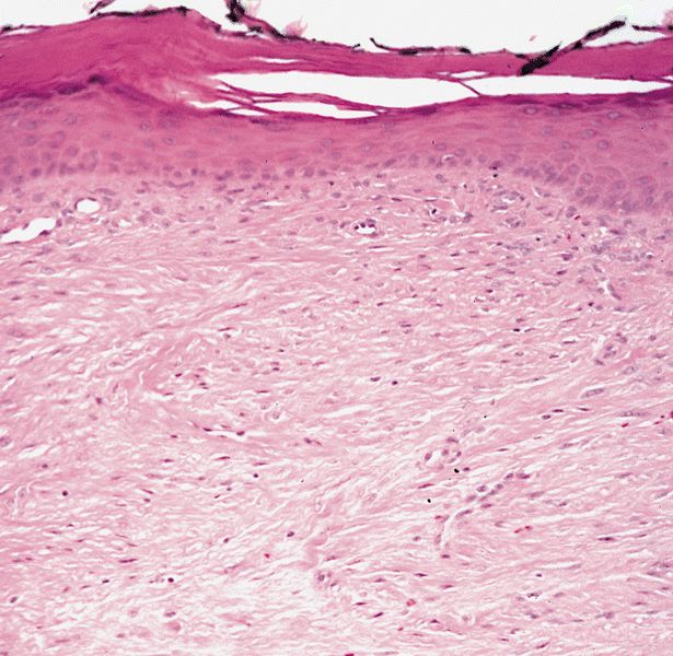
Proliferation
Fibroblastic cells
Cells are bland and monomorphic
Inclusions resemble red blood cells
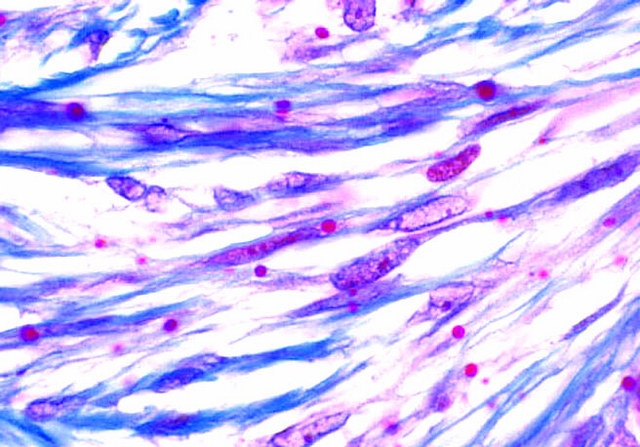

Cytoplasmic inclusions
Various images
Images hosted on other servers:

Various images
Cytology description
Positive stains
Negative stains
- Inclusions: PAS
- Spindle cells: keratin, ER, PR, beta-catenin
Electron microscopy description
- Spindle cells are myofibroblasts with rough endoplasmic reticulum and free lying inclusions composed of compact masses of actin granules and filaments without a limiting membrane (Am J Pathol 1979;94:19)
Differential diagnosis
- Infantile fibrosarcoma:
- Not digits, usually > 2 cm, more cellular, chromatin is denser and more irregular, more mitotic figures, no inclusions
- Infantile desmoid fibromatosis:
- Rare on hand, usually > 2 cm, more cellular, no inclusions
