Cite this page: Shankar V. Rhabdomyoma. PathologyOutlines.com website. https://www.pathologyoutlines.com/topic/softtissueadultrhabdomyoma.html. Accessed January 18th, 2025.
Adult type rhabdomyoma
Definition / general
Clinical features
Case reports
Treatment
Clinical images
Contributed by Mark R. Wick, M.D.
Gross description
Gross images
Contributed by Mark R. Wick, M.D.
Microscopic (histologic) description
Microscopic (histologic) images
Contributed by Mark R. Wick, M.D. and AFIP
Cytology description
Cytology images
Images hosted on other servers:
Positive stains
Negative stains
Electron microscopy description
Differential diagnosis
- Benign tumor of mature skeletal muscle
- Extracardiac rhabdomyomas are divided into fetal, adult and genital histologic types (eMedicine: Rhabdomyomas [Accessed 26 January 2023])
- Extracardiac tumors are not associated with tuberous sclerosis
- Some cases may be due to degeneration / regeneration and not be neoplastic (Am J Surg Pathol 1989;13:791, Head Neck 2006;28:275
Clinical features
- Very rare
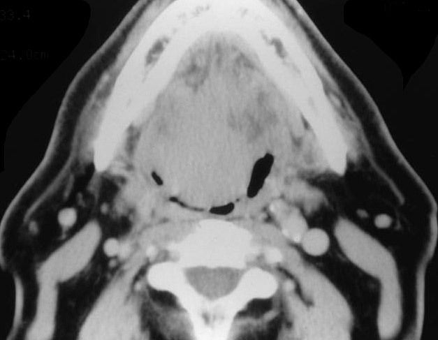
- Usually head and neck, particularly oral cavity
- Median age 60 years, 75% male (Hum Pathol 1993;24:608)
- May be multifocal (25%)
Case reports
- 43 year old man with diagnosis of adult rhabdomyoma by fine needle aspiration cytology (Acta Cytol 2010;54:968)
- Elderly woman with intraoral multifocal adult rhabdomyoma (Arch Pathol Lab Med 1983;107:638)
- Adult rhabdomyoma of the extremity (Hum Pathol 2000;31:1074)
- Rhabdomyoma of the parapharyngeal space presenting with dysphagia (Dysphagia 2008;23:202)
Treatment
- Excision is curative but may recur if incompletely excised
Clinical images
Contributed by Mark R. Wick, M.D.
Gross description
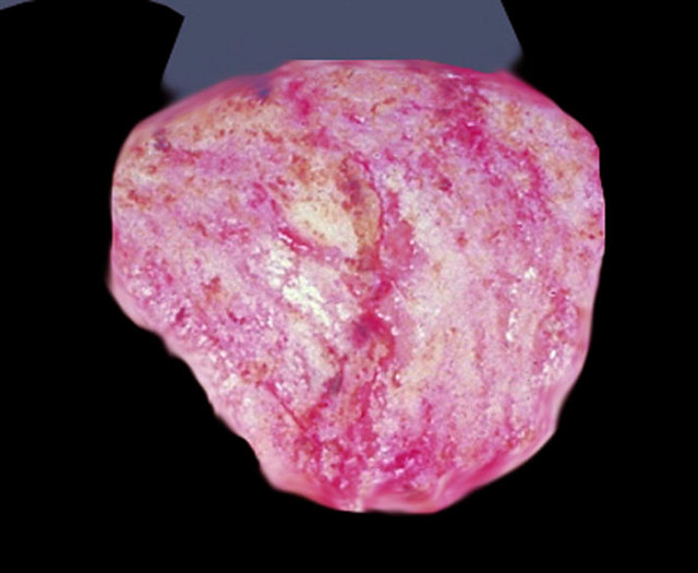
- Median 3 cm, circumscribed, soft, tan-red-brown
- Nodular or lobulated
Gross images
Contributed by Mark R. Wick, M.D.
Microscopic (histologic) description
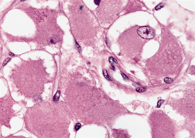
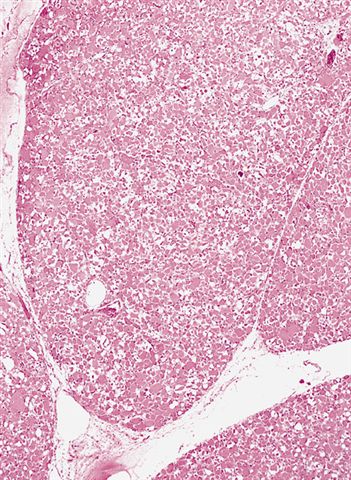
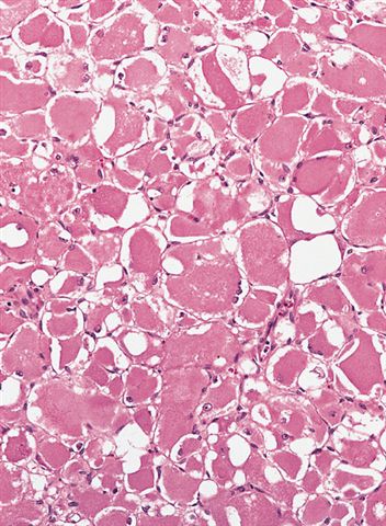
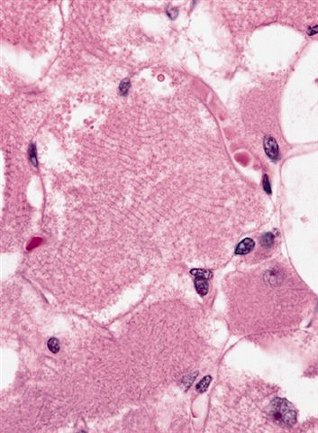
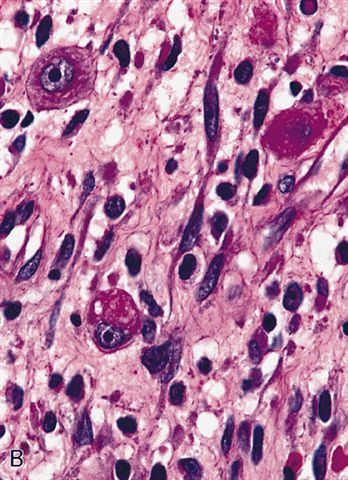
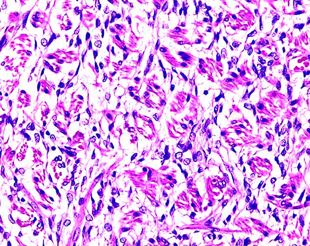
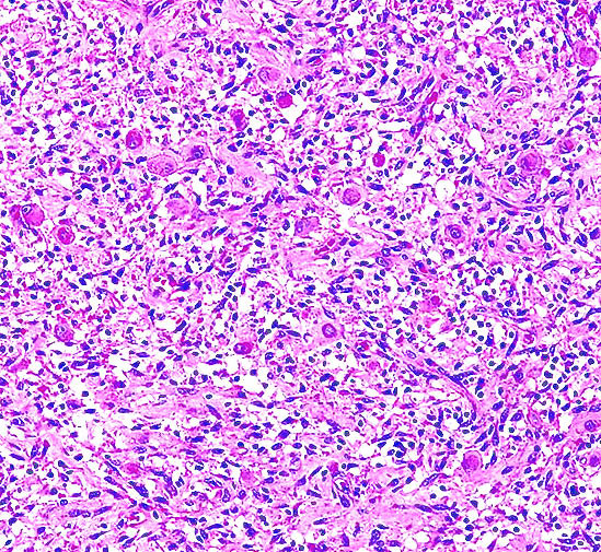
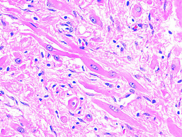
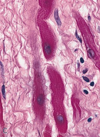
- Well circumscribed, not encapsulated, sheets of large, well differentiated skeletal muscle cells
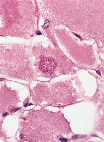
- Cells are round or polygonal with abundant eosinophilic fibrillar or granular cytoplasm with frequent cross striations and intracytoplasmic rod-like inclusions
- Nuclei are small, round and vesicular, may have prominent nucleoli
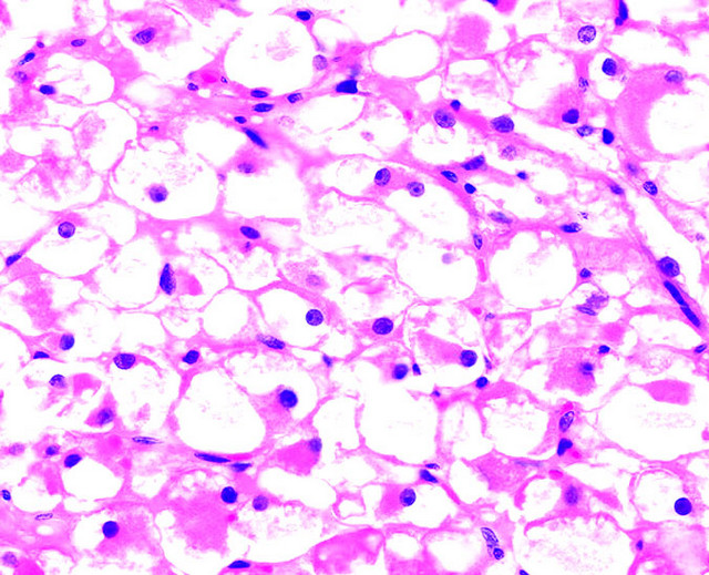
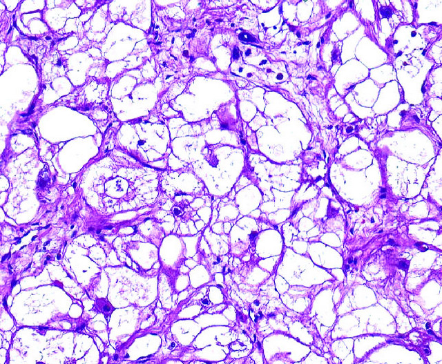
- May have spider cells with vacuolated cytoplasm (cells resemble spider webs)
- Variable glycogen and lipid
- No mitotic activity, no atypia
Microscopic (histologic) images
Contributed by Mark R. Wick, M.D. and AFIP
Cytology description
- Fragments of tumor cells, which are large, polygonal cells
- Cytoplasm is eosinophilic and finely granular, may resemble granular cell tumor, which is S100+, muscle markers- (Diagn Cytopathol 2009;37:483)
- Eccentrically placed nuclei
- Cross striations and inclusions are not conspicuous (Acta Cytol 2010;54:968)
Cytology images
Images hosted on other servers:
Positive stains
- Muscle specific actin, desmin and myoglobin (100%)
- PAS+, diastase sensitive (detects glycogen)
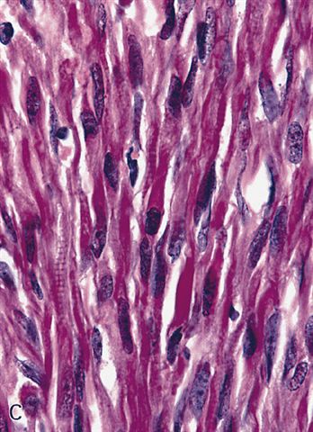
- PTAH and Masson trichrome highlight cross striations and rod-like inclusions
Negative stains
Electron microscopy description
- Myofilaments, Z bands, glycogen granules
Differential diagnosis
- Alveolar soft part sarcoma
- Crystal storing histiocytosis
- Granular cell tumor:
- No skeletal muscle differentiation, no glycogen, smaller cells have poorly defined cell borders, often overlying pseudoepitheliomatous hyperplasia
- S100+
- Hibernoma:
- No skeletal muscle differentiation, no glycogen
- Paraganglioma:
- NSE+, synaptophysin+, chromogranin+
- Well differentiated rhabdomyosarcoma
Fetal type rhabdomyoma
Definition / general
Epidemiology
Sites
Case reports
Treatment
Gross description
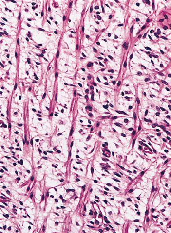
Microscopic (histologic) description
Microscopic (histologic) images
Contributed by Mark R. Wick, M.D. and AFIP
Cytology description
Positive stains
Negative stains
Electron microscopy description
Differential diagnosis
- Rare benign tumor of immature skeletal muscle differentiation, usually in the head and neck
- Retroauricular in ages 0 - 3 years
Epidemiology
- 70% male, median age 4.5 years, range 3 - 58 years (Hum Pathol 1993;24:754)
- M > F
- Multiple cases of fetal rhabdomyoma have been reported in patients with nevoid basal cell carcinoma syndrome (Virchows Arch 2011;459:235)
Sites
- Usually head and neck; post auricular region is the most common site
Case reports
- 1 month old infant girl with swelling in submandibular region (Indian Pediatr 2004;41:839)
- 1 year old girl with skin tumor (Am J Surg Pathol 2008;32:485)
- 9 year old boy with cellular fetal rhabdomyoma of thigh (Pediatr Blood Cancer 2009;52:881)
- Congenital tumor with recurrence (Int J Pediatr Otorhinolaryngol 2006;70:1115)
Treatment
- Complete excision
- Only rare recurrences (Pediatr Pathol Lab Med 1996;16:673)
- No metastases
Gross description
- Median 3 - 5 cm
- Solitary, well circumscribed mass of soft tissue or mucosa
- Gray-white-tan-pink, soft with glistening cut surface
Microscopic (histologic) description
- Circumscribed but not encapsulated
- Myxoid:
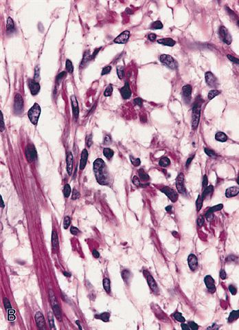
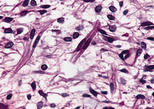
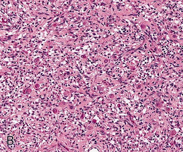
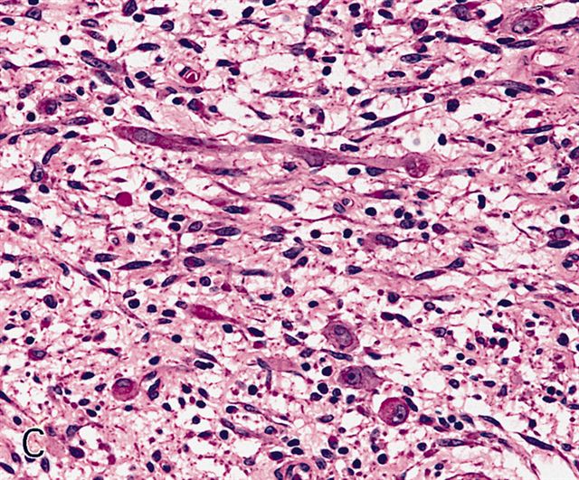
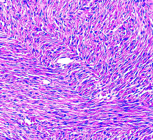
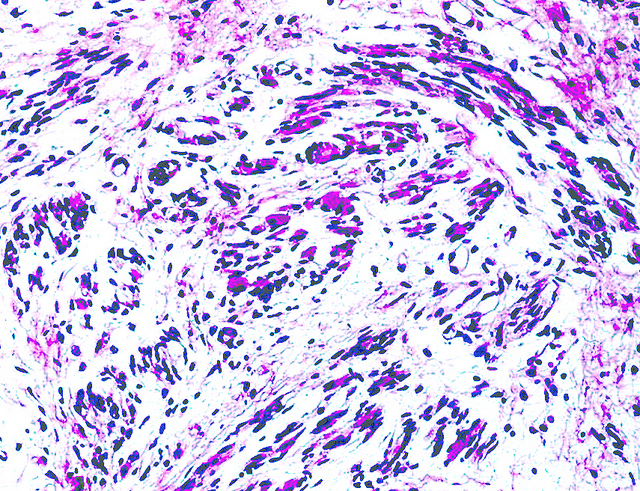
- Bundles or fascicles of immature slender skeletal muscle with delicate cytoplasmic cross striations and thin tapering eosinophilic processes, resembling myotubules at week 7 - 12 of gestation
- Also undifferentiated round / oval or spindled mesenchymal cells
- Stroma is myxoid or fibromyxoid
- Skeletal muscle cells mature towards periphery, may have pseudocambium layer of plasma cells and lymphocytes under mucosal epithelium
- Cellular:
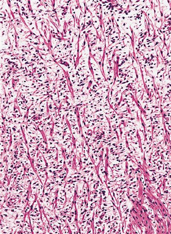
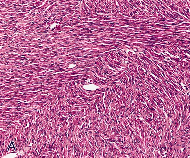
- Bundles or fascicles of cells in parallel or plexiform patterns
- Sparse collagenous or myxoid stroma
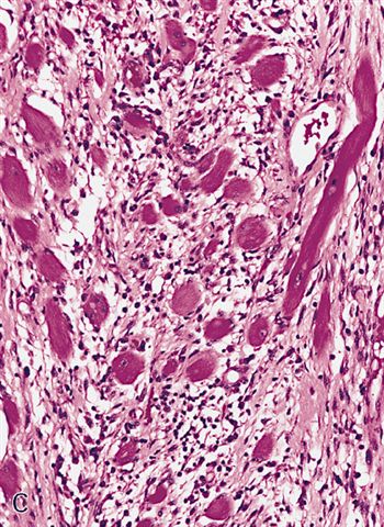
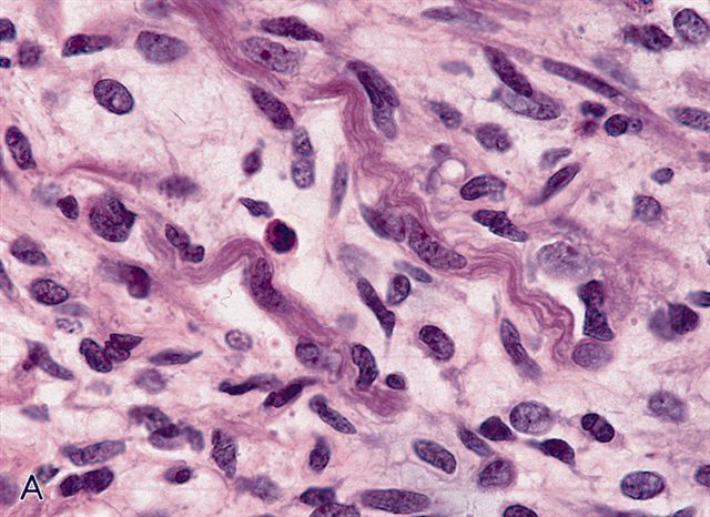
- Cells have variable skeletal muscle differentiation ranging from immature cells of myxoid pattern (but in larger numbers) to ganglion cell-like rhabdomyoblasts with prominent nucleoli or strap cells with abundant basophilic or eosinophilic cytoplasm and prominent cross striations
- Infiltration of skeletal muscle may make margins difficult to determine
- Variable glycogen containing vacuoles
- No / rare mitotic figures
Microscopic (histologic) images
Contributed by Mark R. Wick, M.D. and AFIP
Cytology description
- Spindle cells and rhabdomyoblasts with abundant eosinophilic cytoplasm (Singapore Med J 2009;50:e138)
Positive stains
- Muscle specific actin, desmin and myoglobin (100%), GFAP (40%)
Negative stains
Electron microscopy description
- Hypertrophied Z band material, thick and thin filaments, numerous mitochondria, some with inclusions
Differential diagnosis
- Botryoid variant of embryonal rhabdomyosarcoma:
- Resembles myxoid variant of fetal rhabdomyoma but has deep location, true cambium layer, atypia, numerous mitotic figures, tumor cell necrosis, infiltrative margins, no maturation of cells at periphery
- Infantile fibromatosis:
- Deep location, fascicles of spindle cells, no cross striations, no undifferentiated cells
- Neuromuscular hamartoma:
- S100+ nerve fibers and skeletal muscle in same perimysial sheath
- Spindle cell variant of embryonal rhabdomyosarcoma:
- Resembles cellular variant of fetal rhabdomyoma but has cellular pleomorphism and tumor cell necrosis
Genital type rhabdomyoma
Definition / general
Epidemiology
Case reports
Treatment
Gross description
Microscopic (histologic) description
Microscopic (histologic) images
AFIP images
Positive stains
Negative stains
Differential diagnosis
- Rare benign tumor with skeletal muscle differentiation in vagina, vulva, cervix and rarely urethra, usually seen in middle aged women
Epidemiology
- Rarely occurs in men in the paratesticular region, epididymis or in the tunica vaginalis of the testis or prostate
- Mean age is 42 years
Case reports
- Infant girl with genital rhabdomyoma of the urethra (Hum Pathol 2012;43:597)
- 17 year old boy with locally invasive tumor (J Pediatr Surg 2007;42:E5)
- 19 year old man with testicular tumor (Mod Pathol 1997;10:608)
- 20 year old man with epididymal tumor (Arch Pathol Lab Med 2000;124:1518)
- 48 year old woman with lower abdominal pain and vaginal bleeding (Hum Pathol 2005;36:433)
Treatment
- Local excision is curative
Gross description
- Well circumscribed, solitary, up to 3 cm
- Resembles polyp
- Covered by smooth mucosa
Microscopic (histologic) description
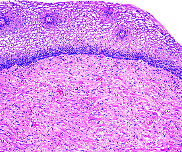
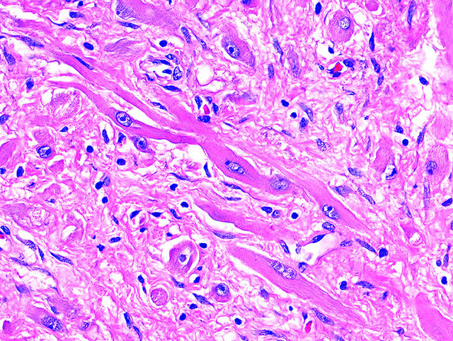
- Submucosal, polypoid, well circumscribed, no capsule
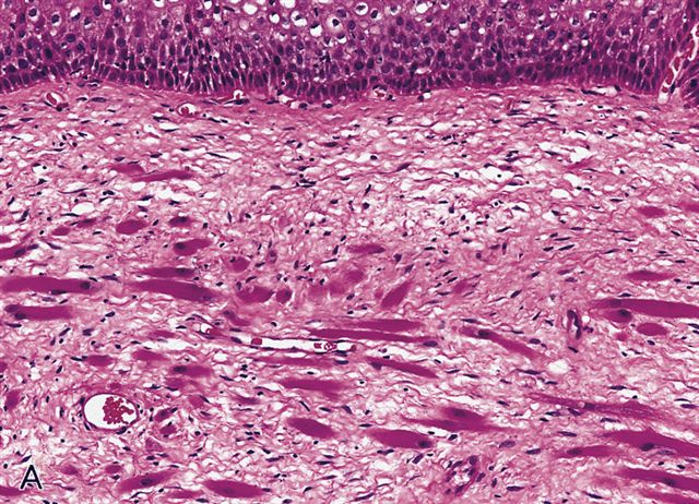
- Haphazard strap-like or round striated muscle fibers in fibrous stroma with dilated vessels
- Cells have abundant eosinophilic cytoplasm with glycogen, cross striations, longitudinal myofibrils
- Nucleus is round, vesicular, central and uniform
- May have binucleated or multinucleated cells
- No / rare mitotic figures, no cambium layer, no spider cells, no spindle cells or rhabdomyoblasts, no necrosis, no nuclear pleomorphism
Microscopic (histologic) images
AFIP images
Positive stains
- Muscle specific actin and desmin highlight rod-like structures; myoglobin (100%)
Negative stains
Differential diagnosis
- Botryoid variant of embryonal rhabdomyosarcoma:
- Usually < 25 years old with rapidly growing mass, cambium layer, atypia, mitotic activity
- Vaginal polyp:
- May have atypical cells but no cross striations


