

Soft tissue
Fibroblastic / myofibroblastic
Fibrosarcoma
Adult fibrosarcoma
Author: Komal Arora, M.D.

Last author update: 1 July 2012
Last staff update: 12 November 2024
Copyright: 2002-2024, PathologyOutlines.com, Inc.
PubMed Search: Fibrosarcoma adult

Table of Contents
Definition / general | Treatment | Gross description | Gross images | Microscopic (histologic) description | Microscopic (histologic) images | Positive stains | Negative stains | Electron microscopy description | Molecular / cytogenetics description | Differential diagnosis | Additional referencesCite this page: Arora K. Adult fibrosarcoma. PathologyOutlines.com website. https://www.pathologyoutlines.com/topic/softtissuefibrosarcoma.html. Accessed December 17th, 2024.
Definition / general
- Malignant tumor of fibroblasts with herringbone architecture and variable collagen
- Rare (up to 3% of adult sarcomas)
- Some limit diagnosis to those age 10+ years, most patients are ages 40 - 55 years
- Many cases formerly called fibrosarcoma are actually dedifferentiated liposarcoma, fibromatosis, fibrosarcomatous DFSP, low-grade fibromyxoid sarcoma, MPNST, synovial sarcoma or MFH-pleomorphic
- Usually deep soft tissue of lower extremities or trunk, only rarely in retroperitoneum or mediastinum
- 50% recur, 25% metastasize (lung, bone)
- More metastases if more cellular and higher mitotic activity
- Survival:
- 5 year - 41%, 10 year - 29%
- Better if tumor is superficial and better differentiated, low mitotic rate, no necrosis
- See also peripheral ameloblastic fibrosarcoma, sclerosing epithelioid fibrosarcoma
Treatment
- Radical excision, radiation if residual tumor or positive margins
- Possibly chemotherapy if high grade
Gross description
- May appear well circumscribed but nonencapsulated
- Fleshy, hemorrhagic, necrotic, white-tan
Gross images
Contributed by Mark R. Wick, M.D. and AFIP

Various images
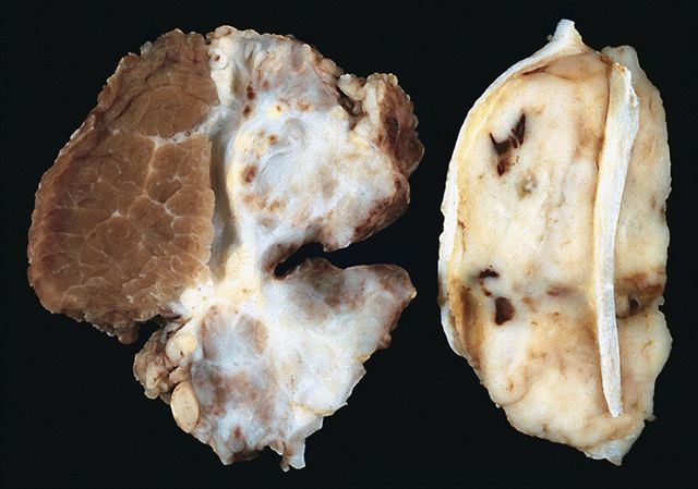
High grade fibrosarcoma (right)
Microscopic (histologic) description
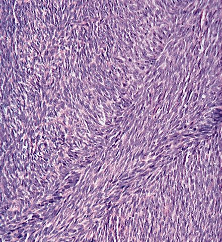
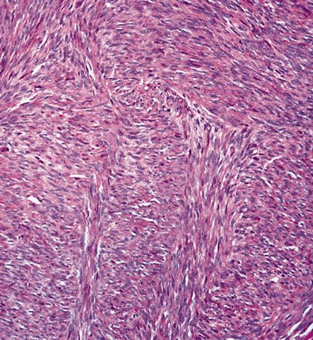
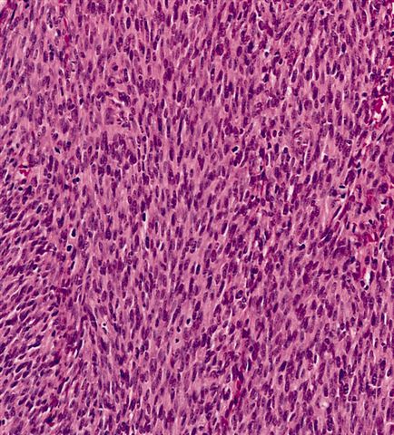
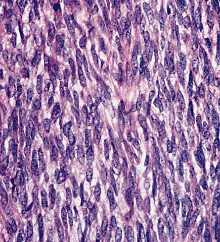
- Highly cellular fibroblastic proliferation in herringbone pattern (cells in columns of short parallel lines with all the lines in one column sloping one way and lines in adjacent columns sloping the other way)
- Cells have scant cytoplasm, tapering elongated dark nuclei with increased granular chromatin, variable nucleoli
- Mitotic activity present, often with abnormal forms
- Variable collagen
- Usually no giant cells
- No pleomorphism (or call pleomorphic MFH), no other distinct cell types
- Patterns:
- Keloid-like (thick hyalinized collagen fibers), loose fascicular, focally myxoid
Microscopic (histologic) images
Contributed by Mark R. Wick, M.D. and AFIP


Adult type
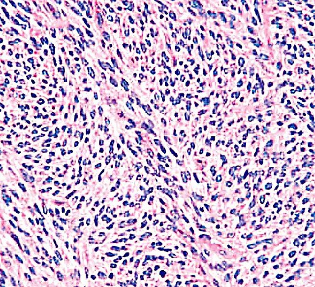
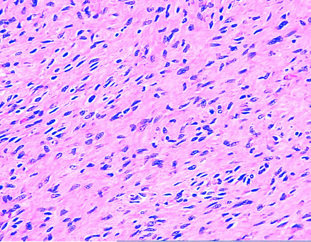
Adult type
Atypical uniform cells in herringbone pattern
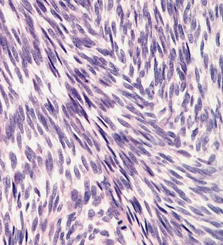
Minimal pleomorphism

Grade I tumor

Grade II tumor
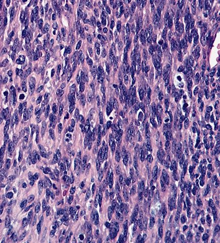
Grade III tumor
Positive stains
Electron microscopy description
- Fibroblasts with prominent rough endoplasmic reticulum but no myofilaments, no external lamina, no intercellular junction
- No distinct myofibroblasts (if present, call myofibrosarcoma)
Molecular / cytogenetics description
- Aneuploid
Differential diagnosis
- Other tumors with fibrosarcomatous areas:
- Dedifferentiated liposarcoma
- Fibromatosis: less cellular, less hyperchromasia, no atypia, < 1 mitotic figure/HPF
- Low grade fibromyxoid sarcoma
- MFH-pleomorphic
- MPNST
- Synovial sarcoma
Additional references

