Table of Contents
Definition / general | Essential features | ICD coding | Epidemiology | Sites | Etiology | Clinical features | Diagnosis | Radiology description | Radiology images | Prognostic factors | Case reports | Treatment | Clinical images | Gross description | Gross images | Microscopic (histologic) description | Microscopic (histologic) images | Virtual slides | Positive stains | Negative stains | Molecular / cytogenetics description | Videos | Sample pathology report | Differential diagnosis | Additional references | Board review style question #1 | Board review style answer #1 | Board review style question #2 | Board review style answer #2Cite this page: Kunzler E, Rohr BR. Leiomyoma. PathologyOutlines.com website. https://www.pathologyoutlines.com/topic/skintumornonmelanocyticleiomyoma.html. Accessed January 2nd, 2025.
Definition / general
- Cutaneous leiomyomas are benign tumors originating from arrector pili (piloleiomyoma), vascular (angioleiomyoma), periareolar and genital smooth muscle cells (genital leiomyoma)
Essential features
- Cutaneous leiomyomas are benign dermal tumors of smooth muscle cells arranged in interlacing fascicles
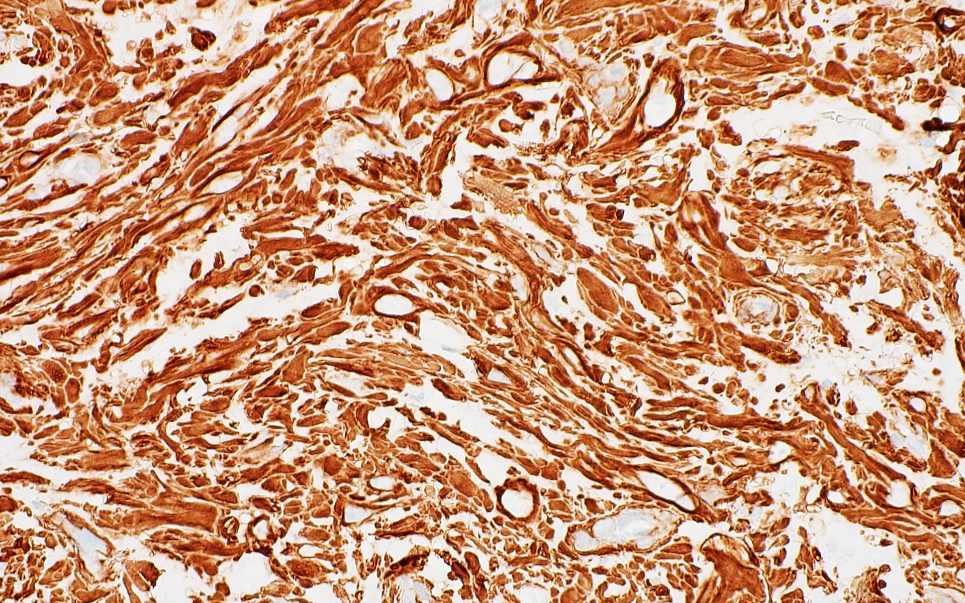
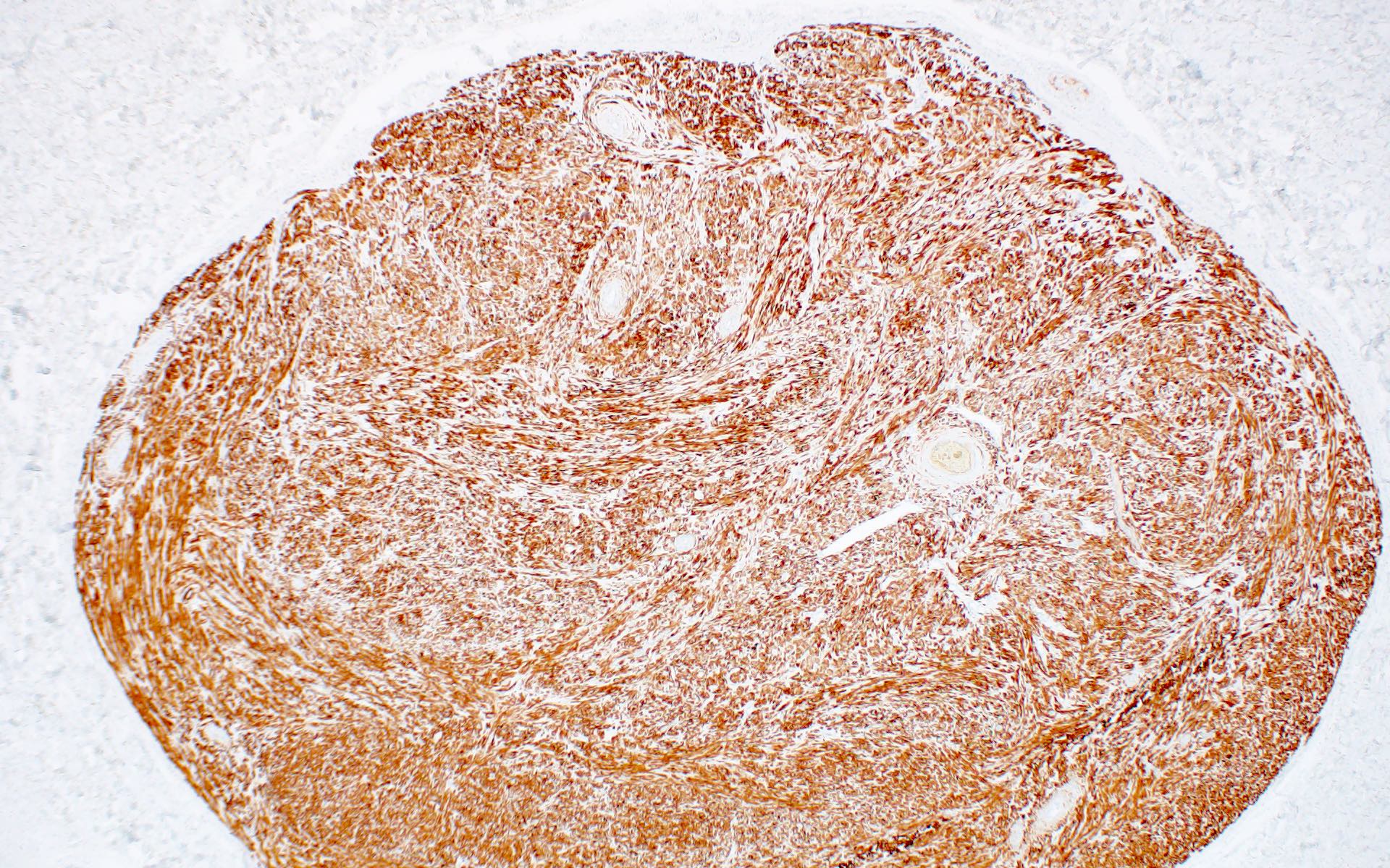
- Leiomyomas may be differentiated from other spindle cell tumors due to their positivity for both SMA and desmin
- Cutaneous piloleiomyomas can be sporadic or inherited in association with multiple cutaneous and uterine leiomyomatosis (also known as Reed syndrome or hereditary leiomyomatosis and renal cell cancer)
ICD coding
- ICD-10: D21.9 - benign neoplasm of connective and other soft tissue, unspecified
Epidemiology
- Leiomyomas are typically diagnosed in young and middle aged adults; however, they can occur in children
- Frequency of piloleiomyomas is about equal in males and females and it is unknown whether a sex predominance exists for genital leiomyomas; there is no known racial predilection for cutaneous leiomyomas (StatPearls: Cutaneous Leiomyomas [Accessed 10 April 2024])
- Angioleiomyomas have been further split by some authors into solid (more common in females), cavernous and venous subtypes (cavernous and venous being more common in males) (Laryngoscope 2004;114:661)
Sites
- Cutaneous leiomyomas can occur anywhere on the body
Etiology
- Germline heterozygous mutations of the fumarate hydratase gene are found in cutaneous piloleiomyomas associated with the autosomal dominant syndrome multiple cutaneous and uterine leiomyomatosis (also known as Reed syndrome or hereditary leiomyomatosis and renal cell cancer)
- Fumarate hydratase missense mutations are associated with decreased enzyme activity, suggesting a tumor suppressor role (J Mol Diagn 2005;7:437)
- Little is known about the etiology of sporadic cutaneous leiomyomas (Int J Mol Med 2004;13:13, Cancer Genet Cytogenet 2006;170:58, Cancer Genet Cytogenet 2010;199:21)
- Recurrent losses in chromosome 22 and recurrent gains in Xq have been reported in few cases of sporadic angioleiomyomas
- Few studies have examined cytogenetics of genital leiomyomas
- No consistent mutations have been identified in sporadic piloleiomyomas to date
Clinical features
- Cutaneous leiomyomas can be solitary or multiple; when multiple, they may be clustered or linear in distribution
- They present as skin colored or red-brown, firm papules and nodules (Am J Clin Dermatol 2015;16:35)
- Pain and cold sensitivity are common complaints
- Genital leiomyomas may be pedunculated and are less likely to be painful compared to other subtypes
Diagnosis
- Diagnosis is often made by histopathology
- Imaging including computed tomography (CT), magnetic resonance imaging (MRI) and ultrasound can be performed
- Germline mutations of the fumarate hydratase gene are diagnostic of multiple cutaneous and uterine leiomyomatosis (also known as Reed syndrome or hereditary leiomyomatosis and renal cell cancer) (Semin Cancer Biol 2020;61:158)
Radiology description
- Imaging is not routinely performed for cutaneous leiomyomas
- Characteristic imaging findings of angioleiomyomas have been described
- Calcifications due to degenerative change can be seen on CT
Prognostic factors
- Cutaneous leiomyomas are slow growing and can increase in number over time
- Excised solitary, sporadic cutaneous leiomyomas have an excellent prognosis with low recurrence rates (J Oral Maxillofac Pathol 2013;17:281)
- Malignant transformation is rare in cutaneous leiomyomas
- Leiomyosarcomas have been described in patients with multiple cutaneous and uterine leiomyomatosis (also known as Reed syndrome or hereditary leiomyomatosis and renal cell cancer); subcutaneous extension is a poor prognostic factor in leiomyosarcoma (JAAD Case Rep 2015;1:150, J Am Acad Dermatol 2014;71:919)
- Presence of multiple cutaneous piloleiomyomas is cause for further work up due to the association of the following malignancies with multiple cutaneous and uterine leiomyomatosis (also known as Reed syndrome or hereditary leiomyomatosis and renal cell cancer)
- Renal cell cancers, most commonly type II papillary renal cell carcinoma
- Cutaneous and uterine leiomyosarcomas
- Cases have been reported of breast cancer, bladder cancer, gastrointestinal stromal tumors, adrenal tumor, testicular and ovarian tumors; however, it is unknown whether these are coincidental or related (GeneReviews: FH Tumor Predisposition Syndrome [Accessed 10 April 2024])
Case reports
- 24 year old woman with painful lesions of the back and left shoulder (J Clin Aesthet Dermatol 2011;4:37)
- Woman in her 30s with painful nodules of the head, neck and lower extremities (JAMA Dermatol 2016;152:1041)
- 36 year old woman with multiple papules of the right upper extremity and chest (Clin Case Rep 2023;11:e6904)
- 41 year old woman with a tender nodule on the right heel (Cureus 2018;10:e3419)
Treatment
- Surgical excision can be performed when practical for symptomatic lesions or for elective removal
- Conservative but complete excision should be advised if atypical features are present on histopathology
- Alternative destructive interventions include CO2 laser, electrodessication and cryosurgery
- Topical, intralesional and systemic treatments for symptomatic relief may be indicated (J Am Acad Dermatol 2017;77:149)
- Topical options include nitroglycerin, lidocaine and capsaicin
- Use of intralesional botox has been reported
- Systemic treatment options include gabapentin, pregabalin, NSAIDs, calcium channel blockers, doxazosin, nitroglycerin, phenoxybenzamine and duloxetine
Clinical images
Gross description
- Grossly, cutaneous leiomyomas can be rubbery or firm and grey, white or brown in color
Microscopic (histologic) description
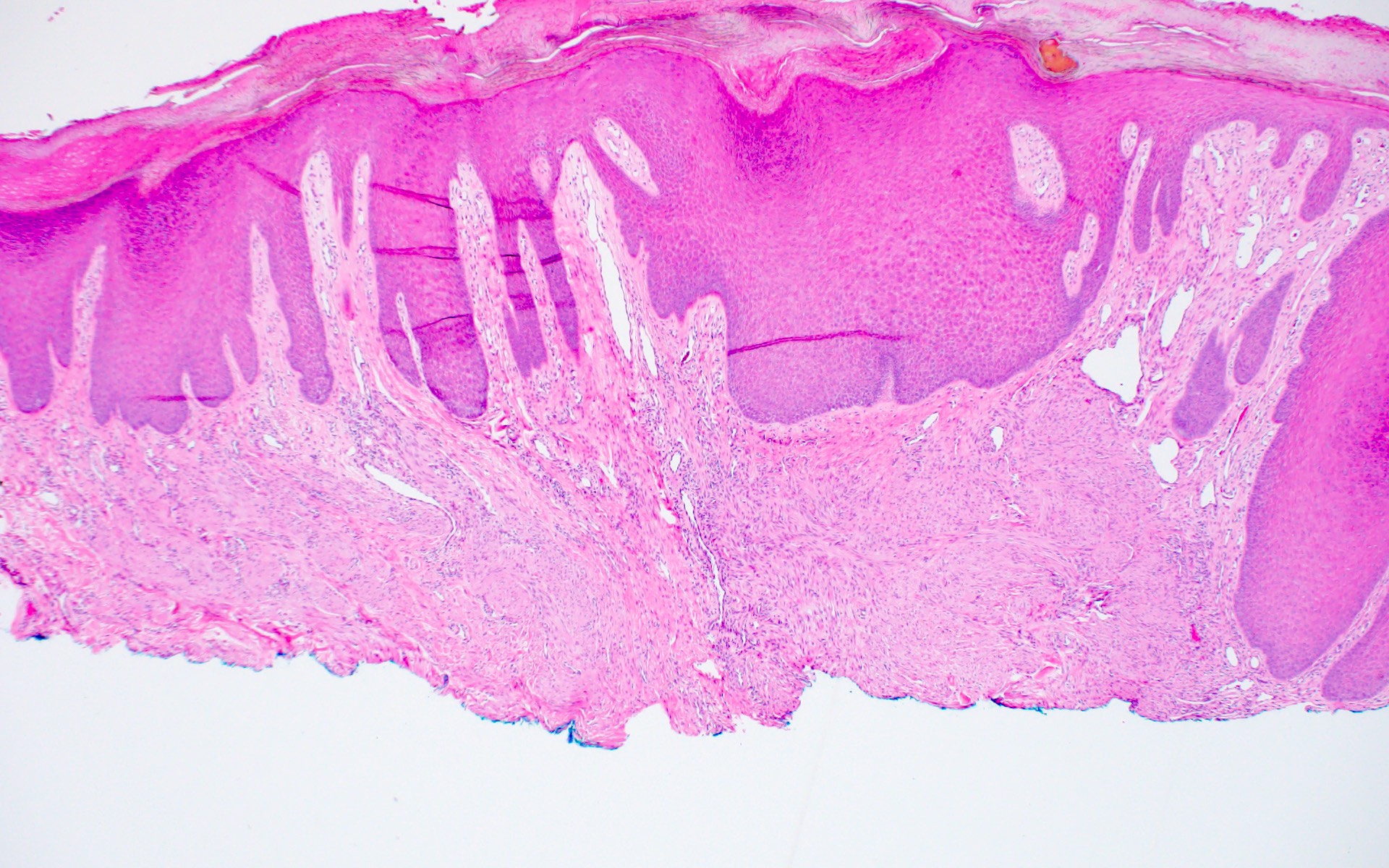
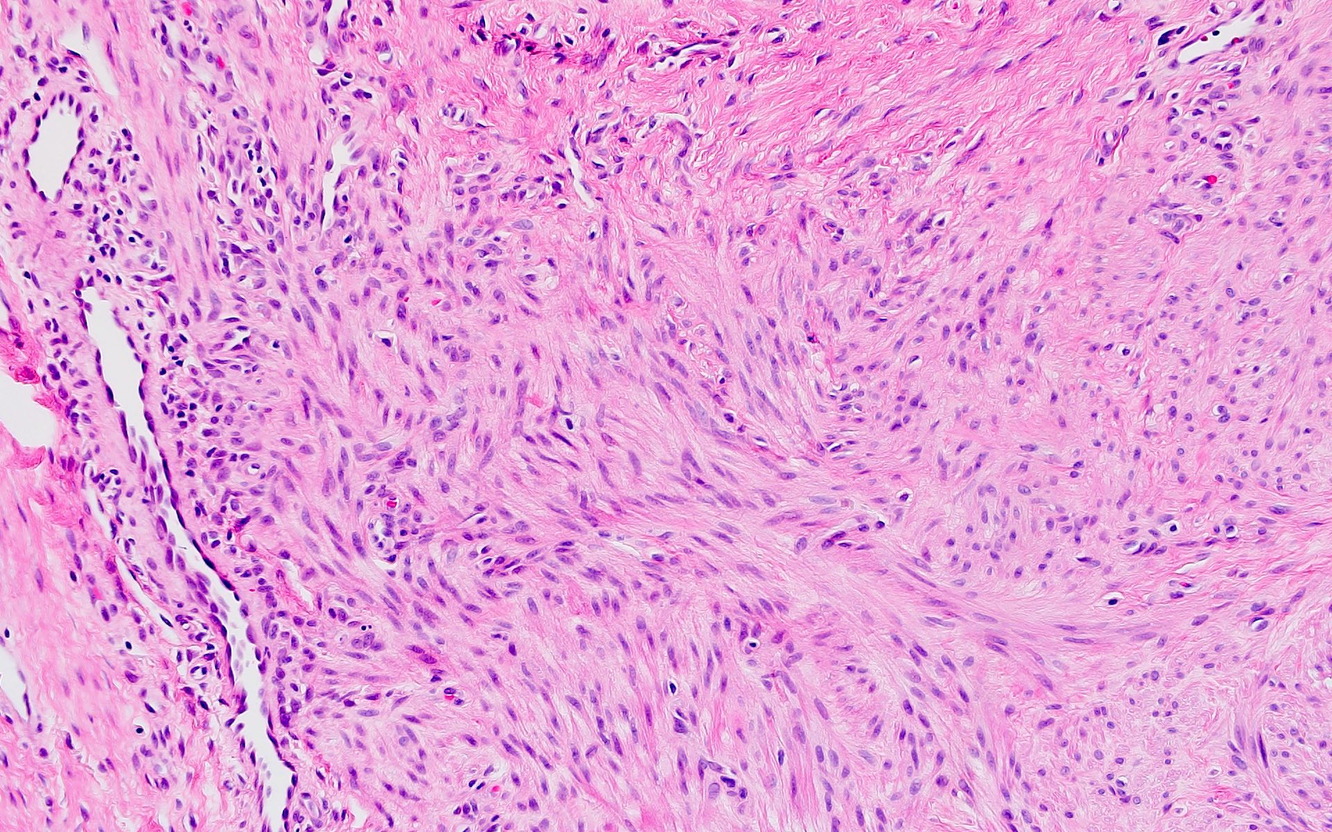
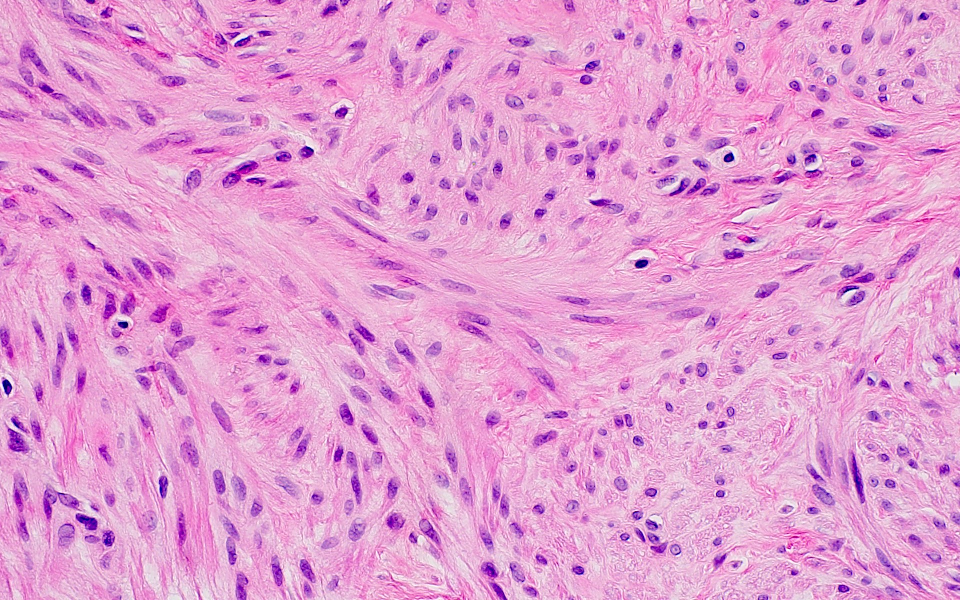
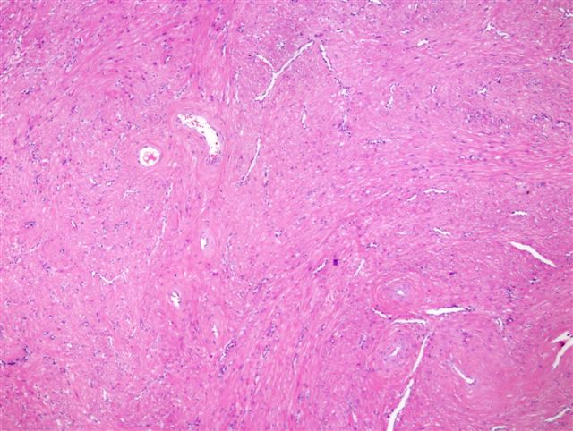
- Smooth muscle cells have elongated, thin nuclei with blunt ends; they are often described as cigar shaped with pink, eosinophilic cytoplasm
- Perinuclear vacuoles made of glycogen may be seen (Am J Dermatopathol 1997;19:2)
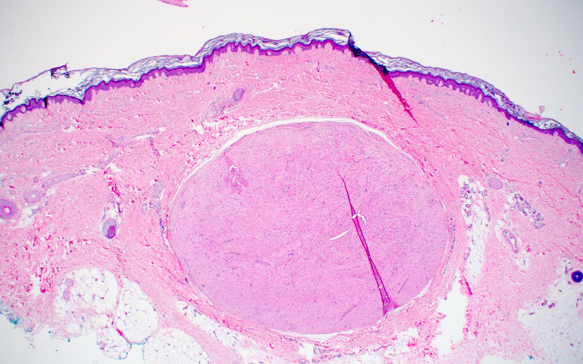
- Well circumscribed tumor consisting of fascicles and interlacing smooth muscle bundles
- Piloleiomyomas are nonencapsulated while angioleiomyomas can be encapsulated
- Angioleiomyomas have prominent vessels described as slit-like or round; smooth muscle layers surrounding the vessels interlace with surrounding smooth muscle fascicles
- Overlying epidermal changes such as acanthosis and hyperkeratosis can be seen
- Cytologic atypia is usually absent and mitotic activity is negligible
- Presence of nuclear atypia and degenerative features have been described in symplastic leiomyomas (Dermatol Surg 2004;30:1249)
Microscopic (histologic) images
Positive stains
Negative stains
- S100
- SOX10
- HMB45
- CD34
- Fumarate hydratase: loss of fumarate hydratase expression can be seen in piloleiomyomas associated with multiple cutaneous and uterine leiomyomatosis (also known as Reed syndrome or hereditary leiomyomatosis and renal cell cancer); sensitivity 70%, specificity 97.6% (Am J Surg Pathol 2017;41:801)
Molecular / cytogenetics description
- For piloleiomyomas, fumarate hydratase gene mutations can be identified using a variety of methods, including sequencing and copy number analyses; a variant specific test can be used if there is a known germline pathogenic variant
- Fumarate hydratase activity can be measured in fibroblasts or leukocytes (GeneReviews: Fumarate Hydratase Deficiency [Accessed 10 April 2024])
Videos
Pilar leiomyoma video
by Dr. Jerad Gardner
Angioleiomyoma video
by Dr. Jerad Gardner
Sample pathology report
- Skin, right shoulder, shave biopsy:
- Piloleiomyoma (see comment)
- Comment: This is a dermal tumor consisting of interlacing fascicles of smooth muscle cells. Cellular atypia and mitoses are not identified. The tumor is positive for SMA.
Differential diagnosis
- Leiomyosarcoma:
- Marked cytologic atypia
- Frequent mitoses
- Necrosis
- Subcutaneous extension
- Congenital smooth muscle hamartoma:
- Presentation in infancy; can see overlying epidermal changes
- May see hypertrichosis
- Becker nevus:
- May see hyperpigmentation
- Variable hypertrichosis
- Spindled squamous cell carcinoma:
- Pleomorphism
- Cytokeratin+
- Variable SMA
- Atypical fibroxanthoma (AFX):
- Spindle cell melanoma:
- Dermatomyofibroma:
- Horizontal arrangement of spindled cells
- Desmin negative
- Neurofibroma:
- S100+
- Cells have scant cytoplasm and are loosely arranged
- Dermatofibroma:
- Myoepithelioma:
- S100+
- EBV associated smooth muscle tumor:
- EBER ISH +
- Myopericytoma:
- Negative for desmin
Additional references
Board review style question #1
A 25 year old man presents with a painful nodule on the right lower leg. On histopathology, there is a dermal nodule with fascicles of spindle cells that have eosinophilic cytoplasm. Tumor cells are positive for SMA and desmin and are negative for S100. What is the diagnosis?
- Angioleiomyoma
- Atypical fibroxanthoma
- Dermatomyofibroma
- Neurofibroma
Board review style answer #1
A. Angioleiomyoma. The photomicrograph depicts a well circumscribed dermal tumor made of smooth muscle cells, confirmed by positive SMA and desmin staining. These findings, along with the clinical presentation of a painful nodule on the extremity, are compatible with angioleiomyoma. Answer D is incorrect because neurofibromas are positive for S100 and negative for SMA and desmin. Answer C is incorrect because the spindled cells are horizontal, not arranged in fascicles as in dermatomyofibroma. Answer B is incorrect because the well circumscribed dermal tumor and patient age point toward a benign diagnosis. In atypical fibroxanthoma, cytologic atypia is more apparent.
Comment Here
Reference: Leiomyoma
Comment Here
Reference: Leiomyoma
Board review style question #2
A 36 year old woman presents for excision of a painful, slowly enlarging nodule on the thigh present for many years. Her father has history of papillary type 2 renal cell carcinoma. Mutation in which of the following enzymes is implicated in this genetic syndrome?
- Alpha galactosidase A
- Ferrochelatase
- Fumarate hydratase
- Uroporphyrinogen decarboxylase
Board review style answer #2
C. Fumarate hydratase. Fumarate hydratase is mutated in multiple cutaneous and uterine leiomyomatosis (also known as Reed syndrome or hereditary leiomyomatosis and renal cell cancer). Answer A is incorrect because alpha galactosidase A is mutated in Fabry disease. Answer B is incorrect because ferrochelatase is mutated in erythropoietic protoporphyria. Answer D is incorrect because decreased uroporphyrinogen decarboxylase function is associated with porphyria cutanea tarda.
Comment Here
Reference: Leiomyoma
Comment Here
Reference: Leiomyoma


