

Soft tissue
Adipose tissue tumors
Lipoma and variants
Chondroid lipoma
Editorial Board Member: Jose G. Mantilla, M.D.
Deputy Editor-in-Chief: Borislav A. Alexiev, M.D.


Last author update: 27 April 2022
Last staff update: 27 April 2022
Copyright: 2002-2024, PathologyOutlines.com, Inc.
PubMed Search: Chondroid lipoma
Table of Contents
Definition / general | Essential features | Terminology | ICD coding | Epidemiology | Sites | Pathophysiology | Etiology | Clinical features | Diagnosis | Radiology description | Radiology images | Prognostic factors | Case reports | Treatment | Gross description | Gross images | Microscopic (histologic) description | Microscopic (histologic) images | Cytology description | Cytology images | Positive stains | Negative stains | Electron microscopy description | Molecular / cytogenetics description | Molecular / cytogenetics images | Videos | Sample pathology report | Differential diagnosis | Additional references | Board review style question #1 | Board review style answer #1 | Board review style question #2 | Board review style answer #2Cite this page: Tahir U, Qureshi MB, Ud Din N. Chondroid lipoma. PathologyOutlines.com website. https://www.pathologyoutlines.com/topic/softtissueadiposechondroid.html. Accessed April 19th, 2024.
Definition / general
- Benign adipose tissue tumor composed of lipoblasts intermixed with mature adipocytes in a myxohyaline chondroid matrix (Ann Diagn Pathol 2012;16:230)
Essential features
- Rare variant of lipoma with features of immature fat and immature cartilage
- Rare, slow going, painless mass in adult women
- Lobulated, circumscribed, deep seated tumor
- Mostly affects the proximal extremities and limb girdles of adult women
- Nests and cords of uni and multivacuolated lipoblasts with variable adipose tissue in a myxochondroid matrix
- Characterized by t(11;16)(q13;p13) translocation (Histopathology 2013;62:925)
- Surgical excision is curative with rare recurrence
Terminology
- Extraskeletal chondroma with lipoblast-like cells (first described terminology, subsequently disapproved) (Hum Pathol 1986;17:1285)
ICD coding
- ICD-O: 8862/0 - chondroid lipoma
- ICD-11: 2E80.0Z & XH7WX8 - lipoma, unspecified & chondroid lipoma
Epidemiology
- Rare neoplasm
- Primarily affects adult females
- M:F = 1:4
- Age range: 20 - 40 years
- Reference: Ann Diagn Pathol 2012;16:230
Sites
- Most common sites: proximal extremities and limb girdles
- Less common sites: trunk, distal extremities, head and neck (J Clin Diagn Res 2017;11:ED17)
- Deep seated tumor involves skeletal muscles, deep fibrous connective tissue or deep subcutaneous fat
- Superficial in 20% of cases
Pathophysiology
- Shows recurrent t(11;16)(q13;p13) translocation with resultant C11orf95 and MRTFB fusion (Genes Chromosomes Cancer 2010;49:810)
- Megakaryoblastic leukemia 2 (MKL2) / myocardin related transcription factor B (MRTFB) acts as coactivator of transcription factor serum response factor (SRF)
- SRF controls cellular processes including cytoskeleton organization, cell migration, growth and differentiation
- C11orf95 encodes protein of unknown function
Etiology
- Not known
Clinical features
- Benign, painless, slow growing tumor of variable duration
- History of recent increase in size in 50% of cases
- Reference: Exp Ther Med 2021;22:1087
Diagnosis
- Requires presence of classic histologic features (i.e., presence of lipoblasts, chondroid cells and adipocytes in a myxohyaline matrix in a circumscribed lobulated tumor)
- Diagnosis can also be made on cytology (Am J Surg Pathol 1999;23:1300)
Radiology description
- CT typically shows a well defined, heterogeneously enhancing mass with variable areas of fat attenuation
- Can exhibit calcification and ossification on radiographs (Australas Med J 2012;5:355, Skeletal Radiol 2004;33:670)
- MRI usually shows a well defined mass with lobulated areas of T2 hyperintense or fluid-like signal intensity (Balkan Med J 2015;32:107, Skeletal Radiol 1996;25:592)
- Appears isointense or hypointense to muscle, with focal hyperintense areas (fat areas) on T1 weighted MR images
- May show hypermetabolic activity at PET / CT
Radiology images
Contributed by Nasir Ud Din, M.B.B.S.
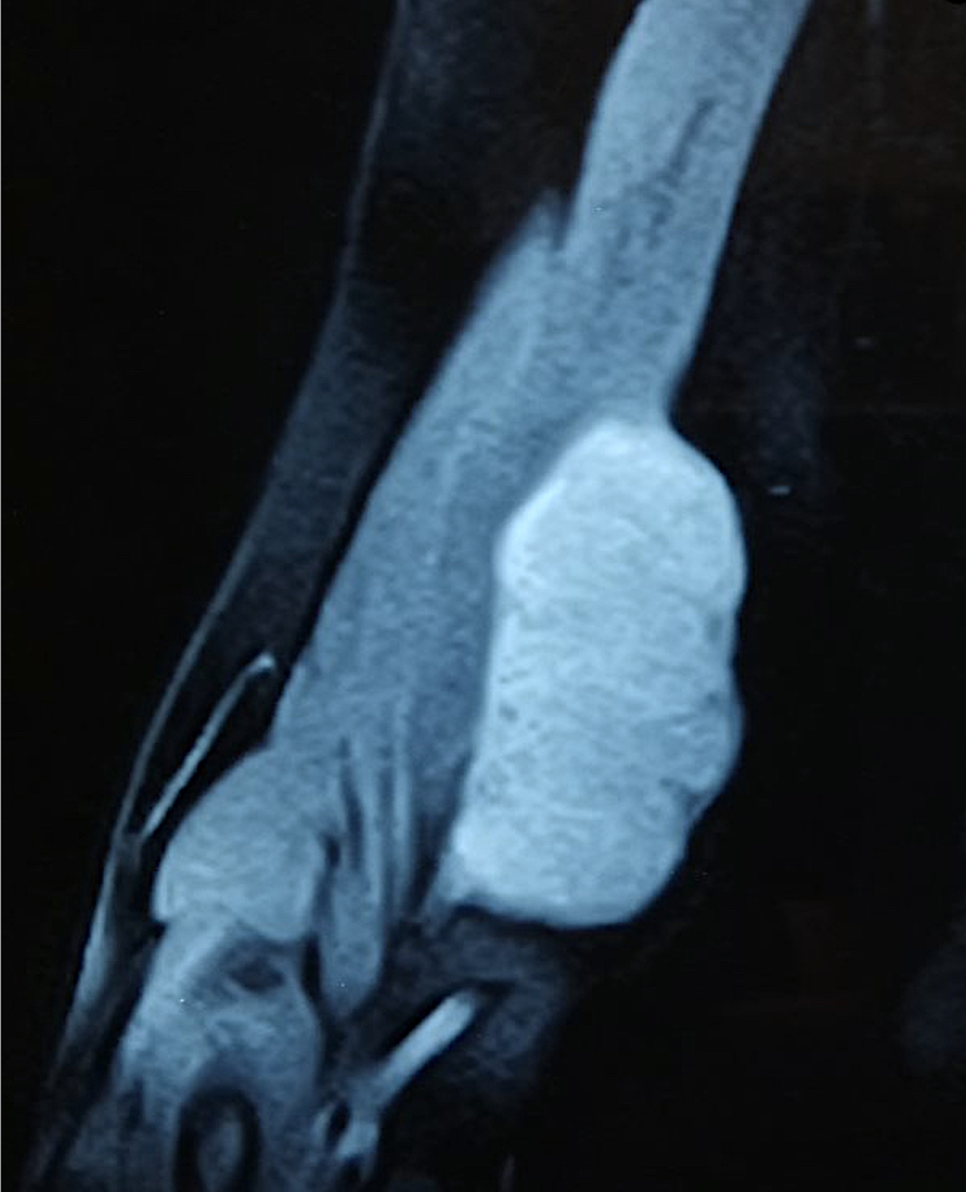
Right upper limb, coronal view
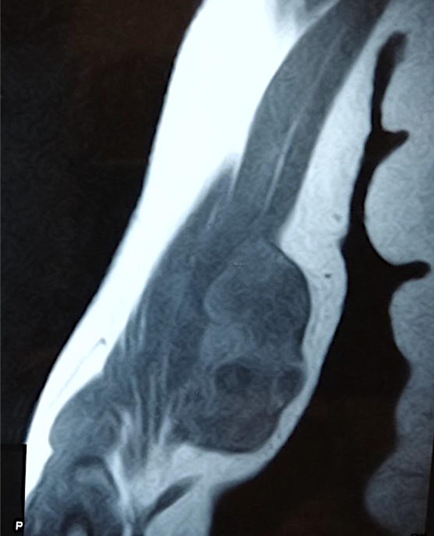
Right upper limb, T1
Prognostic factors
- Does not recur or metastasize if completely excised
- Rare local recurrence
Case reports
- 9 year old girl with enlarging left gluteal mass (Skeletal Radiol 2020;49:161)
- 37 year old woman with painful left buttock (Skeletal Radiol 2008;37:475)
- 39 year old man with large left neck mass (BMC Res Notes 2018;11:415)
- 40 year old woman with painless left thigh mass (J Clin Diagn Res 2017;11:ED17)
- 43 year old woman with painless growth on dorsum of forearm (Oman Med J 2008;23:116)
Treatment
- Complete simple surgical resection is curative (StatPearls: Lipoma Pathology [Accessed 16 March 2022])
- Does not recur or metastasize
Gross description
- Well delineated, frequently encapsulated
- Multilobulated (33%)
- Cut surface: yellowish tan, gelatinous
- Hemorrhagic foci can be seen
- 1.5 - 11 cm, hemorrhage is associated with size increase
- Reference: Medicine (Baltimore) 2019;98:e15587
Gross images
Contributed by Nasir Ud Din, M.B.B.S.
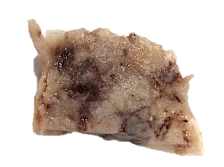
Cut surface
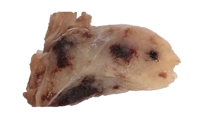
Gelatinous appearance
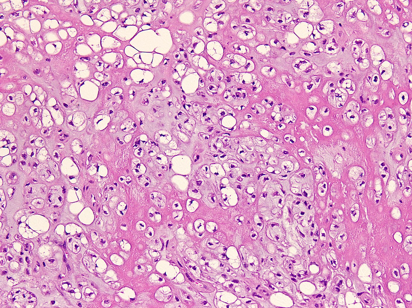
Microscopic (histologic) description
- Encapsulated with occasional lobulations
- Lobulations are formed by fibrous septa
- Composed of 3 components arranged in nests, cords and sheets in variable proportion:
- Mature adipose tissue
- Cells displaying lipoblastic differentiation
- Myxohyaline chondroid matrix
- Mature adipocytes contain eccentrically placed nuclei and vacuolated cytoplasm
- Cells with lipoblastic differentiation may show various patterns:
- Undifferentiated bland cells with minimal cytoplasm
- Small univacuolated to multivacuolated lipoblasts with fat droplets scalloping bland nuclei
- Cells with granular eosinophilic cytoplasm may be seen
- Rich vascular network: thick walled blood vessels alternate with large, gaping thin walled vascular spaces
- No significant nuclear atypia or mitotic activity
- Hemorrhage and hemosiderin deposition can be present (Hum Pathol 1995;26:706)
- Fibrosis, calcification and metaplastic bone formation may be seen (Skeletal Radiol 2008;37:475)
Microscopic (histologic) images
Contributed by Nasir Ud Din, M.B.B.S.
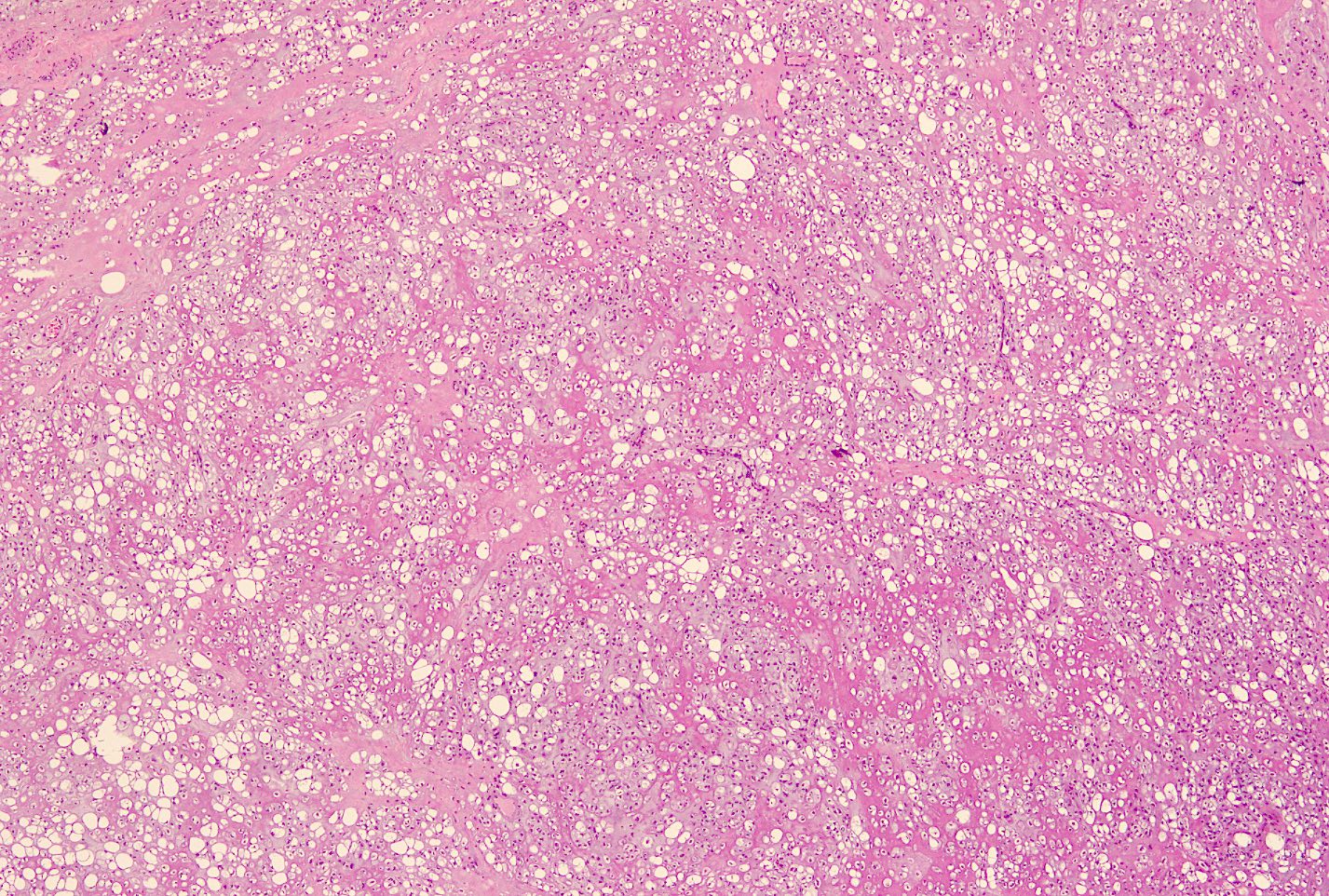
Lobulated appearance
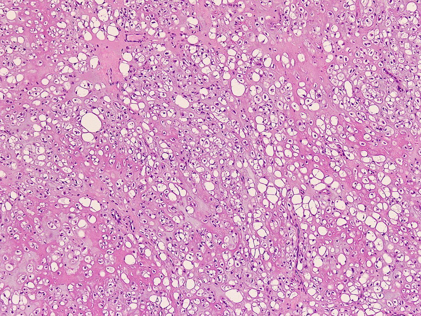
Cellular morphology
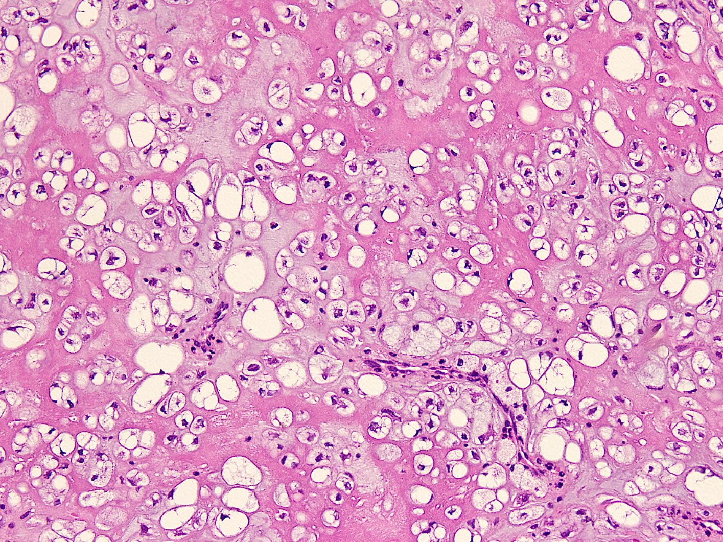
Prominent matrix
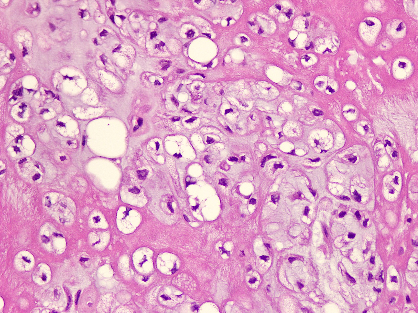
Multivacuolated lipoblasts
Cytology description
- Cohesive clusters and aggregates of mature adipocytes (Arch Pathol Lab Med 2001;125:1224)
- Lipoblasts of variable size
- Myxochondroid background matrix
Cytology images
Images hosted on other servers:

Cell block sections
Positive stains
- Immunostains are not very helpful in differential diagnosis
- S100:
- Positive in mature adipocytes (strong)
- Lipoblasts (weak)
- Negative in cells that do not show lipoblastic differentiation
- Keratins: rarely positive
- Cyclin D1: positive (Sarcoma 2011;2011:638403)
- PAS special stain highlights intracytoplasmic glycogen
- Toluidine and Alcian blue special stains highlight chondroitin sulfate in matrix at low pH
Negative stains
- EMA
- SMA
- HMB45
- CD34
- Reference: Hum Pathol 1995;26:706
Electron microscopy description
- Mature adipose tissue (Am J Surg Pathol 1995;19:1272)
- Small embryonal cells with characteristics of lipoblasts, chondroblasts or both
- Matrix shows thin filament network, thin collagen fibers and copious proteoglycan particles
Molecular / cytogenetics description
- Chromosomal translocation t(11;16)(q13;p12-13) (C11orf95-MKL2) (Histopathology 2013;62:925)
- Rare 3 way translocation involving chromosomes 1, 2 and 5
Molecular / cytogenetics images
Images hosted on other servers:

t(11;16) translocation
Videos
Chondroid lipoma pathology
Sample pathology report
- Brachialis muscle lump, excision:
- Chondroid lipoma (see comment)
- Comment: Chondroid lipoma is a rare variant of lipoma characterized by admixture of chondroid cells, lipoblasts and adipocytes in a myxohyaline matrix. The tumor is benign and excision is curative.
Differential diagnosis
- Soft tissue chondroma:
- Occurs in hands and feet
- True hyaline cartilage is present
- Lacks adipocytic component
- Atypical lipomatous tumor:
- Adipocytes in sheets with variation in size and shape
- Thick fibrous septa that have atypical stromal cells
- May exhibit myxoid stroma
- May show metaplastic cartilage
- MDM2 amplification present
- Myxoid liposarcoma:
- Spindle to stellate cells with myxoid stroma
- Plexiform vasculature
- Lipoblasts of signet type are typically present
- Round cell component shows uniform round cells with moderate pleomorphism and scant cytoplasm
- t(12;16), t(12;22) translocation
- Extraskeletal myxoid chondrosarcoma:
- Larger infiltrative mass
- More lobulated architecture
- Anastomosing and reticular pattern of uniform spindle to epithelioid cells
- Prominent myxoid stroma
- Lacks adipocytes and lipoblasts
- Characteristic translocation t(9;17), t(9;22)
- Myoepithelioma of soft tissue:
- Chondrolipoma:
- Typical lipoma with mature hyaline cartilage
Additional references
Board review style question #1
A 30 year old woman presented with a painless lump in right thigh. Excision specimen showed an encapsulated lesion of 4 x 4 cm. Histology showed a circumscribed adipocytic lesion composed of admixture of chondroid cells with vacuolated to eosinophilic cytoplasm, multivacuolated lipoblasts and mature adipocytes in a chondromyxoid background. Mature hyaline cartilage, necrosis and atypia were absent. Occasional mitotic figures were present. What is the most likely diagnosis?
- Chondroid lipoma
- Chondrolipoma
- Extraskeletal myxoid chondrosarcoma
- Myxoid liposarcoma
- Well differentiated liposarcoma
Board review style answer #1
Board review style question #2
Which of the following lipomas shows the presence of lipoblasts?
- Angiolipoma
- Chondroid lipoma
- Chondrolipoma
- Myelolipoma
- Osteolipoma
Board review style answer #2
