

Skin nonmelanocytic tumor
Adnexal tumors
Sweat gland derived (apocrine & eccrine glands)
Hidradenoma
Editorial Board Members: Robert E. LeBlanc, M.D., Kiran Motaparthi, M.D.


Last author update: 30 November 2021
Last staff update: 30 November 2021
Copyright: 2002-2024, PathologyOutlines.com, Inc.
PubMed Search: Eccrine acrospiroma [title]
Table of Contents
Definition / general | Essential features | Terminology | Epidemiology | Sites | Clinical features | Case reports | Treatment | Gross description | Microscopic (histologic) description | Microscopic (histologic) images | Positive stains | Negative stains | Molecular / cytogenetics description | Sample pathology report | Differential diagnosis | Board review style question #1 | Board review style answer #1Cite this page: Saleh J, Speiser J. Hidradenoma. PathologyOutlines.com website. https://www.pathologyoutlines.com/topic/skintumornonmelanocyticeccrineacrospiroma.html. Accessed April 24th, 2024.
Definition / general
- Benign dermal nodule of eccrine / apocrine gland derivation (Middle East Afr J Ophthalmol 2010;17:374)
Essential features
- Presents as a slow growing, nodular, solid or cystic cutaneous mass usually measuring up to 3 cm in diameter (Dermatology 2016;232:78)
- Characterized by variably sized nests and nodules of epithelial cells within the upper or mid dermis, typically with no overlying connection to the epidermis (J Clin Pathol 2007;60:145)
Terminology
- Also known as nodular hidradenoma, eccrine acrospiroma, clear cell hidradenoma, eccrine sweat gland adenoma and solid cystic hidradenoma
Epidemiology
- Frequently occurs in the third to fifth decade of life with a slight predominance in females (Case Rep Oncol 2018;11:298)
Sites
- Typically located on the head, neck and limbs (although any site may be affected) (Case Rep Oncol 2018;11:298)
Clinical features
- Presents as a slow growing, nodular, solid or cystic cutaneous mass usually measuring up to 3 cm in diameter (Dermatology 2016;232:78)
- Overlying skin can be skin colored, erythematous or blue and may exhibit superficial ulceration or serous discharge (Indian Dermatol Online J 2016;7:410)
Case reports
- 13 year old boy with nodular hidradenoma on the upper arm (Australas J Dermatol 2017;58:e135)
- 20 year old woman with nodular hidradenoma on the breast (Lab Med 2019;50:320)
- 61 year old woman with clear cell hidradenoma on the chin (J Craniofac Surg 2017;28:e454)
Treatment
- Recurrence rate of 12% if not completely excised (J Clin Pathol 2007;60:145)
Gross description
- Solitary well circumscribed dermal nodule (Int J Gynecol Pathol 2008;27:457)
Microscopic (histologic) description
- Well circumscribed but unencapsulated, lobulated / cystic mass with variably sized nests and nodules of epithelial cells within the upper or mid dermis, typically with no overlying connection to the epidermis (J Clin Pathol 2007;60:145)
- Shows both solid and cystic components (Arch Pathol Lab Med 2005;129:e113)
- Solid portion composed of 2 types of cells: polyhedral cells with basophilic cytoplasm and glycogen containing clear cells with eccentric round nucleus
- Cystic areas secondary to degeneration of tumor cells
- Tubular lumina are lined by cuboidal or columnar secretory cells (J Oral Maxillofac Pathol 2013;17:136)
- Fibrovascular, collagenous or hyalinized stroma (J Cutan Pathol 2007;34:497, Arch Pathol Lab Med 2005;129:e113)
- Focal apocrine decapitation secretion, squamous differentiation, squamous eddy formation, keratinization, mucinous change or sebaceous differentiation can be seen (Am J Dermatopathol 2012;34:461, J Cutan Pathol 2007;34:801, J Cutan Pathol 2007;34:497)
- Malignant hidradenocarcinoma presents with infiltrative growth pattern, nuclear atypia and pleomorphism, predominantly solid cell islands, angiolymphatic invasion, necrosis and ≥ 4 mitoses per 10 high power fields (Middle East Afr J Ophthalmol 2010;17:374, Mod Pathol 2009;22:600)
- Variants:
- Clear cell hidradenoma (Cutis 2014;94:268)
- Has a biphasic cellular population: (1) round, fusiform, or polygonal cells with vesicular nuclei and eosinophilic cytoplasm and (2) cells with clear cytoplasm and often eccentrically located nuclei
- Solid cystic hidradenoma (Arch Pathol Lab Med 2005;129:e113)
- Shows both solid and cystic components
- Cysts probably represent cystic degeneration
- Mucinous hidradenoma (J Cutan Pathol 2007;34:497)
- Demonstrates diffuse and prominent mucinous cell proliferation
- Poroid hidradenoma (Ann Dermatol 2021;33:289)
- Has architectural features of the apocrine hidradenoma and cytological features of poroid and cuticular cells
- Poroid cells are uniform, small cuboidal cells with an oval to round nuclei
- Cuticular cells have an abundant eosinophilic cytoplasm with a larger nucleus that shows occasional multinucleation
- Pigmented nodular hidradenoma (Arch Dermatol 1971;104:117)
- Shows melanin pigmentation
- Clear cell hidradenoma (Cutis 2014;94:268)
Microscopic (histologic) images
Contributed by Jasmine Saleh, M.D., M.P.H. and Jodi Speiser, M.D.
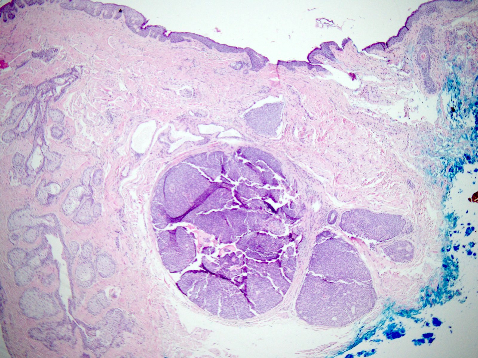
Intradermal nodule
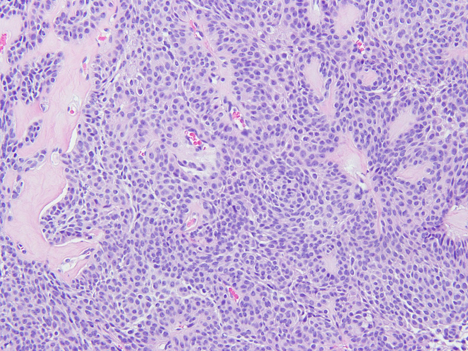
Polyhedral basophilic cells
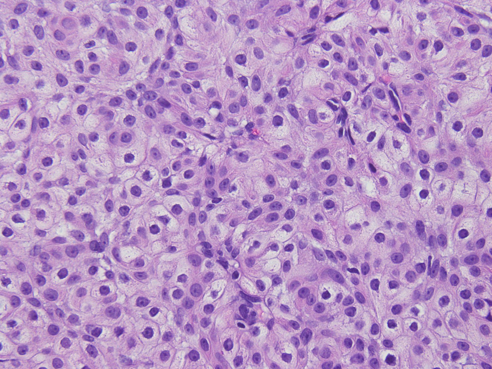
Clear cells
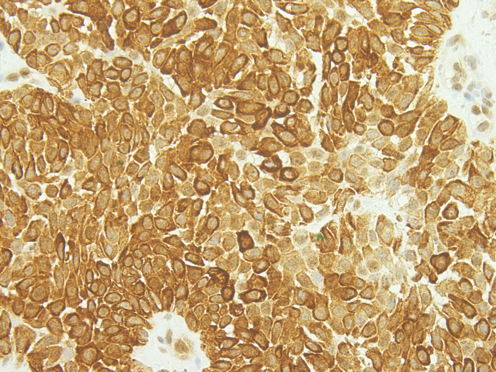
CK8/18
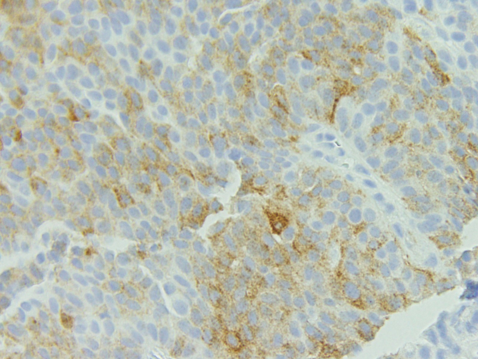
CEA
Positive stains
- AE1 / AE3 and other low molecular weight cytokeratins, EMA, CEA (J Cytol 2011;28:235, J Clin Pathol 2007;60:145)
- Some cases show partial staining with SMA, p63, actin-muscle specific (J Cytol 2011;28:235)
- Stains are not particularly helpful in the diagnosis of this entity and can be misleading
Negative stains
- Some cases show partial staining with SMA, p63, actin - muscle specific (J Cytol 2011;28:235)
- CD10 is typically negative
Molecular / cytogenetics description
- t(11;19) translocation has been reported (Am J Dermatopathol 2007;29:457)
Sample pathology report
- Skin, neck, excision:
- Hidradenoma, extending to the deep margin
Differential diagnosis
- Metastatic renal cell carcinoma
- Absence of ductal differentiation/sweat production
- Positive for PAX8 and CD10
- Negative for p63
- Basal cell carcinoma with eccrine differentiation:
- Peripheral palisading and no cystic areas
- Stromal epithelial retraction
- Epidermal connection
- Hidradenocarcinoma:
- Greater cytologic atypia, mitoses and infiltrative
- Squamous cell carcinoma:
- Epidermal connection, keratin pearls and infiltrative
- Absence of secretory differentiation
- Trichilemmoma:
- Broad connection with the epidermis
- CD34, thickened hyalinized basement membrane and peripheral cell palisading
- Poroma:
- Epidermal connection and broad, anastomosing cords
- Cells are more basaloid while cells of hidradenoma are more eosinophilic
- Recurrent YAP1-MAML2 and YAP1-NUTM1 fusions identified in a subset
- Cylindroma:
- Jigsaw puzzle pattern
- Hyaline globules and thickened basement membranes
Board review style question #1
- Which of the following is true about this identity?
- Always expresses CD10
- Has no connection to the epidermis
- Is typically encapsulated
- Presents as a fast growing mass
Board review style answer #1
