

Skin nonmelanocytic tumor
Vascular tumors
Benign lymphangioendothelioma / acquired progressive lymphangioma
Authors: Joel Tjarks, M.D., Sara C. Shalin, M.D., Ph.D.


Last author update: 1 August 2018
Last staff update: 9 June 2023
Copyright: 2001-2024, PathologyOutlines.com, Inc.
PubMed Search: Benign lymphangioendothelioma
Table of Contents
Definition / general | Essential features | Terminology | ICD coding | Epidemiology | Sites | Pathophysiology | Etiology | Clinical features | Case reports | Treatment | Microscopic (histologic) description | Microscopic (histologic) images | Positive stains | Negative stains | Differential diagnosis | Additional references | Board review style question #1 | Board review style answer #1 | Board review style question #2 | Board review style answer #2Cite this page: Tjarks J, Shalin SC. Benign lymphangioendothelioma / acquired progressive lymphangioma. PathologyOutlines.com website. https://www.pathologyoutlines.com/topic/skintumornonmelanocyticbenignlymphangioendothelioma.html. Accessed April 26th, 2024.
Definition / general
- Rare benign lymphovascular proliferation of unknown etiology, initially described in 1990 (J Am Acad Dermatol 1990;23:229)
Essential features
- Delicate, thin walled, endothelium lined dilated vascular spaces involving the superficial dermis
- Intravascular papillary stromal projections resemble papillary endothelial hyperplasia
- Deeper in dermis, vascular spaces collapse and dissect dermal collagen in angiosarcoma-like pattern
Terminology
- Also called acquired progressive lymphangioma
ICD coding
- ICD-10: D18.1 - lymphangioma, any site
Epidemiology
- Uncommon; around 50 reported cases in English literature
- Wide age distribution
- No sex predilection
Sites
- Trunk / limbs usually; other sites have been described
Pathophysiology
- Favored to be a benign vascular malformation rather than a true neoplasm
Etiology
- Some cases possibly related to trauma
Clinical features
- Bruise-like red-brown patch or plaque with indolent growth
- May be nodular, fluctuant and rubbery
- Drainage of serous fluid has been reported (Clin Exp Dermatol 2009;34:e341, Ann Acad Med Singapore 2011;40:106, J Cutan Pathol 2015;42:217)
Case reports
- 1 year old boy with extensive acquired progressive lymphangioma (Pediatr Dermatol 2018;35:486)
- 2 year old patient with alopecia lesions in scalp (Cir Pediatr 2000;13:170)
- 32 year old man with tumor in hypogastric region (Actas Dermosifiliogr 2010;101:792)
- 75 year old man with giant benign lymphangioendothelioma (J Cutan Pathol 2012;39:950)
- HIV+ man with acquired progressive lymphangioma (J Cutan Pathol 2007;34:882)
- 4 cases of benign lymphangioendothelioma (J Cutan Pathol 2013;40:945)
Treatment
- Complete excision is usually curative but not necessary
- May recur
- Reports of regression with sirolimus (J Am Acad Dermatol 2014;71:e221), pulsed dye laser (Dermatol Surg 2014;40:218), 5% imiquimod (Pediatr Dermatol 2017;34:e302)
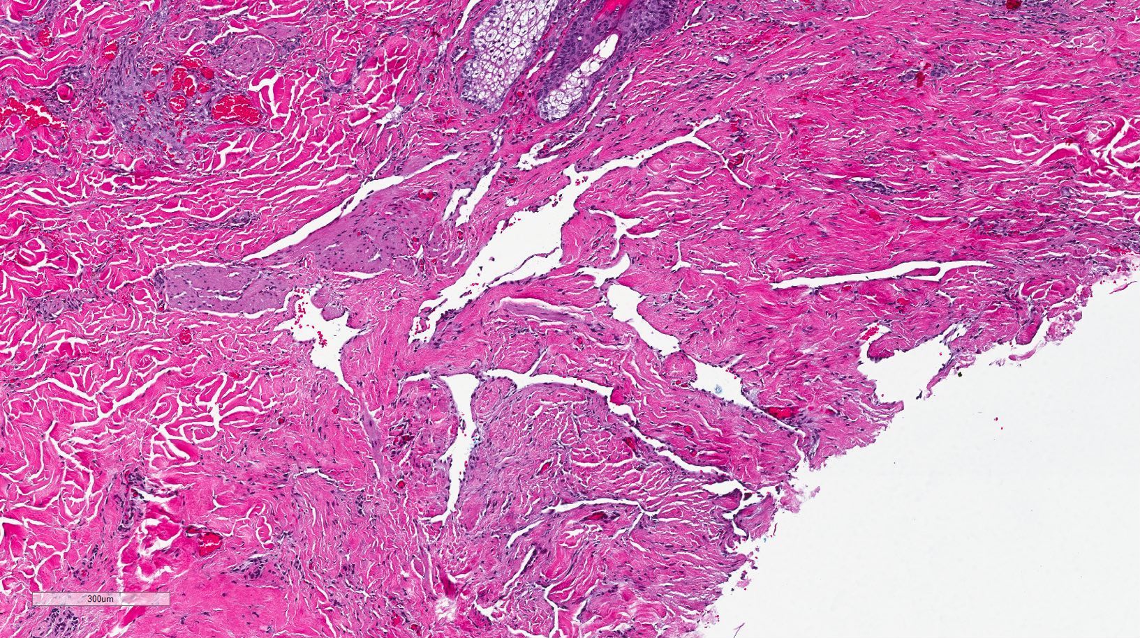
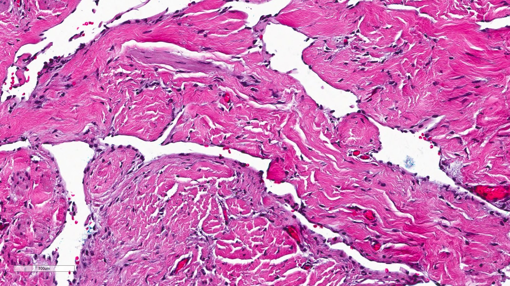
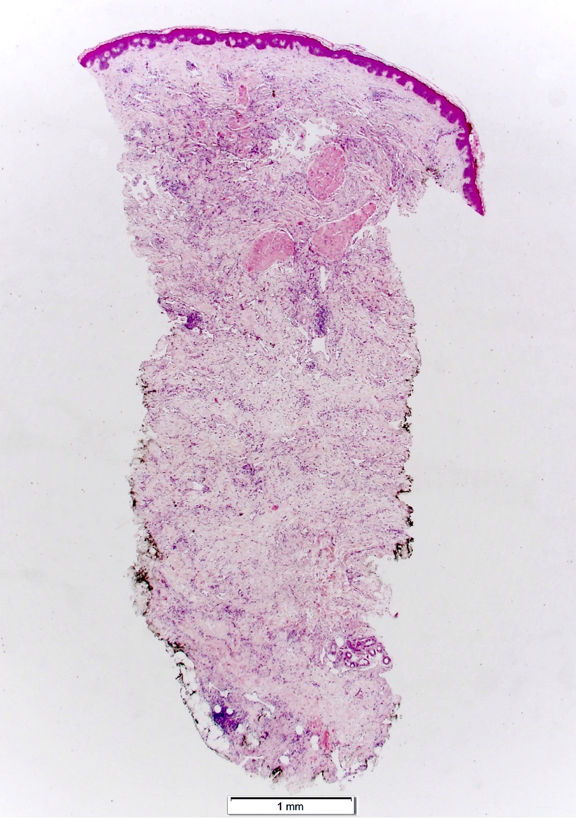
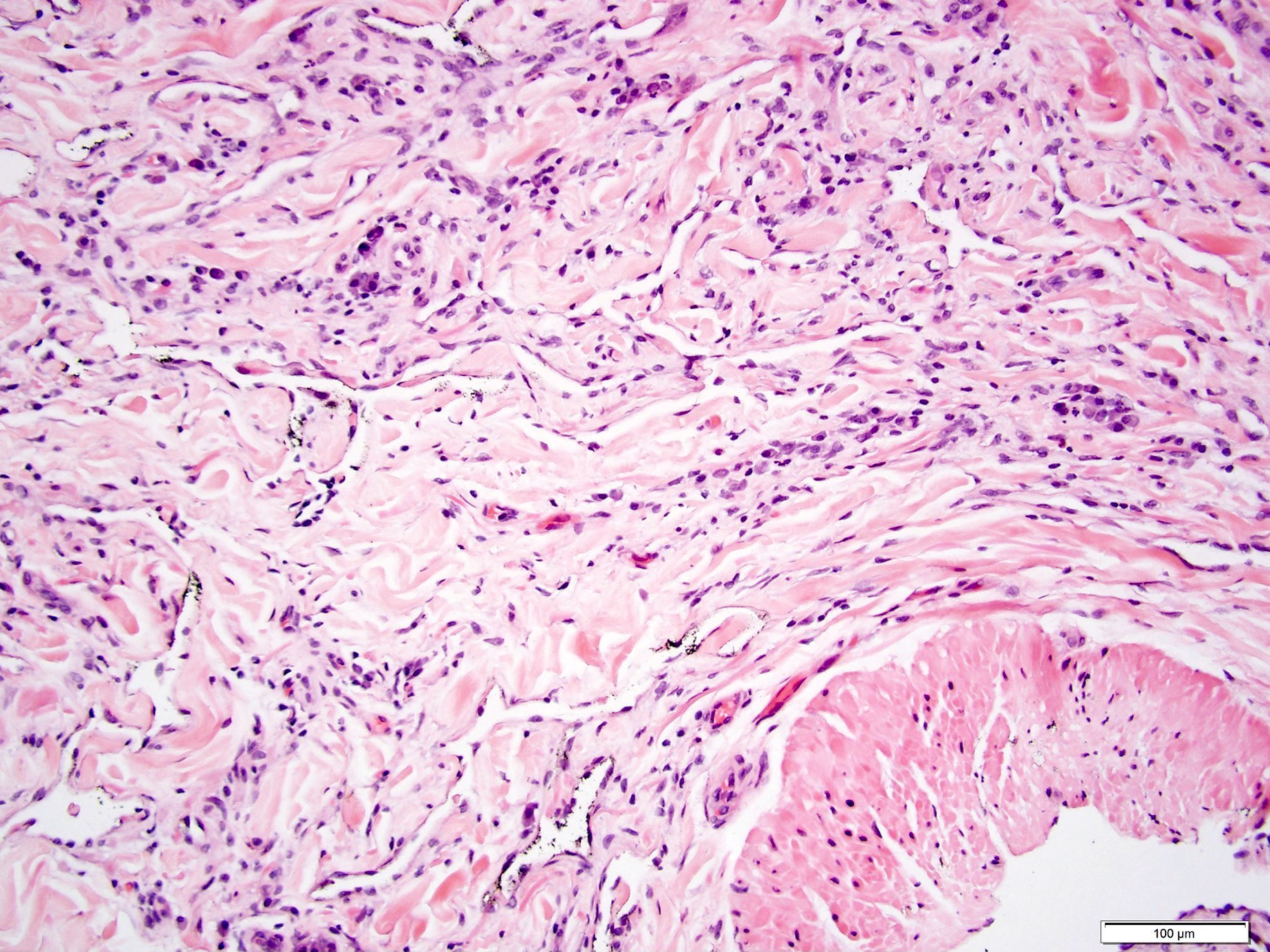
Microscopic (histologic) description
- Delicate, thin walled, endothelium lined dilated vascular spaces involving the superficial dermis
- Intravascular papillary stromal projections resemble papillary endothelial hyperplasia
- Deeper portion of lesions have vascular space collapse and dissect collagen bundles, mimicking patch stage Kaposi sarcoma
- Preexisting vessels and adnexal structures of the dermis also appear dissected by newly formed vascular channels
- Smooth muscle often focally present around vascular spaces
- Endothelial cells may hobnail, may form morula resembling giant cells
- Crowding of endothelial cells present but no endothelial atypia
- Vascular spaces lack erythrocytes and hemosiderin deposits
- No mitotic figures
Microscopic (histologic) images
Contributed by Joel Tjarks, M.D. and Sara C. Shalin, M.D., Ph.D.
6.8x
20x
2x

10x
20x

CD31
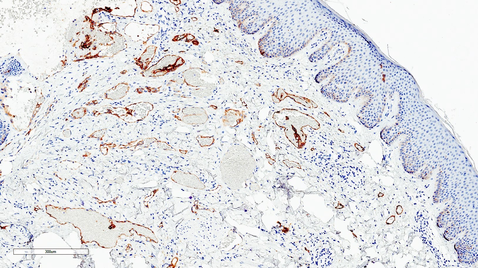
D2-40
Differential diagnosis
- Kaposi sarcoma: HHV8+; usually demonstrates slit-like vasculature without marked dilation of vessels and plasma cell infiltrate
- Atypical vascular lesion: demonstrates more endothelial prominence and atypia; associated with radiation
- Angiosarcoma: dissects through dermal collagen with significant atypia, atypical intraluminal cells, endothelial stacking (multilayering) and mitoses
- Superficial lymphangioma (lymphangioma circumscriptum): congenital hamartoma often associated with cytogenetic abnormalities; typically does not dissect dermal collagen but may show dilated superficial dermal vessels
- Targetoid hemosiderotic hemangioma (hobnail hemangioma): may have similar infiltration pattern to benign lymphangioendothelioma; endothelial cells protruding into vascular lumen (hobnail cells); deep stromal hemosiderin deposition
Additional references
Board review style question #1
Which of the following immunohistochemical stains is negative in benign lymphangioendothelioma?
- CD31
- CD34
- D2-40
- ERG
- HHV8
Board review style answer #1
Board review style question #2
Which of the following histologic features argues against the diagnosis of benign lymphangioendothelioma?
- Delicate thin walled vessels
- Frequent mitotic figures
- Hobnail endothelial cells
- Infiltration into the deep dermis
- Intravascular papillary stromal projections
Board review style answer #2
B. Frequent mitotic figures
Comment Here
Reference: Benign lymphangioendothelioma / acquired progressive lymphangioma
Comment Here
Reference: Benign lymphangioendothelioma / acquired progressive lymphangioma
