

Skin nonmelanocytic tumor
Fibrous, fibrohistiocytic and myofibroblastic neoplasms
Acquired digital fibrokeratoma
Author: Hillary Rose Elwood, M.D.

Last author update: 1 March 2015
Last staff update: 4 November 2020
Copyright: 2002-2024, PathologyOutlines.com, Inc.
PubMed Search: Acquired digital fibrokeratoma [title]
Table of Contents
Definition / general | Terminology | Epidemiology | Sites | Etiology | Clinical features | Case reports | Treatment | Clinical images | Gross description | Microscopic (histologic) description | Microscopic (histologic) images | Negative stains | Differential diagnosisCite this page: Elwood HR. Acquired digital fibrokeratoma. PathologyOutlines.com website. https://www.pathologyoutlines.com/topic/skintumornonmelanocyticacquireddigitalfibrokeratoma.html. Accessed April 25th, 2024.
Definition / general
- Acquired benign lesion of acral (limb or other extremity) skin
Terminology
- Also known as acral fibrokeratoma and acquired periungual fibrokeratoma
Epidemiology
- Middle aged adults
- Men > women
Sites
- Classically located on fingers and toes but can occur elsewhere on acral skin
Etiology
- Etiology unknown
- Trauma has been implicated but no studies have substantiated that hypothesis
Clinical features
- Skin colored, slow growing firm nodule, from a few millimeters to over 1 cm in size
- Rare giant variants have been described (Ann Dermatol 2011;23:64)
- Hyperkeratotic collarete at base is characteristic
- May have prominent verruciform surface resembling a verruca or cutaneous horn
Case reports
- 15 year old boy with acquired fibrokeratoma presenting as multiple plantar nodules (Dermatol Online J 2010;16:5)
- 18 year old woman with giant acquired digital fibrokeratoma occurring on left great toe (Ann Dermatol 2011;23:64)
- 35 year old man with acquired digital fibrokeratoma accompanied by pyogenic granuloma (Dermatol Online J 2009;15:8)
- 50 year old man with acquired fibrokeratoma presenting as a giant pedunculated lesion (Dermatol Online J 2008;14:10)
Treatment
- Benign, although appearance or discomfort may prompt treatment
- Excision is curative
Clinical images
Images hosted on other servers:

Left great toe
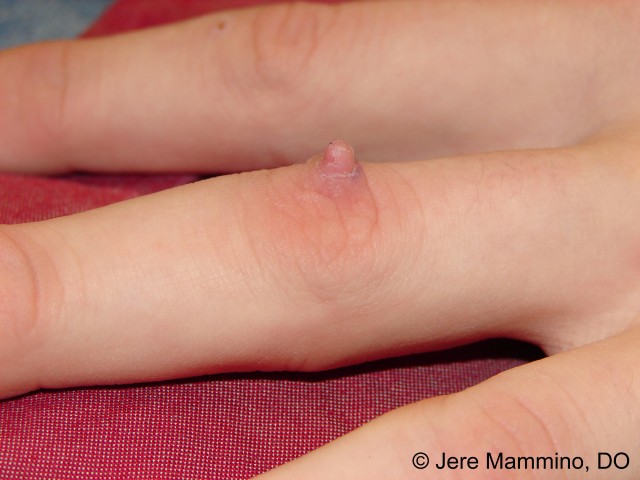
Middle finger


Collarette of epidermis surrounds nodules


Solitary, projecting keratotic mass


Skin colored, pedunculated firm nodule protruding from right heel toward sole
Gross description
- Pedunculated to dome shaped nodule on acral skin
Microscopic (histologic) description
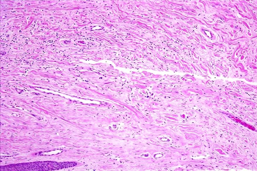
- Polypoid lesion with variably hyperplastic epidermis covering a dermal proliferation composed of dense collagen fibers and variable amounts of mature fibroblasts, small blood vessels and elastic tissue (J Am Acad Dermatol 1985;12:816)
- Thickened collagen in dermis is oriented predominantly in the vertical direction
- Stellate stromal cells may be present
- Covered by variably acanthotic epidermis with hyperkeratotic orthokeratosis
- Lesion merges with adjacent normal dermis
- Neural structures are absent or inconspicious
- Lacks adnexal structures
Microscopic (histologic) images
Contributed by Hillary Rose Elwood, M.D.

Polypoid dermal proliferation
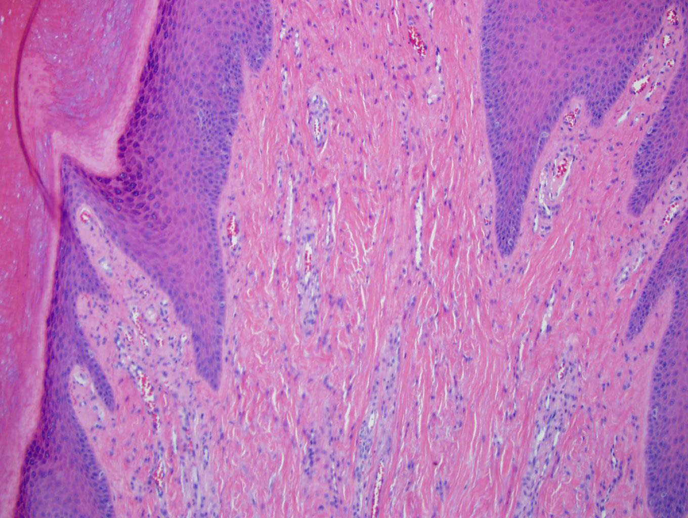
Prominent dense collagen
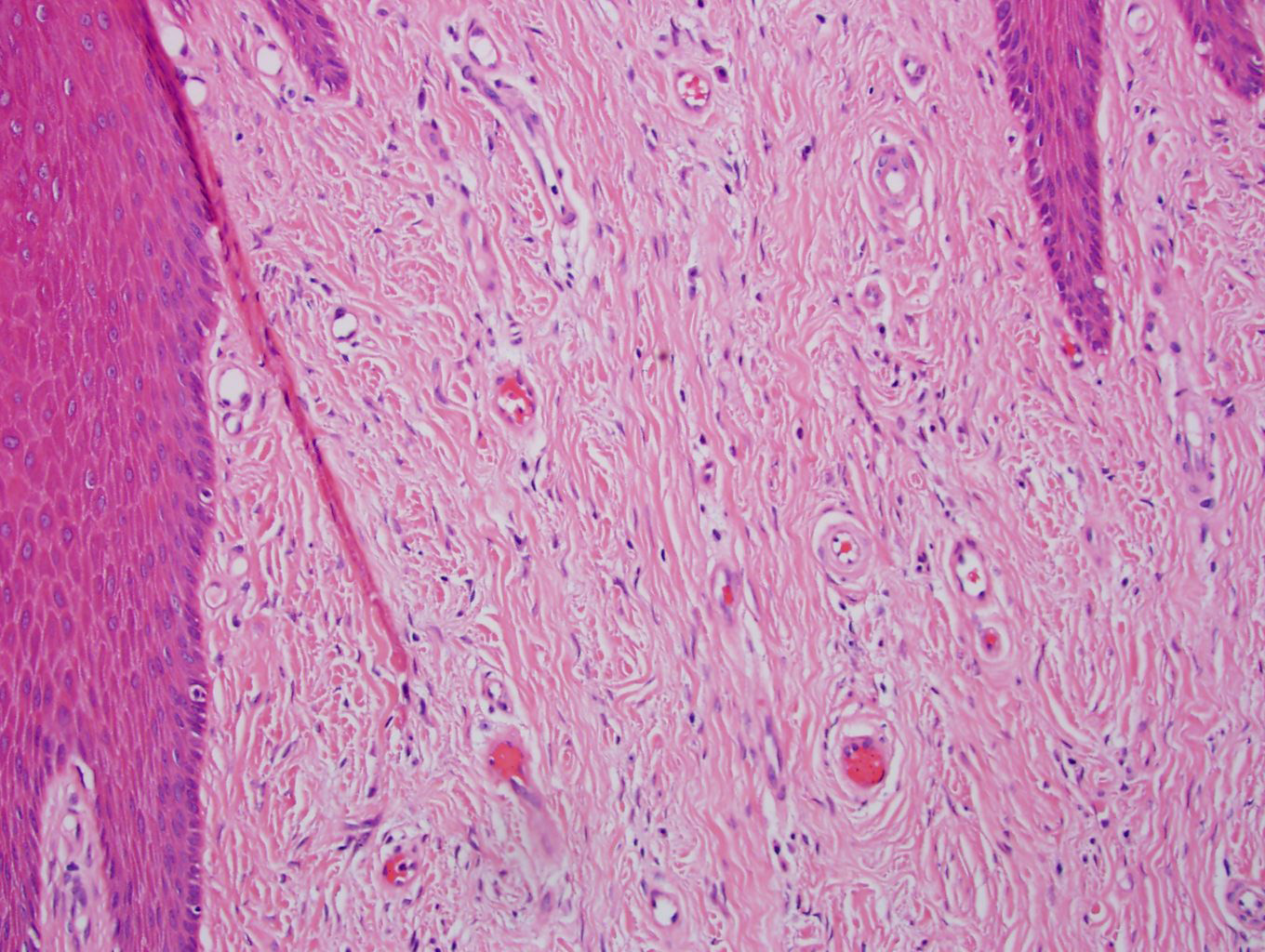
Vertically oriented collagen fibers
Images hosted on other servers:

Dome shaped tumor


Collagen fibers perpendicularly arranged

Acanthosis and massive orthokeratosis


Thickened collagen

Hyperkeratosis and acanthosis


Proliferating fibroblasts and capillaries

Various images
Negative stains
- Noncontributory
Differential diagnosis
- Acrochordon: non acral location, pedunculated, less hyperkeratotic, less dense connective tissue
- Hypertrophic scar: normal or atrophic epidermis, dermal band of fibroblasts and dense collagen, blood vessels oriented perpendicular to epidermis
- Periungual fibroma (Koenen tumor): similar/identical histology, distinction is predominantly based on clinical findings (i.e. location, multiple lesions, patient with tuberous sclerosis); some have noted that periungual fibromas can have prominent stellate atypical myofibroblasts, may have a more prominent vascular component, and may lack the epidermal changes of digital fibrokeratoma (Arch Dermatol 1995;131:1465)
- Supernumerary digit: has prominent neural structures (i.e. peripheral nerves or tactile corpuscles), sometimes cartilage/bone is present; most are related to the fifth digit
