Revised: 21 November 2017 Copyright: (c) 2001-2017, PathologyOutlines.com, Inc.
Dermoscopy
Reviewer: Christopher Hale, M.D. (see Reviewers page) Revised: 30 March 2013, last major update November 2012 Copyright: (c) 2002-2013, PathologyOutlines.com, Inc.
General
=========================================================================
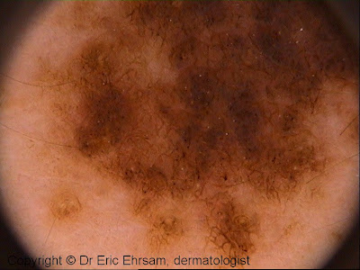
Method of skin examination using an optical instrument with a light source
Often combined with immersion fluid to reduce light reflection from skin surface; common immersion fluids include oil, ultrasonographic gel, alcohol-based disinfectants, water; recommended to use alcohol based gel as immersion fluid to prevent nosocomial infections (Dermatol Surg 2006;32:552)
Allows visualization of pigmentation patterns and deeper skin structures than can be seen with naked eye
Dermoscopy is a link between clinical and histologic examination, permitting an earlier diagnosis (Int J Dermatol 2008;47:712)
Uses by pathologists / other physicians
=========================================================================
Analyze pigmented skin lesions - benign lesions tend to have symmetrical dermoscopic structures and colors, but malignant lesions tend to have irregular and atypical dermoscopic structures (J Dermatol 2006;33:513)
Improves the ability of primary care physicians to accurately triage lesions suggestive of skin cancer (J Clin Oncol 2006;24:1877)
Nevus associated with skin type I Nevus associated with skin type IV Reticular nevus in white patients
(always burn and never tan) (tan easily and never burn) who may burn or tan
Reviewer: Christopher Hale, M.D. (see Reviewers page) Revised: 16 December 2014, last major update November 2012 Copyright: (c) 2002-2014, PathologyOutlines.com, Inc.
General
=========================================================================
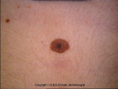
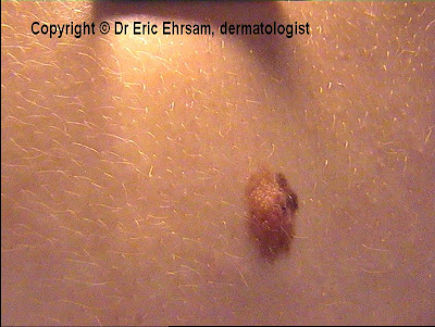
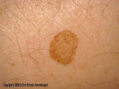
Congenital or acquired benign melanocytic proliferation
Color: due to Tyndall effect (scattering of light as it hits melanin granules, Wikipedia); melanin in stratum corneum appears black, melanin in reticular dermis appears slate-gray or blue
Nevi may regress due to lymphocytic infiltration (see halo nevus)
Clinical features
=========================================================================
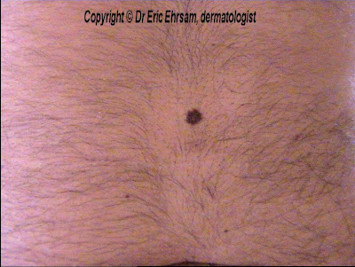
Nevi common on head, neck and trunk in Caucasians, on acral sites in Asians and Afro-Caribbeans
Mostly occur in skin, but also mucosal membranes covered by squamous epithelium
May be neoplastic since many are clonal
Existence of freckles, lentigines (small, pigmented, flat or slightly raised spots with a clearly defined edge, but no nests of melanocytes) and melanocytic nevi increases chance of having melasma (BMC Dermatol 2008;8:3)
Often accompanied by keratinous cysts, abscess or folliculitis
Incidental microscopic aggregates of nevus cells occur in 1% of skin excisions (Am J Dermatopathol 2008;30:45); also occur in clusters in lymph node capsules (intracapsular nevus), particularly in axilla (see lymph nodes chapter)
Increasing numbers of nevi are associated with neonatal phototherapy (Arch Dermatol 2006;142:1599), sun exposure on hot holidays (J Invest Dermatol 2005;124:56) and number of nevi in parents (Cancer 2003;97:628), although this does not necessarily mean that these factors are risk factors for melanoma
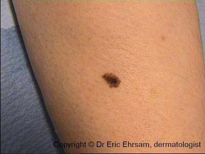
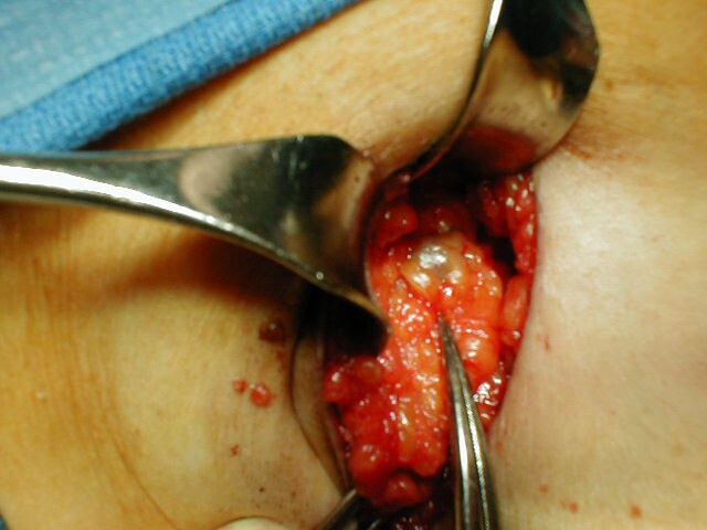
Biopsy any clinically atypical melanocytic lesions in adults, such as nevi causing chronic mechanical irritation, itching, bleeding, ulceration or oozing of serum, nevi with rapid growth, deepening pigmentation, pigmentation beyond outline of lesion, flat areas of depigmentation or erythema
Pathologically confirmed banal nevi and mildly atypical nevi do not require additional treatment
Nevi with moderate and severe atypia usually are excised with negative margins
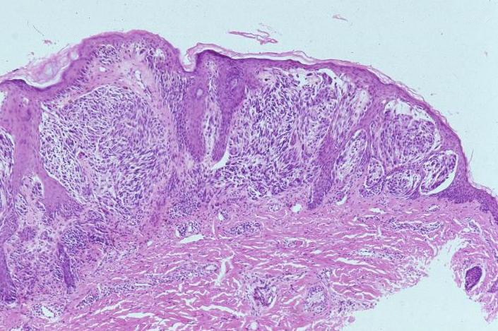
Junctional nests of melanocytes uniform in size, distributed at the tips of the rete ridges
Dermal component: Type A morphology:
In superficial dermis
Pigmented epithelioid cells with well-defined cell boundaries
Abundant eosinophilic to amphophilic cytoplasm containing coarse melanin granules
Uniform round / oval nuclei slightly smaller than that of adjacent keratinocytes
Finely dispersed chromatin
Delicate nuclear membrane
No / small distinct eosinophilic nucleoli
Type B morphology:
In intermediate dermis
Cells more lymphoid than epithelioid
Decreased cytoplasm with no melanin
Smaller and slightly hyperchromatic nuclei with dispersed chromatin and no nucleoli
Type C morphology:
In deep dermis
Spindled, fibroblast-like or schwannian cells with oval nuclei and bland chromatin
Single cell infiltration of superficial reticular collagen
Maturation:
Deeper portion of lesion has smaller cells with less pigment and less atypia
Deep cells grow in smaller sized nests or single cells
May resemble neural tissue
Terminal differentiation recapitulates some aspects of Schwann cell development
(Am J Pathol 1999;155:549)
Traumatized nevi:
Features include parakeratosis (92%), dermal telangiectasias (61%), ulceration (51%), dermal inflammation (49%), melanin within stratum corneum (24%), dermal fibrosis (25%), pagetoid spread of melanocytes limited to the site of trauma (20%) or away from areas of trauma (8%, Am J Dermatopathol 2007;29:134)
Reviewer: Christopher Hale, M.D. (see Reviewers page) Revised: 20 April 2013, last major update November 2012 Copyright: (c) 2002-2013, PathologyOutlines.com, Inc.
Acral nevi General
=========================================================================
Usually defined as nevi of palmar or plantar skin including nailbed
Parallel patterns present (Dermatology 2008;216:205); more prominent pigmentation seen in sulci (furrows) of dermatoglyphs, indicative of benign nature
Lentiginous pattern more common than in nevi of non-acral sites
Often (61%) low level pagetoid, single cell migration into stratum spinosum (Am J Surg Pathol 1995;19:792), summarized by "MANIAC" (Melanocytic Acral Nevus with Intraepithelial Ascent of Cells)
Pagetoid cells should not be atypical and should not be present beyond center of lesion
Possible transepidermal elimination of pigment within stratum corneum
Uniformly large melanocytes with abundant, pale, finely granular cytoplasm, large, vesicular nuclei but no prominent nucleoli (J Cutan Pathol 2005;32:40)
May be symmetric with pagetoid spread and moderate / marked atypia with nucleoli, but no mitotic figures or apoptotic melanocytes (Am J Dermatopathol 2005;27:111)
May have irregular nesting pattern
Nests may be poorly circumscribed with lateral extension of junctional component beyond dermal component
Head and neck nevi Clinical features
=========================================================================
Papular, usually compound nevi in children / young adults
May evolve into flesh colored dermal nevi with age
Four common patterns: (a) solid pink, (b) solid brown, (c) "eclipse" with tan center and darker outer ring and (d) "cockade" with darker center (Br J Dermatol 2011;165:137)
Congenital nevus: present at birth, > 1.5 cm, nevus cells follow skin appendages
Nevi Active Nevus
Reviewer: Christopher Hale, M.D. (see Reviewers page) Revised: 30 March 2013, last major update November 2012 Copyright: (c) 2002-2013, PathologyOutlines.com, Inc.
Clinical features
=========================================================================
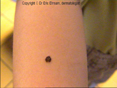
Solitary, small, very dark and papular nevus with prominent junctional component, melanocytic hyperplasia and intraepidermal melanin
Often increased cellularity, dermal inflammation, prominent nucleoli and occasional mitotic figures
Benign behavior
Nevi Balloon cell nevus
Reviewer: Christopher Hale, M.D. (see Reviewers page) Revised: 30 March 2013, last major update November 2012 Copyright: (c) 2002-2013, PathologyOutlines.com, Inc.
Clinical features
=========================================================================
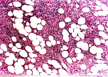
Nevus in which most melanocytes are large, pale with foamy or finely vacuolated cytoplasm, but no atypia
Balloon cell melanoma: has radial growth phase, mitoses and other melanoma cells in vertical growth component
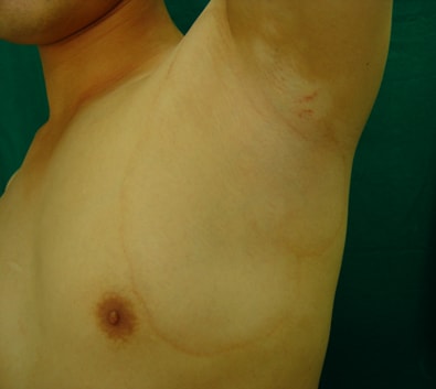
Nevi Becker's nevus (Becker's melanosis)
Reviewer: Christopher Hale, M.D. (see Reviewers page) Revised: 30 March 2013, last major update November 2012 Copyright: (c) 2002-2013, PathologyOutlines.com, Inc.
General
=========================================================================
Sharply demarcated pigmented macule, often with localized hypertrichosis (eMedicine)
May be associated with hypoplasia of ipsilateral breast (Arch Iran Med 2006;9:68), aplasia of ipsilateral pectoralis major muscle, limb shortening, lipoatrophy (Clin Exp Dermatol 2002;27:27), spina bifida and scoliosis
Occasionally associated with melanoma, but overall no known increased risk (Dermatologica 1991;182:77)
Increased basal layer pigmentation, mild acanthosis, hyperkeratosis and regular elongation of rete ridges
Variable hypertrichosis
Areas associated with smooth muscle hamartoma have more pronounced smooth muscle bundles irregularly dispersed within the dermis and unrelated to either hair follicles or vascular channels
Reviewer: Christopher Hale, M.D. (see Reviewers page) Revised: 30 March 2013, last major update November 2012 Copyright: (c) 2002-2013, PathologyOutlines.com, Inc.
General
=========================================================================
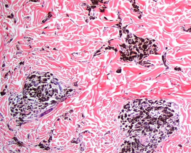
Collection of benign type C melanocytes (Schwann cell-like morphology) in dermis (eMedicine)
Blue color due to Tyndall effect of selective absorption of parts of the light spectrum by deeply located (dermal) melanin pigment, which is usually abundant
No relationship to blue rubber bleb nevus syndrome (eMedicine)
May be due to arrested migration of immature melanocytes in dermis
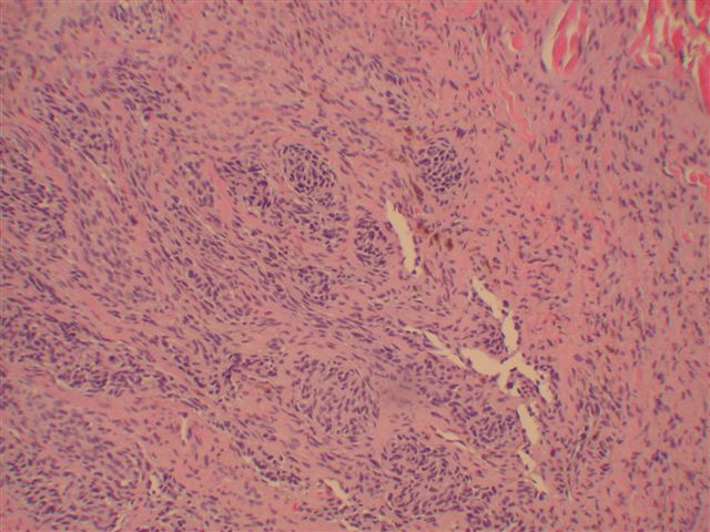
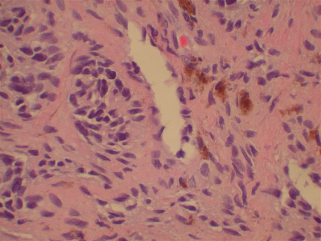
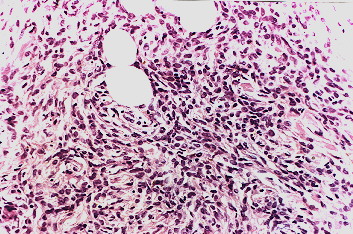
Ill defined deep dermal proliferation of spindled (Type C) melanocytes with abundant pigment and melanophages, dissecting dermal collagen and often extending into subcutis
Reviewer: Christopher Hale, M.D. (see Reviewers page) Revised: 29 March 2013, last major update November 2012 Copyright: (c) 2002-2013, PathologyOutlines.com, Inc.
Atypical cellular blue nevus General
=========================================================================
Atypia insufficient for definitive diagnosis of malignancy
No agreed upon standard, criteria include size (> 5-10 cm), ulceration, nuclear pleomorphism, > 3-4 mitotic figures per mm2 and pushing or infiltrating borders (Histopathology 2004;45:433, J Cutan Pathol 1998;25:252
Cellular blue nevus General
=========================================================================
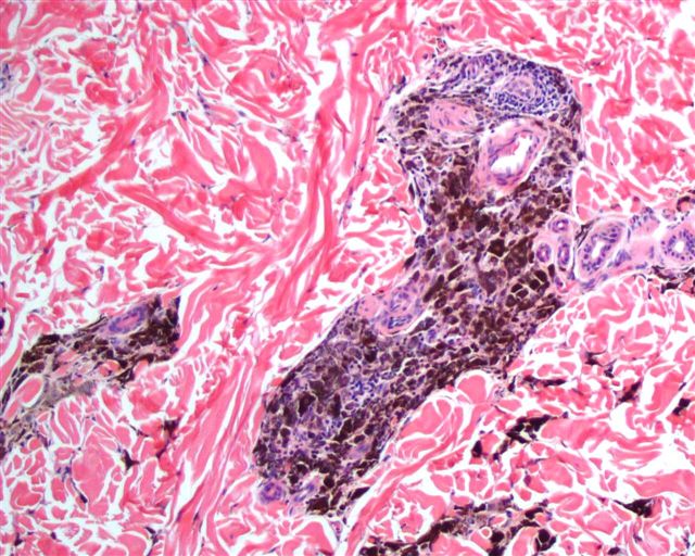
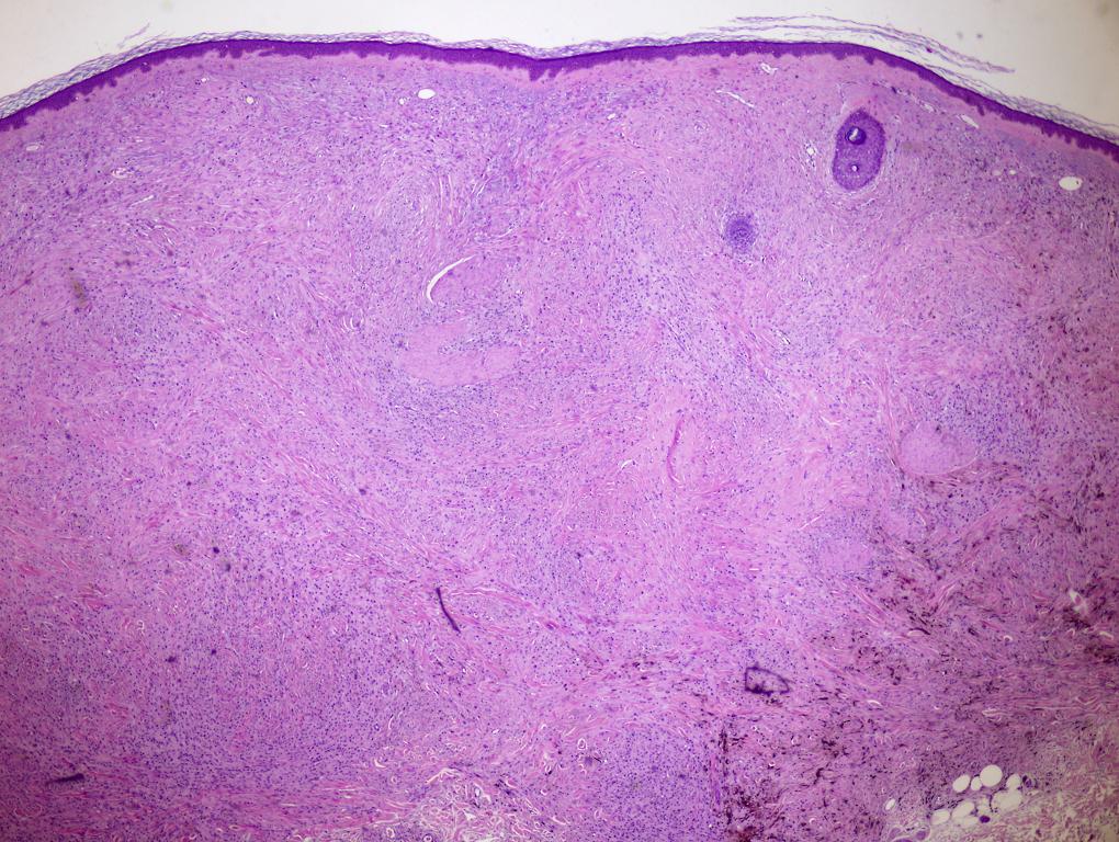
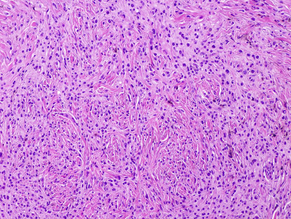
Usually pigmented, biphasic tumor with component of classic blue nevus and distinct cellular areas composed of spindled to oval melanocytes with clear or finely-pigmented cytoplasm (Arch Pathol Lab Med 2011;135:327)
Well-circumscribed collection of interweaving fascicles with increased cellularity and extension into subcutis
Heavily pigmented spindle cells alternate with clear cells
Have pushing margins and variable fasciculation and neural structures
No / minimal atypia; no junctional activity, no epidermal invasion, no peripheral inflammation, no necrosis and no / rare mitotic figures
Scalp lesions may have intracranial extension
"Ancient" blue nevi show stromal changes of large dilated vessels with pseudoangiomatous features, hyaline angiopathy, myxoid changes, sclerosis or hyalinization of stroma and variable edema, similar to ancient melanocytic nevi (Am J Dermatopathol 2008;30:1)
Note: benign cellular blue nevi may involve lymph node parenchyma and sinuses in a metastatic-like pattern; as a result, some tumors are best classified as having "uncertain biologic behavior"
Malignant blue nevus: scalp or heel lesion with marked nuclear atypia, numerous mitotic figures, some atypical and necrosis; variable epithelioid tumor cells
Some authors use "pigmented epithelioid melanocytoma" (PEM) to describe epithelioid blue nevi that is not associated with Carney complex (Arch Pathol Lab Med 2011;135:327)
May be part of Carney complex, which includes cardiac myxoma, psammomatous melanotic schwannoma, multicentric blue nevi and endocrine overactivity (Orphanet J Rare Dis 2006;1:21); may also occur by itself (Am J Dermatopathol 2000;22:473)
May be a low grade melanoma; 60% have nodal metastases, but clinical course is otherwise indolent
Case reports
=========================================================================
2 year old boy with congenital giant melanocytic nevus with pigmented epithelioid cells on back and no evidence of Carney complex (Am J Dermatopathol 2002;24:30)
Poorly circumscribed, but heavily symmetric pigmented dermal lesion
Short fascicles, small nests and single cells
Two populations of melanocytes: (a) intensely pigmented, globular and fusiform and (b) lightly pigmented, polygonal with large cytoplasm (J Cutan Pathol 2000;27:218)
Expansile asymmetric nodule with benign component OR low power benign features plus infiltrative borders, necrosis, mitoses or atypical cytologic features
May have epithelioid features with large hyperchromatic nuclei, prominent nucleoli and cytoplasmic melanin
Note: benign cellular blue nevus may involve lymph node parenchyma and sinuses in a metastatic-like pattern; some blue nevi are best classified as having "uncertain biologic behavior"
One study showed different allelic patterns at hOGG-1 locus between melanoma and control skin, with varying heterozygous allelic patterns in benign and malignant blue nevus (J Cutan Pathol 2008;35:651), but another study of eight genes (MTS1, MXI1, CMM1, p53, NF1, L-myc hOGG1 and MCC) showed no loss of heterozygosity (Am J Dermatopathol 2003;25:21)
Pigmented epithelioid melanocytoma: infiltrative, aggregates along follicles and cells resemble melanophages, but all cells are melanoma cells after bleaching
Cellular blue nevus and related entities
Nevi Cockarde nevus
Reviewer: Christopher Hale, M.D. (see Reviewers page) Revised: 21 November 2014, last major update November 2012 Copyright: (c) 2002-2014, PathologyOutlines.com, Inc.
General
=========================================================================
Reviewer: Christopher Hale, M.D. (see Reviewers page) Revised: 30 March 2013, last major update November 2012 Copyright: (c) 2002-2013, PathologyOutlines.com, Inc.
General
=========================================================================
Reviewer: Christopher Hale, M.D. (see Reviewers page) Revised: 30 March 2013, last major update November 2012 Copyright: (c) 2002-2013, PathologyOutlines.com, Inc.
General
=========================================================================
Features of both junctional and intradermal nevi (i.e. epidermal and dermal components)
Features of both junctional and intradermal nevi (i.e. epidermal and dermal components)
Junctional component is similar to junctional nevus, with nests regularly distributed at bases of rete ridges, occasional lentiginous pattern, no pagetoid spread, no atypia, symmetry diminishes with patient age
Dermal component consists of nests (may be very large) or linear pattern of melanocytes, cells are small with scant cytoplasm and regular nuclei and mature with depth by becoming more slender / spindled with less pigment
Dermal melanocytes or nests are separated by collagenous stroma
Often clusters of chronic inflammatory cells at base of nevus
Reviewer: Christopher Hale, M.D. (see Reviewers page) Revised: 30 March 2013, last major update November 2012 Copyright: (c) 2002-2013, PathologyOutlines.com, Inc.
General
=========================================================================
Most rigidly definition is nevi present at birth
Disagreement regarding whether nevi presenting very early in life but not at birth can be considered congenital (Acta Derm Venereol 2012;92:586)
In practice, nevi often labeled congenital if they have "congenital features" of melanocytes clustered around follicles, adnexae, nerves and between collagen fibers at base of lesion
Agminate / agminated nevus: nevi that are "clustered", and may be confined to a developmental segment (J Invest Dermatol 2011;131:788)
Nevus spilus / speckled lentiginous nevus: common type of agminate nevus with multiple pigmented macules or papules within a pigmented patch (Cutis 2007;80:465)
Neuronevus: compound nevi with prominent neural features, Masson’s neuronevus (cellular blue nevus with neural / schwannian differentiation) or congenital nevus with prominent neural features
Tends to involve reticular dermis, subcutis, skin adnexa, arrector pili muscles and nerves with single cell permeation of collagen
Also neural differentiation with Wagner-Meissner-like corpuscles
Frequent proliferative nodules
Lesions of infants may have pagetoid melanocytic proliferation
Proliferative nodules:
Dermal nodules of large epithelioid or spindled melanocytes that merge with surrounding nevus cells
Often prominent nucleoli, cellular areas, focal hemorrhage and ulceration, but no necrosis, no destructive growth, minimal inflammation and 0-4 mitotic figures/10 HPF
Symmetrical broad proliferation of melanocytes in papillary and reticular dermis with maturation, splaying between collagen bundles, permeation of muscles of hair erection, blood vessels, adnexa
Lesions present at birth usually have NRAS but not BRAF mutations
Lesions with congenital type histologic features but not present at birth more commonly have BRAF but not NRAS mutations (J Invest Dermatol 2007;127:179)
Germline Melanocortin-1-Receptor (MC1R) genotype is associated with severity of cutaneous phenotype in congenital melanocytic nevi (J Invest Dermatol 2012;132:2026)
~2.5% risk of malignant transformation, usually before adolescence (Br J Dermatol 2006;155:1); may give rise to cutaneous or CNS melanoma or related neuroectodermal tumors (malignant peripheral nerve sheath tumor, cutaneous malignant melanotic neurocristic tumor, rhabdomyosarcoma, liposarcoma)
Reviewer: Christopher Hale, M.D. (see Reviewers page)
Revised: 19 September 2012, last major update August 2012 Copyright: (c) 2001-2012, PathologyOutlines.com, Inc.
General
=========================================================================
● Rare connective tissue hamartoma derived from cells of mesodermal origin
● May be syndromic (Proteus syndrome, tuberous sclerosis, others) or isolated (collagenoma)
Case reports
=========================================================================
Reviewer: Christopher Hale, M.D. (see Reviewers page) Revised: 21 November 2014, last major update November 2012 Copyright: (c) 2002-2014, PathologyOutlines.com, Inc.
General
=========================================================================
Very cellular melanocytic nevus consisting of spindled melanocytes extending into reticular dermis or subcutis (Arch Dermatol 1993;129:328)
Case reports
=========================================================================
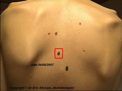
25 year old man with linear arrangement of multiple dark pigmented lesions in right periauricular area, above and behind ear (Arch Dermatol 2003;1319:1608)
Reviewer: Christopher Hale, M.D. (see Reviewers page) Revised: 30 March 2013, last major update November 2012 Copyright: (c) 2002-2013, PathologyOutlines.com, Inc.
General
=========================================================================
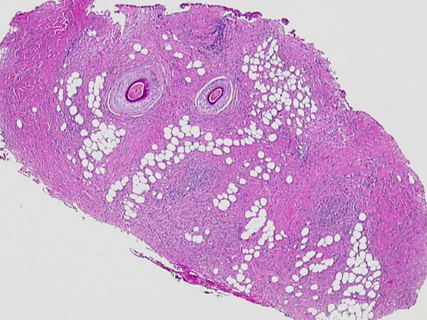
Small nests of melanocytes in upper dermis, often around pilosebaceous units, with variable pigmentation and cellularity
May have multinucleated melanocytes; deeper portion is usually less pigmented and less cellular and may have Wagner-Meissner corpuscles (representing neural portion of nevus)
Rarely nevus giant cells, balloon cells, infiltration by fat cells or osseous metaplasia
No junctional component
Can also be classified as Unna’s pattern (purely adventitial lesion confined to expanded papillary dermis and often to perifollicular dermis, usually neck, trunk or limbs) or Miescher’s pattern (melanocytes diffusely infiltrate adventitial and reticular dermis in wedge shaped pattern, usually on face, Am J Dermatopathol 2007;29:141)
Contributed by Angel Fernandez-Flores, MD, PhD, Hospital El Bierzo and Clinica Ponferrada, Spain: Unna’s pattern
Nevi Divided / kissing nevus
Reviewer: Christopher Hale, M.D. (see Reviewers page) Revised: 7 April 2013, last major update November 2012 Copyright: (c) 2002-2013, PathologyOutlines.com, Inc.
General
=========================================================================
Rare type of congenital nevus usually involving upper and lower eyelids of one eye
Reviewer: Christopher Hale, M.D. (see Reviewers page) Revised: 7 April 2013, last major update November 2012 Copyright: (c) 2002-2013, PathologyOutlines.com, Inc.
General
=========================================================================
Controversial topic, particularly for solitary lesions; better defined in dysplastic nevus syndrome (multiple dysplastic nevi and two family members with melanoma, J Am Acad Dermatol 2012;67:1.e1, eMedicine)
Compound nevi with marked lentiginous proliferation of melanocytes at dermoepidermal junction, extending at least 3 rete ridges beyond lateral margins of dermal component
Nests have cytologic and architectural atypia, including irregular sizes and shapes and bridging of adjacent rete ridges, which are irregular themselves
Papillary dermal lamellar fibroplasia with perivascular infiltrate and vascular dilation
Microarray analysis of four markers (ING4, Cul1, BRG1 and Bim) may distinguish melanoma from dysplastic nevi (PLoS One 2012;7:e45037)
Electron microscopy description
=========================================================================
Cases with severe dysplasia share several features with radial growth phase melanomas, including large cell size, bizarre shaped and pleomorphic nuclei, well-developed Golgi, abundant and deranged mitochondria, aberrant melanosomes with deranged structures and irregular melanization
Reviewer: Christopher Hale, M.D. (see Reviewers page) Revised: 7 April 2013, last major update November 2012 Copyright: (c) 2002-2013, PathologyOutlines.com, Inc.
General
=========================================================================
Large acquired melanocytic nevi that occur in patients with hereditary epidermolysis bullosa
Two theories: 1) repetitive basement membrane disruption => local nevus cell nests or single melanocytes break senescence and proliferate; 2) melanocytes, probably from incipient nevi or subclinical nests of nevus cells, float in fluid-filled cavity of blister, eventually settling down and proliferating in microenvironment of epidermal regeneration (Dermatol Clin 2010;28:179)
Clinical features
=========================================================================
Melanocytic growth factors in blister fluid may promote proliferation, migration and melanogenesis of disconnected melanocytes (Acta Derm Venereol 2003;83:332)
Asymmetrical, irregularly pigmented (initially very dark, then loses pigment)
Foci of stippled pigmentation and scarring
Case reports
=========================================================================
Two female infants with atypical melanocytic lesions in recessive dystrophic epidermolysis bullosa (Clin Exp Dermatol 2005;30:636)
Some features of melanoma are common: irregular pigmentation (96%), multicomponent pattern (87%), atypical pigment network (74%), irregular dots / globules (70%) and atypical vascular pattern (30%)
Other features associated with melanoma progression are rare: irregular streaks, blue-white veil, regression structures (blue-white areas) and black dots (Br J Dermatol 2005;153:97)
Reviewer: Christopher Hale, M.D. (see Reviewers page)
Revised: 20 June 2012, last major update June 2012 Copyright: (c) 2001-2012, PathologyOutlines.com, Inc.
General
=========================================================================
● Rare, congenital, hamartomatous
● Usually on face
Case reports
=========================================================================
● Well-differentiated hair follicles and sebaceous glands
● No cartilage (seen in accessory tragus), central cysts or a central canal (seen in trichofolliculoma)
Electron macroscopy description
=========================================================================
● Follicular germ cells present
● Active fibroblasts around the follicles merge with colloid substance
(J Dermatol 2001;28:324)
Reviewer: Christopher Hale, M.D. (see Reviewers page) Revised: 7 April 2013, last major update November 2012 Copyright: (c) 2002-2013, PathologyOutlines.com, Inc.
General
=========================================================================
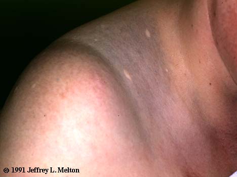
Nevus surrounded by zone of hypopigmented skin (eMedicine)
Clinical features
=========================================================================
Single or multiple
Usually due to regression caused by cell mediated immunity, or less commonly humoral immunity or granulomatous inflammation (Am J Dermatopathol 2008;30:233)
Reviewer: Christopher Hale, M.D. (see Reviewers page) Revised: 9 April 2013, last major update November 2012 Copyright: (c) 2002-2013, PathologyOutlines.com, Inc.
General
=========================================================================
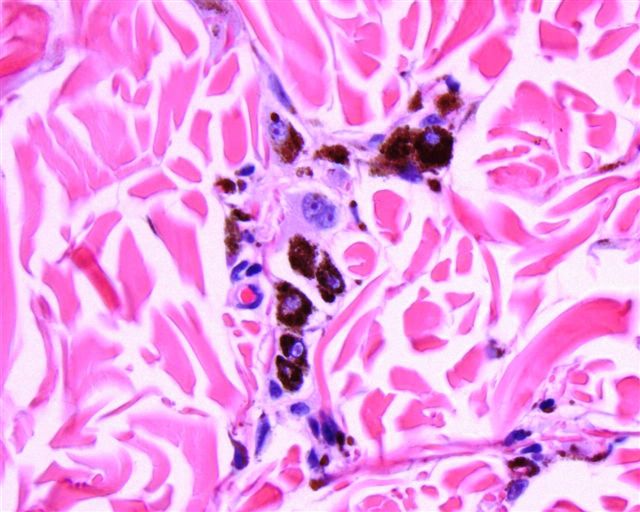
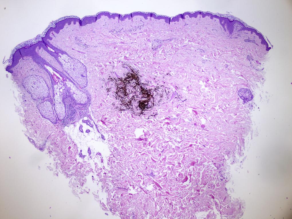
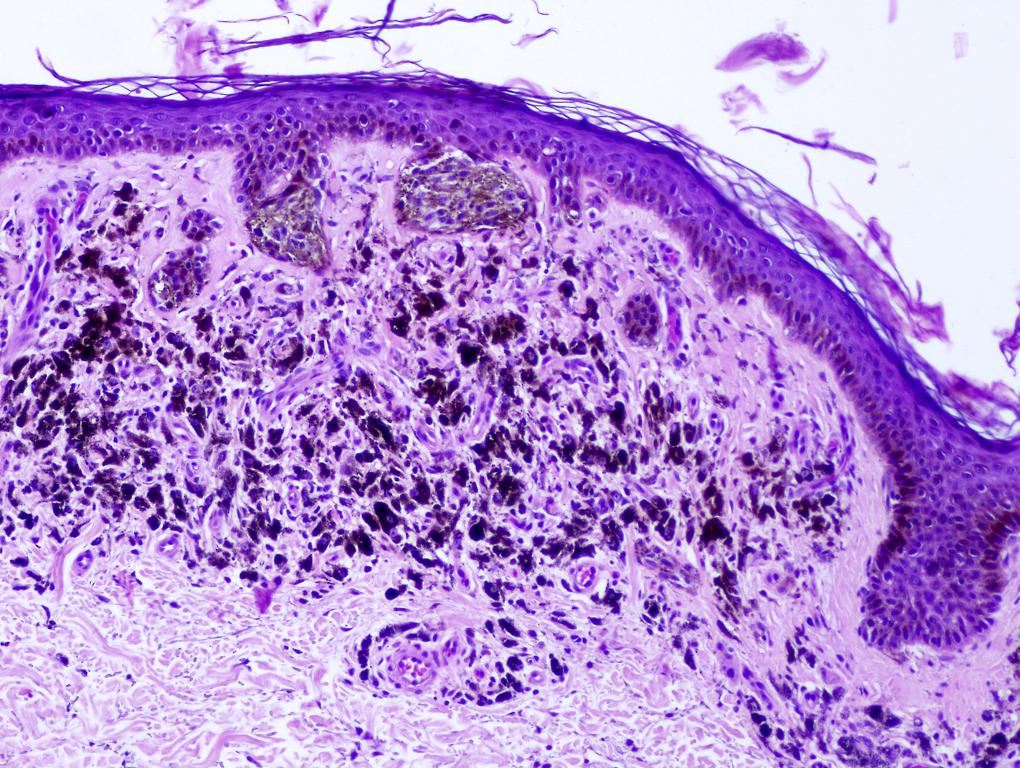
First described by Minor Ito in 1954 (Tohoko J Exper Med. 1954;60:10)
By definition, found in "shawl" or "cloak" distribution
Clinical features
=========================================================================
Macule with irregular blue-gray pigmentation
Similar to nevus of Ota except for location
Ito’s nevus is found in shoulder, side of neck and supraclavicular areas, within the distribution of the lateral (also called posterior) supraclavicular nerve and lateral cutaneous brachial nerves
Nevus of Ota is found around the eyes
Case reports
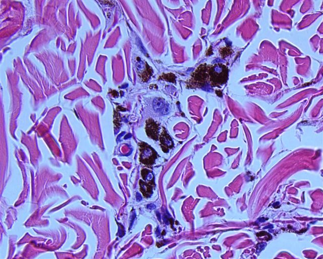
=========================================================================
Deeply pigmented dendritic melanocytes and melanophages dissecting bundles of dermal collagen in reticular dermis
Overlying epidermis is normal
Nevi Junctional nevus
Reviewer: Christopher Hale, M.D. (see Reviewers page) Revised: 9 April 2013, last major update November 2012 Copyright: (c) 2002-2013, PathologyOutlines.com, Inc.
General
=========================================================================
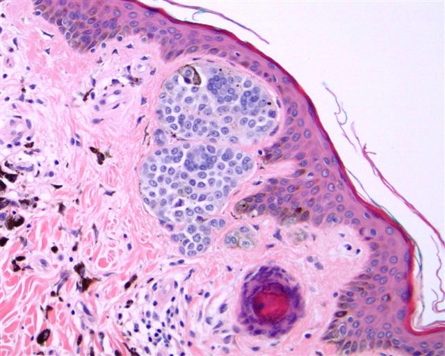
Melanocytic proliferation restricted to basal epidermis (junctional area)
Earliest stage of intraepidermal melanocytic proliferation
Reviewer: Christopher Hale, M.D. (see Reviewers page) Revised: 21 November 2014, last major update November 2012 Copyright: (c) 2002-2014, PathologyOutlines.com, Inc.
General
=========================================================================
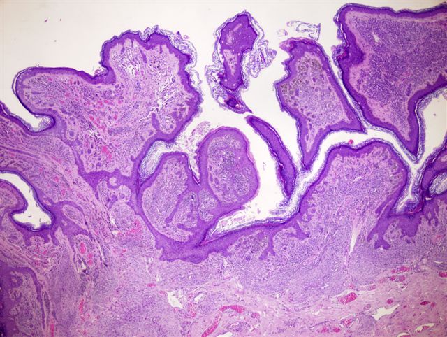
Common type of agminate (clustered) nevus with multiple pigmented macules or papules within a pigmented patch (Cutis 2007;80:465, eMedicine)
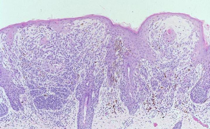
"Shoulder" area of lentiginous junctional melanocytic proliferation beyond lateral border of underlying dermal nevus
Elongation of rete ridges with small nests of melanocytes at tips of rete
Individual unit melanocytes extending along sides of rete, often mild lymphohistiocytic infiltrate with pigment incontinence
No atypia, no pagetoid spread and no dermal fibrosis
Acral lesions: resemble dysplastic nevus due to elongation of rete ridges, continuous proliferation of melanocytes at dermoepidermal junction, single scattered melanocytes or less commonly small clusters within the upper epidermis; poor or absent lateral circumscription, melanocytes with abundant pale cytoplasm and round / oval, sometimes hyperchromatic nuclei and prominent nucleoli present at the dermoepidermal junction; however, unlike dysplastic nevi, they lack anastomosing rete ridges, cytological atypia and well-formed lamellar fibroplasia (Histopathology 1995;27:549)
Variants:
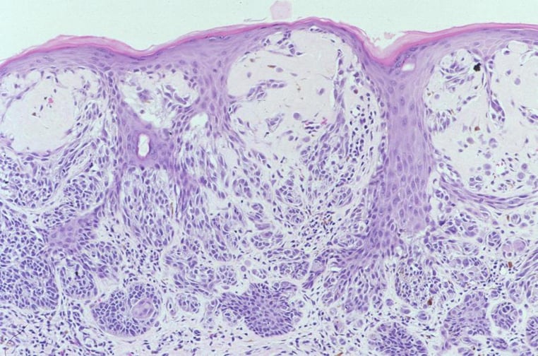
Macular variant:
"jentigo" pattern (lentiginous pattern plus nests of melanocytes at dermal-epidermal junction) in the darker speckles and by some nests of melanocytes at the dermoepidermal junction at the tips of the papillae, but background pigmentation has microscopic features of lentigo; tan-brown background with dark flat speckles in relatively even distribution resembling polka dots; associated with phacomatosis pigmentovascularis
Papular variant: dermal or compound melanocytic nevi; light-brown macule superimposed by multiple melanocytic nevi in the form of papules or nodules that show a more uneven distribution reminiscent of a star map; small dark macules may be present; associated with phacomatosis pigmentokeratotica or speckled lentiginous nevi syndrome (Dermatology 2006;212:53)
A, B: melanocytes are disposed in small nests and single cells; C: epithelioid melanocytes with enlarged nuclei and prominent nucleoli
A: preservation of the retiform epidermis with virtual confluent proliferation of melanocytes at dermoepidermal junction B: Clark level II invasion into the papillary dermis
A, B: melanocytic proliferation and pagetoid spread of melanocytes with Mart-1 stain C: Mitf
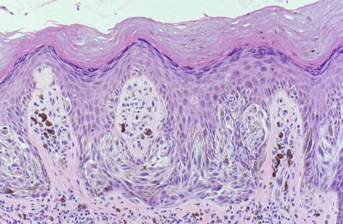
Nests of small and monomorphic melanocytes
at the dermoepidermal junction and within
the reticular dermis, with many melanocytes concentrated
around blood vessels and adnexal structures
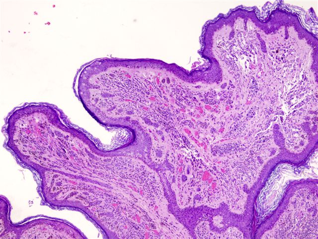
Elongated rete ridges and lentiginous proliferation of melanocytes at the dermal-epidermal junction
Prominent basal layer pigmentation similar to that seen in lentigo simplex
Reviewer: Christopher Hale, M.D. (see Reviewers page) Revised: 9 April 2013, last major update January 2013 Copyright: (c) 2002-2013, PathologyOutlines.com, Inc.
General
=========================================================================
Reviewer: Christopher Hale, M.D. (see Reviewers page) Revised: 5 November 2014, last major update January 2013 Copyright: (c) 2002-2014, PathologyOutlines.com, Inc.
General
=========================================================================
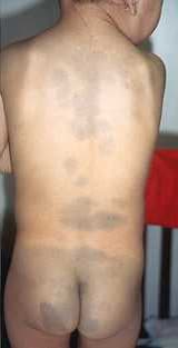
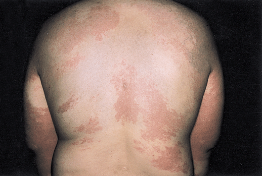
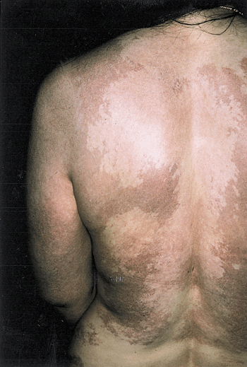
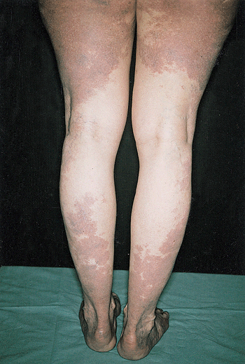
Ill defined area of blue discoloration, up to several centimeters and in lumbosacral region (eMedicine, Wikipedia)
Reviewer: Christopher Hale, M.D. (see Reviewers page)
Revised: 14 July 2012, last major update June 2012 Copyright: (c) 2001-2012, PathologyOutlines.com, Inc.
General
=========================================================================
● Often incorrectly spelled "nevus sebaceous"
● Congenital, organoid epidermal nevus on scalp and face
● Malformed adnexal structures, increased risk of trichoblastomas, basal cell carcinoma, squamous cell carcinoma
● Sex hormone responsive => need prophylactic excision before puberty
Reviewer: Christopher Hale, M.D. (see Reviewers page) Revised: 21 November 2014, last major update January 2013 Copyright: (c) 2002-2014, PathologyOutlines.com, Inc.
General
=========================================================================
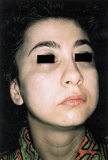
First described by Ota in 1939 (Tokyo Med J 1939;63:1243)
Uncommon hamartoma in periorbital and temporal skin (eMedicine)
Types: see also Jpn J Dermatol 1939;46:435
Type IA: mild orbital type - distribution over upper and lower eyelids, periocular and temple region
Type IB: mild zygomatic type - pigmentation in infrapalpebral fold, nasolabial fold and zygomatic region
Type IC: mild forehead type - involvement of forehead alone
Type ID: involvement of ala nasi alone
Type II: moderate type - distribution over upper and lower eyelids, periocular, zygomatic, cheek and temple regions
Type III: involves scalp, forehead, eyebrow and nose
Also called oculodermal melanosis or nevus fuscoceruleus ophthalmomaxillaris
Similar to nevus of Ito except for location (Ito in shoulder, side of neck and supraclavicular areas, within the distribution of the lateral supraclavicular nerve and lateral cutaneous brachial nerves)
Clinical features
=========================================================================
Tends to persist and extend locally, becoming increasingly prominent with age, puberty and postmenopausal state
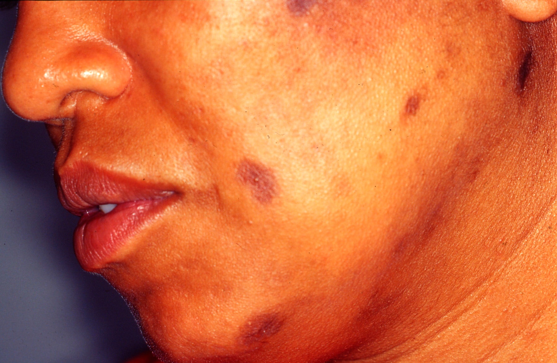
Associated with ipsilateral glaucoma, intracranial melanocytosis; rarely with cutaneous, ocular or intracranial melanoma (Cutis 2008;82:25) and vascular nevus (J Am Acad Dermatol 2008;58:88)
Macule with irregular blue-gray pigmentation in distribution of 1st and 2nd division of trigeminal nerve
May coexist with nevus of Ito
Tanino classification system most common, others include Hirayama system and proposed Chan system (Lasers Surg Med 2001;28:267)
Case reports
=========================================================================
Combined skin abrasion and carbon dioxide snow method (Plast Reconstr Surg 1996;97:544); cryosurgery and microsurgery not recommended due to scarring; chemical bleaching not recommended due to depigmentation
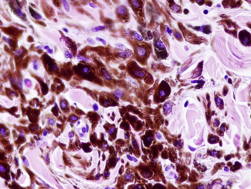
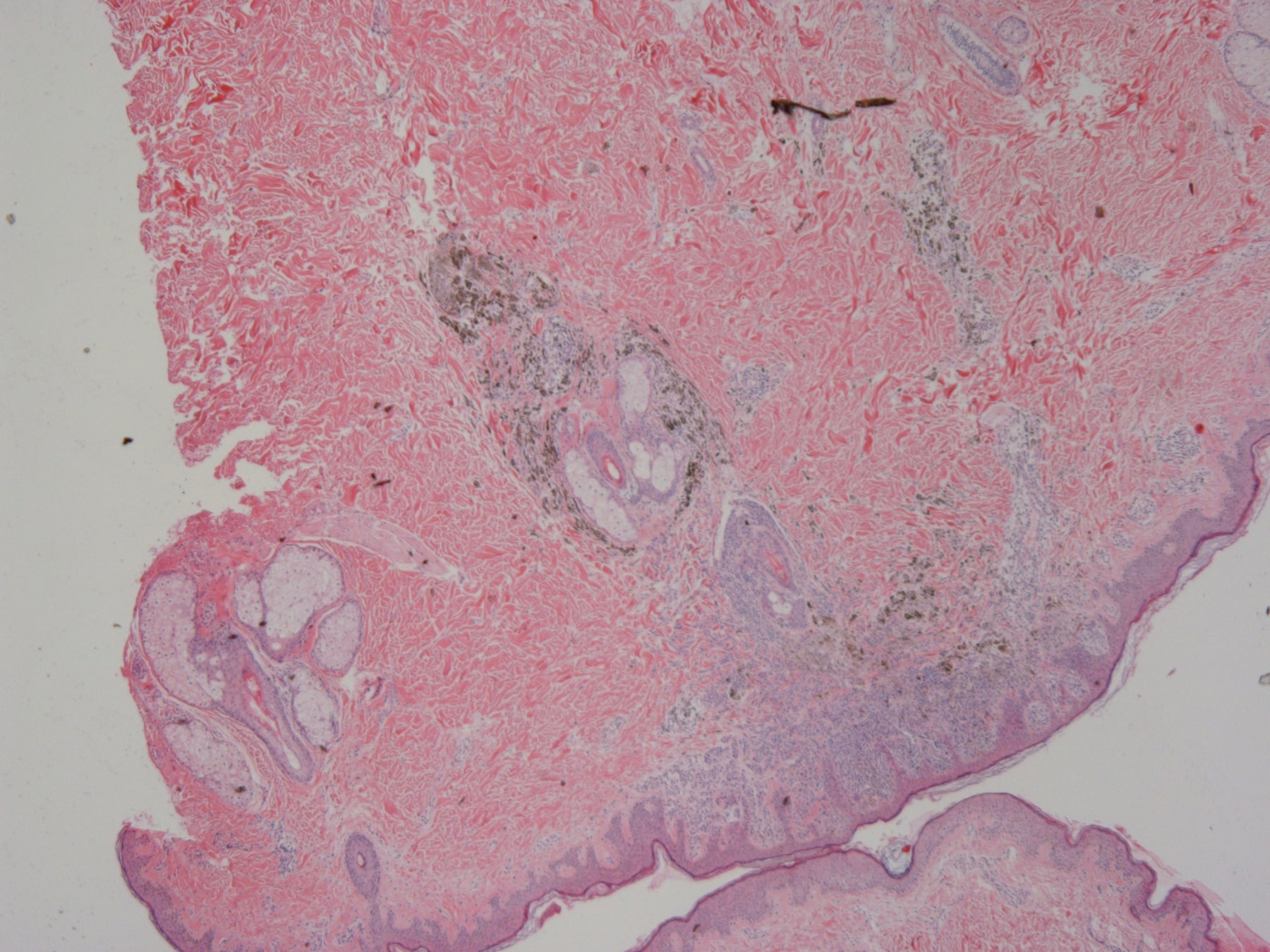
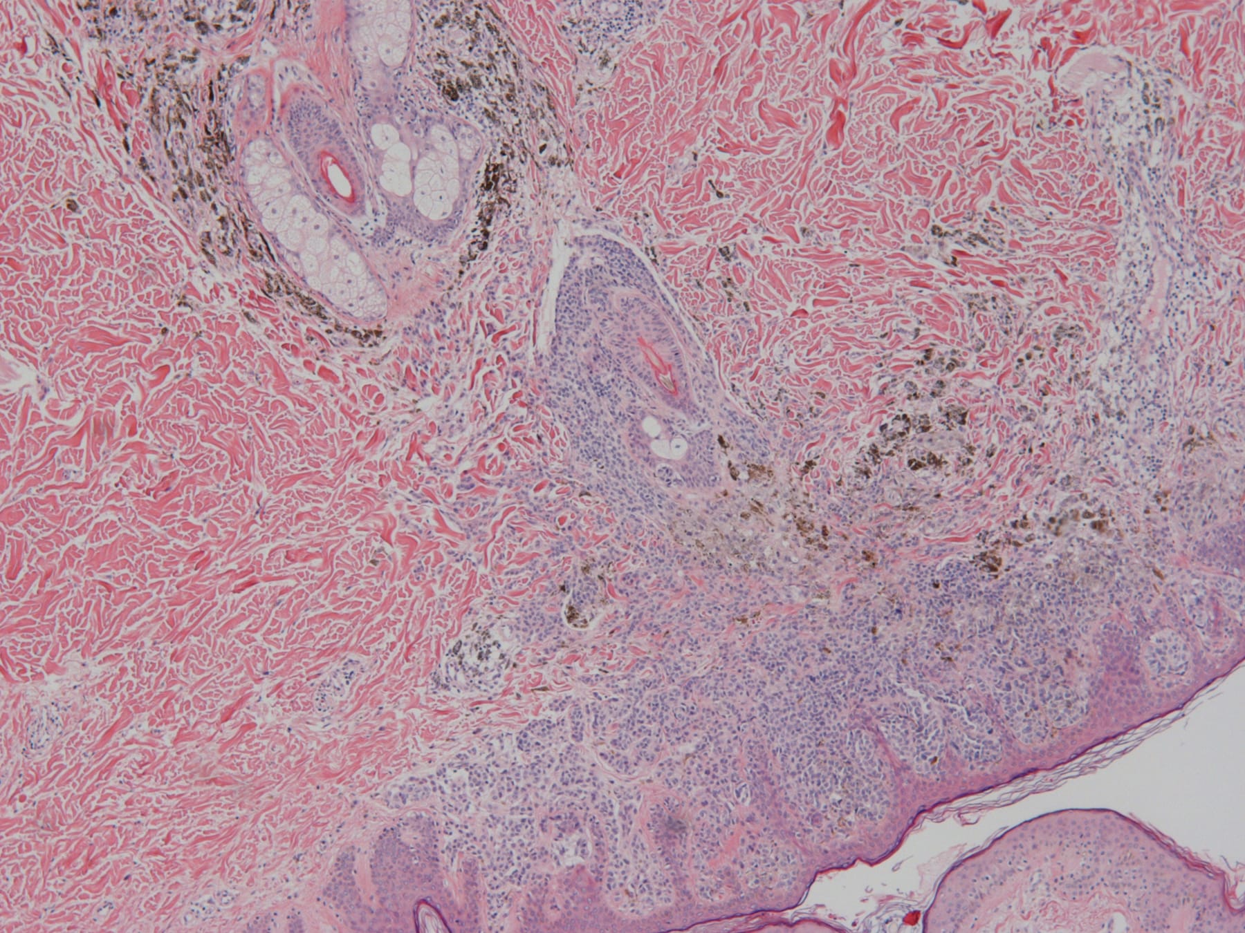
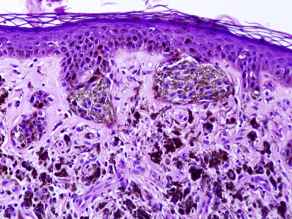
Bipolar dendritic melanocytes in papillary and reticular dermis are arranged as single units between collagen bundles, also numerous pigment-laden macrophages
Contributed by Dr. Asmaa Gaber Abdou, Menofiya University, Egypt:
Elongated dendritic melanocytes scattered with collagen bundles extending around hair follicles
Wavy dendritic cells with evenly dispersed melanin granules distributed in the superficial reticular dermis
Reviewer: Christopher Hale, M.D. (see Reviewers page) Revised: 13 April 2013, last major update January 2013 Copyright: (c) 2002-2013, PathologyOutlines.com, Inc.
General
=========================================================================
Rare syndrome defined by organoid nevus (occasionally with sebaceous differentiation) arranged along Blaschko’s lines plus speckled lentiginous nevus (SLN) of papular type (papular nevus spilus) in checkerboard pattern (Pediatr Dermatol 2011;28:715)
Subdivided based on presence or absence of extracutaneous abnormalities including:
Skeletal: bone cysts, kyphosis and scoliosis, foot and hand deformities, craniofacial defects, dislocation of hip hemihypertrophy and vitamin D resistant rickets
Neurologic: hemimegalencephaly with contralateral motor disease, mental and cognitive deficiency, seizure, hydrocephalus, Dandy-Walker malformation, dysplasia of brain vessels, agenesis of corpus callosum, cerebral heterotropia, cortical agyria, microgyria or pachygyria
Ocular: coloboma, epibulbar lipodermoid, nystagmus, corneal opacity, defects of optic nerve and cortical blindness
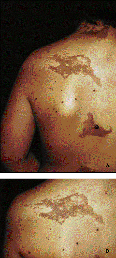
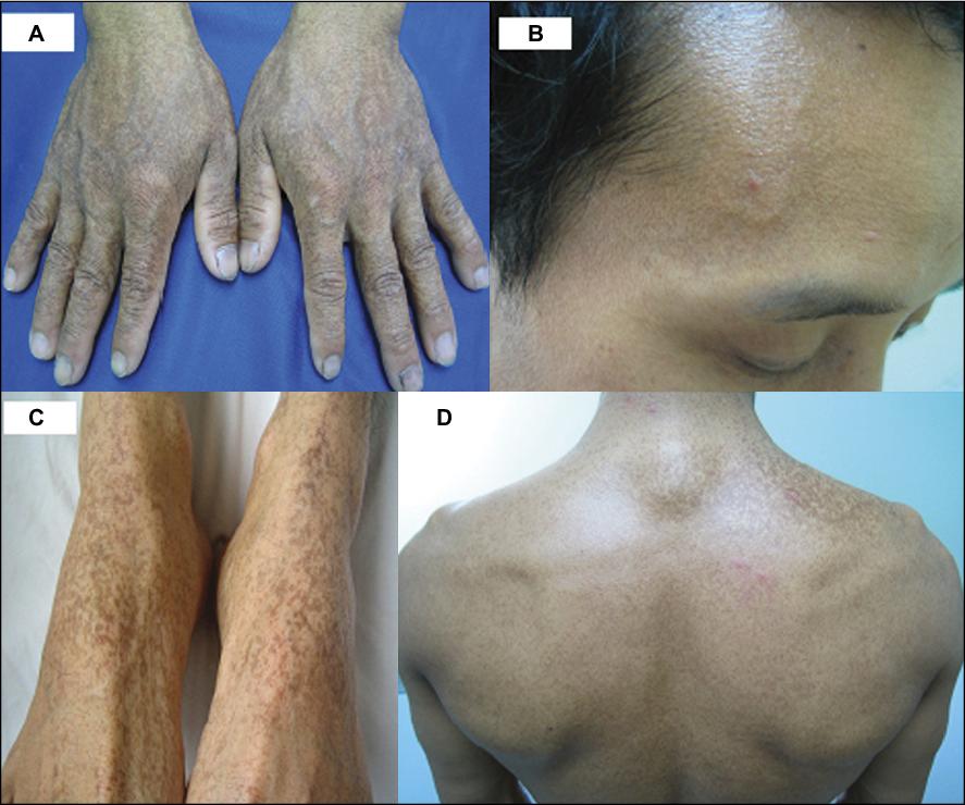
Verrucous epidermal nevus and ipsilateral speckled lentiginous nevus
Papular type speckled lentiginous nevus, telangiectatic nevoid area on upper right side
Sebaceous nevus on left preauricular area
Sebaceous nevus involving face, speckled-lentiginous nevus on trunk
Systematized epidermal nevus pn right side of body, speckled-lentiginous nevus with checkerboard distribution on both sides
Organoid nevus involving eye and mouth
Speckled lentiginous nevus showing a checkerboard distribution and hemiatrophy with postural deviation
Speckled-lentiginous nevus
Linear arrangement of organoid nevus on neck and upper back
Systematized epidermal nevus of left side of body
Epidermal nevus and pigmented lesions right forearm and back
Speckled lentiginous nevus in lumbar area
Epidermal nevus right side of trunk and leg plus epidermal and melanocytic nevus on right hand
With epidermal nevus following Blaschko’s lines and a large light-brown macule on each side of trunk
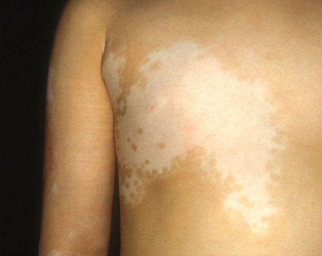
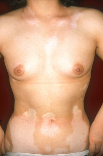
Nevi Phacomatosis pigmentovascularis (PPV)
Reviewer: Christopher Hale, M.D. (see Reviewers page) Revised: 13 April 2013, last major update February 2013 Copyright: (c) 2002-2013, PathologyOutlines.com, Inc.
General
=========================================================================
Phacomatosis cesioflammea: blue spots [caesius = bluish gray] plus nevus flammeus; also called types IIa/IIb; includes nevus of Ota; most common of 3 types (J Am Acad Dermatol 2008;58:88); often associated with Sturge-Weber and Klippel-Trenaunay syndromes
Phacomatosis spilorosea: nevus spilus [speckled lentiginous nevus] plus pale-pink telangiectatic nevus; also called types IIIa/IIIb
Phacomatosis cesiomarmorata: blue spots plus cutis marmorata telangiectatica congenita; also called type V
Subtypes: a - cutaneous involvement only, b - cutaneous and system involvement
Two cases of PPV type IIb with one patient demonstrating concurrent Sturge-Weber syndrome and Klippel-Trenaunay syndrome (Pediatr Dermatol 2010;27:303)
Extensive nevus flammeus involving face, thorax , arms; nevus anemicus on right pectoral area
Aberrant mongolian spots affecting upper part of back
Extensive nevus flammeus involving back and left arm
Blue-black macular pigmentation on periorbital areas (bilateral scleral melanosis)
Extensive nevus flammeus involving lower limbs
Extensive nevus flammeus and livedo reticularis pattern on trunk and extremities, more pronounced on right chest with diffuse involvement of face
Left lower limb 3 cm longer, circumference larger than right side
Diffuse bluish discoloration on back, right lateral chest wall, lower extremities
Phacomatosis cesioflammea
Phacomatosis spilorosea
Phacomatosis cesiomarmorata
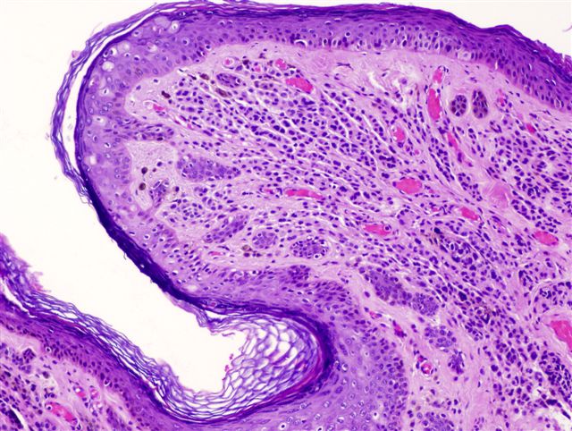
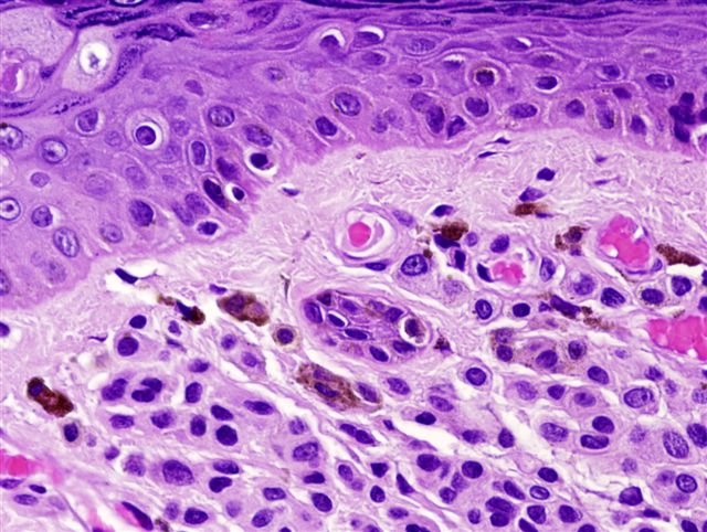
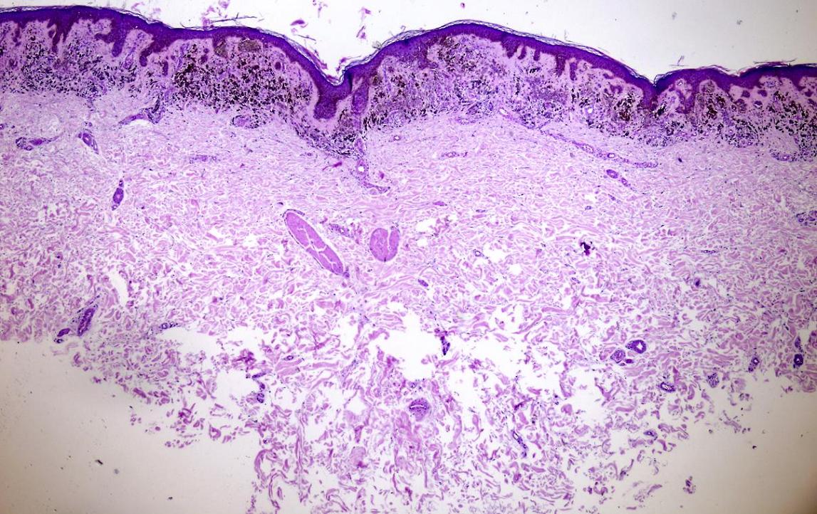
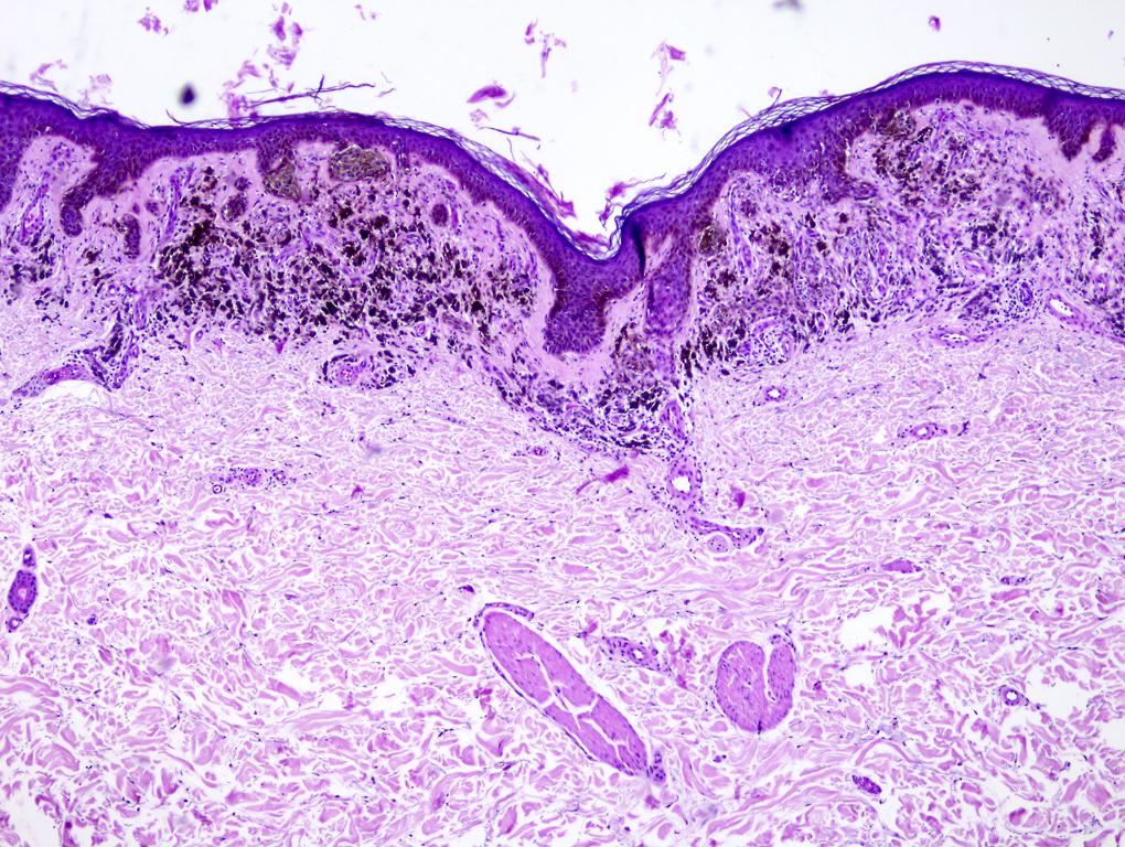
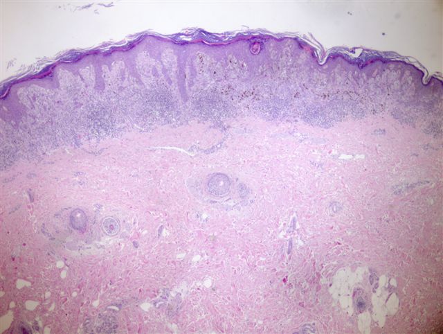
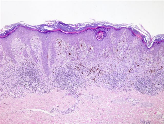
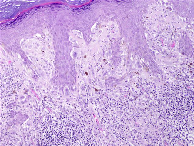
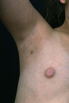
Nevi Pigmented spindle cell nevus
Reviewer: Christopher Hale, M.D. (see Reviewers page) Revised: 14 April 2013, last major update February 2013 Copyright: (c) 2002-2013, PathologyOutlines.com, Inc.
General
=========================================================================
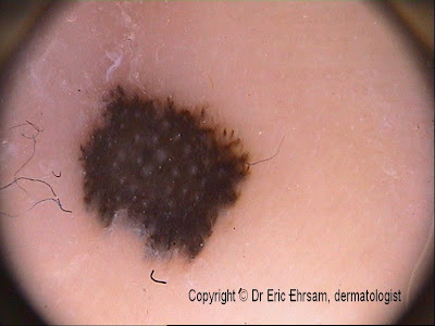
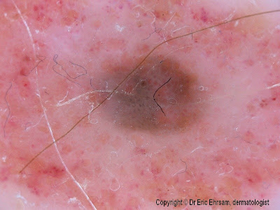
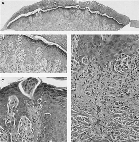
Two patterns reported: (a) brown to blue pigmentation with peripheral rim of large brown globules (globular pattern); (b) dark diffused pigmentation and pseudopods regularly distributed at periphery in stellate or radiate pattern (starbust pattern, Dermatol Online J 2004;10:5)
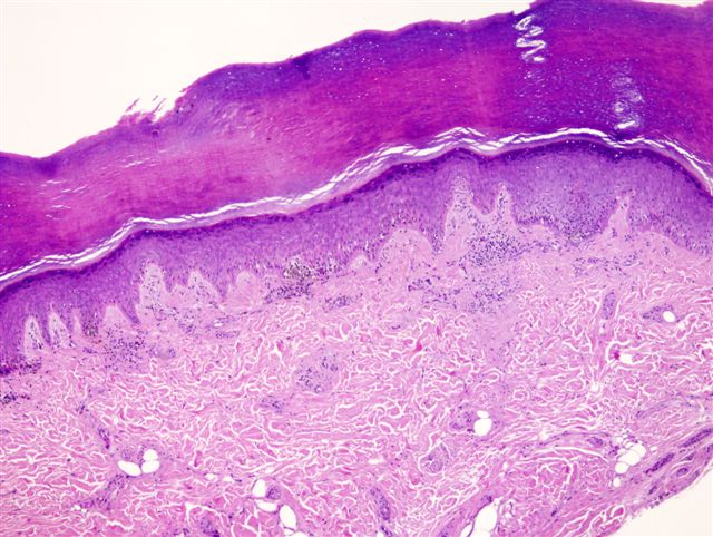
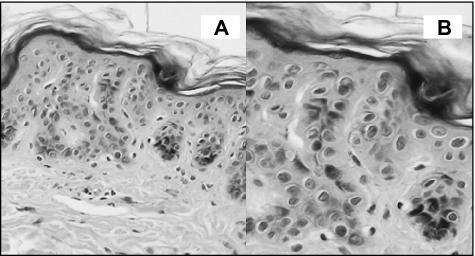
Fig A: bilaterally symmetrical lesion has proliferation of nested melanocytes that "rain down" from epidermis and expand papillary dermis without involving reticular dermis
Fig B: lesional cells mature from superficial to deep
Fig C: the most lateral cells are in the form of a nest rather than single cells (well circumscribed)
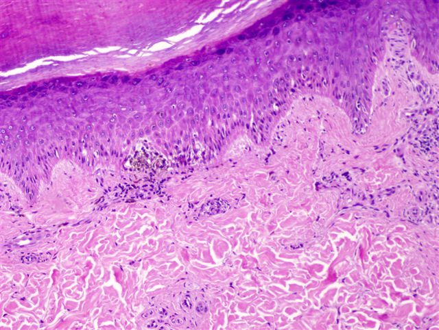
Some intraepidermal nests show clefts similar to Spitz nevi, but they more often blend imperceptibly with keratinocytes in a pigmented spindle cell nevus (fig B);
Mitotic figures in the epidermis are common, but they are not numerous in the dermis;
Maturation towards a small nevoid cell at the base in the papillary dermis, without involvement of the reticular dermis, is typical
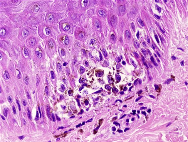
Fig B: bilaterally symmetrical lesion whose cells mature from epidermis to base of papillary dermis, respecting the reticular dermis interface (fig B);
Fig C: nucleolated lesional cells with abundant, coarse and dusty pigment are characteristic
Reviewer: Christopher Hale, M.D. (see Reviewers page) Revised: 20 April 2013, last major update February 2013 Copyright: (c) 2002-2013, PathologyOutlines.com, Inc.
Symmetric with sharp lateral borders, usually compound nevus with prominent intraepidermal component
5% are junctional, 20% are dermal
Composed of spindled and epithelioid cells
Spindle cells may be arranged in fascicles in dermal papillae, are perpendicular to epidermis, cigar-shaped with large nuclei, have prominent nucleoli
Epithelioid cells are dispersed individually, polygonal with abundant eosinophilic cytoplasm, distinct cell borders, large nuclei and prominent nucleoli, have variable mitotic figures, occasional multinucleation and often marked atypia, although most cells appear benign
Cell maturation occurs in deep portion of tumor
Also large and well-formed Kamino bodies (eosinophilic hyaline bodies along dermoepidermal junction)
May have pagetoid growth, lymphatic invasion, pseudoepitheliomatous hyperplasia, "tubular" growth pattern, plexiform growth pattern, halo reaction, prominent vasculature (Am J Dermatopathol 2000;22:135) and lymphocytic infiltrate
Scanty pigmentation
"Consumption of epidermis" (usually associated with melanoma) is seen in 10%, defined as thinning of epidermis with attenuation of basal and suprabasal layers and loss of rete ridges in areas of direct contact with neoplastic melanocytes (Am J Surg Pathol 2004;28:1621)
Lip lesion - superficial nests with spread of cells in deeper areas
Cells demonstrate pleomorphism, hyperchromatism and multinucleation
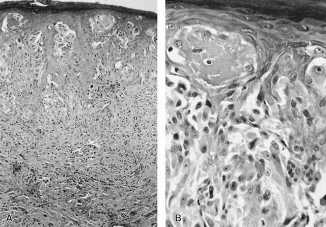
AFIP images:
Bilaterally symmetric lesion has uniform hyperkeratosis with hypergranulosis, fairly uniformly elongated rete ridges
and a proliferation of melanocytes in the epidermis that "rain down" through the papillary dermis into the reticular dermis
as attenuated spindle cells
Fig A: bilaterally symmetrical lesion
Fig B: has lateral cells arranged in nests
Fig C: indicating that the lesion is well circumscribed; "transepidermal elimination" does not indicate pagetoid spread
Fig D: dermal lesional cells show imperfect, but definite maturation with attenuated single cells at the base of the lesion
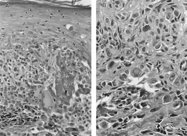
Melanocytes extend focally into epidermis, mostly as nests; the maturation into the dermis, globoid bodies in epidermis and characteristic cell type are attributes of Spitz nevus, not melanoma
Pink eosinophilic globoid Kamino bodies are probably apoptotic cells
Fig A: giant cells are commonly seen in Spitz nevi
Fig B: as are the large intranuclear cytoplasmic invaginations that may also be seen in melanomas
Fig A: mitotic figures, if not numerous and especially when entirely superficial, are not indicative of melanoma
Fig B: maturation from large epithelioid cells to small nevus cells, from superficial to deep, within a Spitz nevus is an extremely important diagnostic feature, as is the permeation of reticular dermis by attenuated single cells at the base
Comparison with melanoma:
Fig A-H: Spitz nevus
Fig I: compared to spitzoid melanoma
Contributed by Angel Fernandez-Flores, MD, PhD, Hospital El Bierzo and Clinica Ponferrada, Spain:
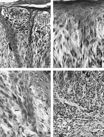
Spitzoid melanoma: asymmetric, irregular lateral borders, uneven base, pagetoid scatter of melanocytes, uneven nests of melanocytes, lack of maturation, spindle cells not perpendicular to surface, epidermal spread, ulcerated surface, most cells appear malignant (Clin Dermatol 2009;27:545)
Nevi Atypical Spitz nevus
Reviewer: Christopher Hale, M.D. (see Reviewers page) Revised: 3 May 2013, last major update March 2013 Copyright: (c) 2002-2013, PathologyOutlines.com, Inc.
General
=========================================================================
Junctional or compound lesion not fulfilling the histopathologic criteria of melanoma but with one of the following features:
Asymmetry
Predominance of single melanocytes over nests in lesions 4 mm or larger
Large epithelioid cells with amphophilic cytoplasm are characteristic of Spitz nevi, and the nuclear atypia observed here should not suggest melanoma in the absence of other features
Buttock tumor:
Multinucleated tumor cells
Sheets of atypical tumor cells with frequent mitotic figures
Reviewer: Christopher Hale, M.D. (see Reviewers page) Revised: 17 April 2013, last major update March 2013 Copyright: (c) 2002-2013, PathologyOutlines.com, Inc.
General
=========================================================================
Spitz (spindle and epithelioid cell) nevus with dense eosinophilic stroma
Other Spitz nevus features present (Kamino bodies, etc.)
Resembles classic Spitz nevus (symmetric with inverted wedge shape, infiltration of dermis by relatively bland epithelioid and spindle cells), but dermal fibrosis encircles individual cells and simulates invasion
Melanocytes with spitzoid appearance, stromal hypereosinophilic bundles of collagen and melanophages in the junctional region and superficial dermis
Near-dermal lesion with laterally sharp border
AFIP images:
Symmetrical lesion shows maturation of lesional cells in the dermis with attenuated single cells among reticular dermis collagen bundles at the base, but unlike desmoplastic melanoma, the
intraepidermal component does not extend substantially beyond the dermal component, and there are no clusters of mitotically active epithelioid melanocytes in the superficial dermis
Desmoplastic melanoma - in situ component present, cells resemble fibroblasts with atypia, negative for MelanA and HMB45
Nevi White sponge nevus
Reviewer: Christopher Hale, M.D. (see Reviewers page) Revised: 20 April 2013, last major update March 2013 Copyright: (c) 2002-2013, PathologyOutlines.com, Inc.
General
=========================================================================
Not a melanocytic entity; benign keratinization defect leading to white plaques on mucosa
Parakeratosis, acanthosis with formation of large blunt rete ridges and spongiosis
Extensive vacuolation of suprabasal keratinocytes
Dyskeratotic cells exhibit dense peri- and paranuclear eosinophilic condensations, which correspond to tonofilament aggregates
Abundant Odland bodies (keratinosome, membrane bound granule in upper stratum spinosum) within keratinocytes, but few are present in the intercellular spaces
Parakeratosis, acanthosis and spongiosis of mucosal epithelium with blunting of rete ridges; vacuolated and dyskeratotic epithelial keratinocytes demonstrate perinuclear eosinophilic condensations
Reviewer: Christopher Hale, M.D. (see Reviewers page) Revised: 20 April 2013, last major update March 2013 Copyright: (c) 2002-2013, PathologyOutlines.com, Inc.
General
=========================================================================
Often only part of lesion is excised, and pathologist report includes recommendation for complete excision (J Clin Pathol 2004;57:1121)
Patterns of benign behavior
=========================================================================
Lentiginous hyperplasia (single cell melanocytic growth along dermoepidermal junction)
Nested proliferation
Melanocyte nuclei are smaller than adjacent keratinocytes
Pagetoid proliferation (discohesive single cell growth throughout entire epidermis) is nonspecific; present in Spitz nevi, acral nevi, melanoma (Am J Surg Pathol 1995;19:792)
Other benign features
=========================================================================
Symmetric pattern of growth and involution
Lateral dimension of dermal component should roughly equal that of original epidermal component ("shoulder" = when epidermal component extends laterally beyond dermal)
Other melanocytic lesions Atypical melanocytic hyperplasia
Reviewer: Christopher Hale, M.D. (see Reviewers page) Revised: 20 April 2013, last major update March 2013 Copyright: (c) 2002-2013, PathologyOutlines.com, Inc.
General
=========================================================================
Atypical single unit melanocytes limited to epidermis, often seen at periphery of classic melanoma
In contrast to melanoma in situ, exhibits only focal confluence at dermoepidermal junction, has limited pagetoid spread and involves only upper hair follicle
Factors favoring atypical melanocytic hyperplasia over melanoma in situ (Mod Pathol 2000;13:857):
Reviewer: Christopher Hale, M.D. (see Reviewers page) Revised: 20 April 2013, last major update April 2013 Copyright: (c) 2002-2013, PathologyOutlines.com, Inc.
General
=========================================================================
Pigmented macule 0.5 mm+, flat, round / oval, sharply demarcated with even pigmentation; long axis is along cutaneous nerve tract
May have irregular borders
Pigment histologically restricted to basal layer of epidermis
In U.S. study of children under 5 years, 19% had one, 0.75% had >2; more present in African-Americans (Arch Dis Child 1966;41:316)
If solitary, usually non-syndromic; multiple associated with neurofibromatosis type 1 - any person with 6+ lesions should be presumed to have neurofibromatosis until proven otherwise
Also associated with McCune-Albright syndrome and other syndromes (eMedicine)
Case reports
=========================================================================
7 week old infant with McCune-Albright syndrome and multiple, bilateral lesions (Pediatr Dermatol 1991;8:35)
Syndromic patients: more DOPA+ melanocytes in lesional skin contrasted to sporadic patients with decreased DOPA+ melanocytes within lesion (Arch Dermatol 1970;102:442)
Pigmented nevus: deeper pigment clinically, melanocytes not restricted to basal epidermis
Other melanocytic lesions Ephelis (freckle)
Reviewer: Christopher Hale, M.D. (see Reviewers page) Revised: 20 April 2013, last major update April 2013 Copyright: (c) 2002-2013, PathologyOutlines.com, Inc.
General
=========================================================================
Benign lesion of sun exposed skin, associated with fair skin and red hair (Wikipedia)
Appears in early childhood; fades in winter, reappears in summer
Reviewer: Christopher Hale, M.D. (see Reviewers page) Revised: 20 April 2013, last major update April 2013 Copyright: (c) 2002-2013, PathologyOutlines.com, Inc.
General
=========================================================================
Common, usually harmless condition, in which patches of skin become darker than normal surrounding skin (Wikipedia)
Due to melanocyte stimulation from drugs (Merck), heat, hormones, inflammation (eMedicine), malignancy, metabolic disease, scars, sunlight, various dermatoses or familial progressive hyperpigmentation (Eur J Dermatol 2006;16:246)
Note: hydroquinone slows production of melanin, so darker areas gradually fade to match surrounding skin; tretinoin and cortisone take 3-6 months to produce improvement
Imipramine: gold brown pigment in upper and mid dermis
Familial Progressive Hyperpigmentation
Electron microscopy images
=========================================================================
Photoexposed pigmented skin
Other melanocytic lesions Linear and whorled nevoid hypermelanosis
Reviewer: Christopher Hale, M.D. (see Reviewers page) Revised: 25 April 2013, last major update April 2013 Copyright: (c) 2002-2013, PathologyOutlines.com, Inc.
General
=========================================================================
Rare skin disorder, often in first weeks of life, with swirls and streaks of macular hyperpigmentation along lines of Blaschko (Wikipedia)
Epidermal hypermelanosis without pigment incontinence in the dermis
Diffuse melanosis of basal layer of epidermis
Other melanocytic lesions Melasma
Reviewer: Christopher Hale, M.D. (see Reviewers page) Revised: 25 April 2013, last major update April 2013 Copyright: (c) 2002-2013, PathologyOutlines.com, Inc.
General
=========================================================================
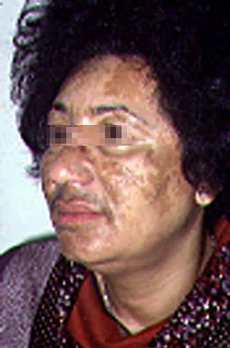
Acquired large areas of darkened skin, usually due to hormonal changes (birth control pills, pregnancy) and usually on both sides of face (eMedicine)
Clinical patterns are centrofacial, malar and mandibular
Classification (based on level of increased melanin in skin determined by Wood’s light examination, J Am Acad Dermatol 1981;4:698):
Epidermal: 70% of cases, increased melanin in basal and suprabasal epidermis; skin pigmentation enhanced under Wood’s light; responds best to bleaching agents
Dermal: 10% of cases, increase in melanophages in upper dermis; no enhancement of skin pigmentation under Wood’s light, responds poorly to bleaching agents
Mixed: 20% of cases, mixture of epidermal and dermal features; patchy enhancement of skin pigmentation under Wood’s light
Indeterminate: 2% of cases; not possible to characterize pigmentation pattern
Hori's nevus: acquired bilateral nevus of Ota-like macules
Lichen planus pigmentosus: uncommon variant of lichen planus characterized by hyperpigmented, dark-brown macules in sun-exposed areas and flexural folds
Pigmented contact dermatitis (Riehl’s melanosis)
Post inflammation pigmentation
Other melanocytic lesions Paraganglioma-like dermal melanocytic tumor
Reviewer: Christopher Hale, M.D. (see Reviewers page) Revised: 25 April 2013, last major update April 2013 Copyright: (c) 2002-2013, PathologyOutlines.com, Inc.
General
=========================================================================
Well demarcated cellular nodule in dermis with normal epidermis
Tumor restricted to deep dermis without any contact with normal overlying epidermis
S100+
Various images
Neoplasm composed of organoid and nested groups of large epithelioid cells, separated by delicate fibrous strands and prominent blood vessels; epithelioid cells have somewhat clear cytoplasm, large nuclei with prominent red nucleoli
Reviewer: Christopher Hale, M.D. (see Reviewers page) Revised: 25 April 2013, last major update April 2013 Copyright: (c) 2002-2013, PathologyOutlines.com, Inc.
General
=========================================================================
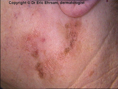
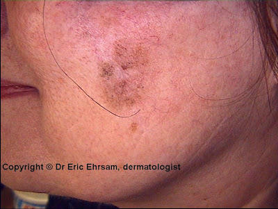
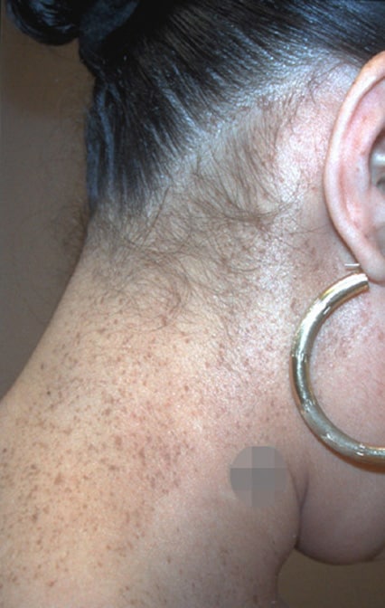
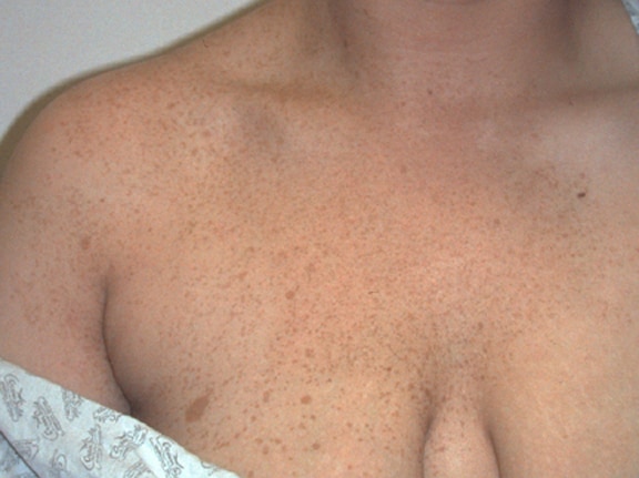
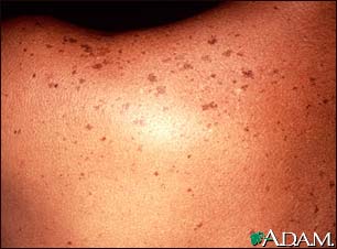
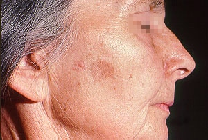
Sun exposed skin of elderly (90% of whites age 60+); also truck drivers on sun-exposed face (J Dermatol 2008;35:146)
Multiple large solar lentigos on upper back and shoulders suggest prior severe sunburn, a risk factor for melanoma (Dermatology 2007;214:25)
PUVA (psoralen + ultraviolet A) treatment for psoriasis cause numerous solar lentigines with atypia; similar findings after severe radiation exposure (Arch Dermatol 1997;133:209)
Clinical features
=========================================================================
Reviewer: Christopher Hale, M.D. (see Reviewers page) Revised: 25 April 2013, last major update April 2013 Copyright: (c) 2002-2013, PathologyOutlines.com, Inc.
General
=========================================================================
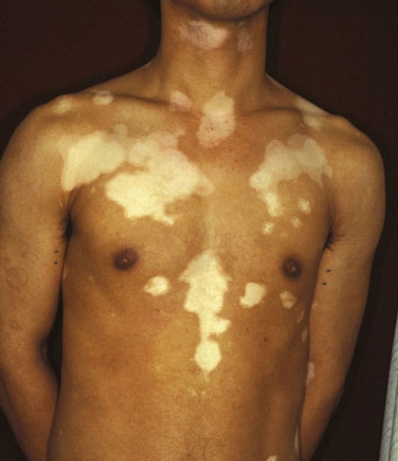
Partial or complete loss of pigment producing melanocytes within epidermis (Wikipedia, eMedicine)
Patterns:
Focal (only a few areas)
Segmented (one side of body only)
Generalized (most common, both sides of body)
Trichrome (patient has three shades of skin color)
Affects 1% of world’s population; more noticeable in dark skinned individuals
Usually hands / wrists, axilla, perioral, periorbital or anogenital skin
Clinical features
=========================================================================
Asymptomatic, flat and well-demarcated zones of pigment loss, due to an autoimmune disorder associated with pernicious anemia, Addison’s disease and Hashimoto’s thyroiditis
Leukoderma: chemical, melanoma-related, scleroderma-related; acquired condition with localized loss of skin pigmentation associated with inflammatory skin conditions, burns, intralesional steroid injections, postdermabrasion (Wikipedia)
Oculocutaneous albinism: melanocytes present, but no melanin due to defect in tyrosinase enzyme or melanogenesis
Melanoma Melanoma in situ
Reviewer: Christopher Hale, M.D. (see Reviewers page) Revised: 25 April 2013, last major update April 2013 Copyright: (c) 2002-2013, PathologyOutlines.com, Inc.
General
=========================================================================
Malignant melanocytes in epidermis, without dermal invasion
Variants include lentigo maligna, superficial spreading and acral melanoma
Large pigmented lesions, irregular margins and irregular pigmentation
Hutchinson's sign: periungual extension of brown-black pigmentation from longitudinal melanonychia (pigmented stripe within length of nail bed) onto the proximal and lateral nailfolds
Case reports
=========================================================================
Reviewer: Christopher Hale, M.D. (see Reviewers page) Revised: 29 May 2013, last major update May 2013 Copyright: (c) 2002-2013, PathologyOutlines.com, Inc.
General
=========================================================================
Malignancy of melanocytes, predominantly in skin, but also eyes, ears, GI tract, leptomeninges, mucous membranes (Wikipedia, eMedicine)
Only 4% of skin cancers but majority of skin cancer deaths
In US in 2000, were 48,000 cases and 9,200 deaths; in 2013, estimated 76,690 cases and 9,480 deaths (Cancer Facts & Figures 2013)
Populations at higher risk:
Whites with fair skin, red hair, tendency to burn or freckle from sun exposure, large number of melanocytic nevi, xeroderma pigmentosum, familial dysplastic nevi, melanosis, vitiligo, frequent sunburns at any age (Ann Epidemiol 2008;18:614); 5-10% are familial (Surg Clin North Am 2008;88:897)
Blacks and Hispanics in US have low risk, their common melanoma sites are palms, soles, nail beds or mucous membranes; often poorer prognosis (Cancer Control 2008;15:248)
Usually occurs after puberty, occasionally children - all have same morphology
Regional lymph nodes, liver, lungs, GI tract, bone, CNS, heart, skin and other sites
Isolated tumor cells or tumor deposits > 0.1 mm (within lymph nodes) that meet the criteria for histologic or IHC detection of melanoma should be scored as node positive (J Clin Oncol 2009;27:6199)
Satellite tumors are considered intralymphatic metastases within 2 cm of primary tumor; termed in transit metastases if > 2 cm from primary tumor, but before the first echelon of regional lymph nodes (Br J Dermatol 2002;147:62)
Overall 5 year survival is 60%, but behavior is variable, with occasional late deaths or long survival even with widespread satellite nodules
5 year and 10 year survival rates (TNM classification) range from 97% and 93% for T1aN0M0 melanomas to 53% and 39% for T4bN0M0 (J Clin Oncol 2009;27:6199)
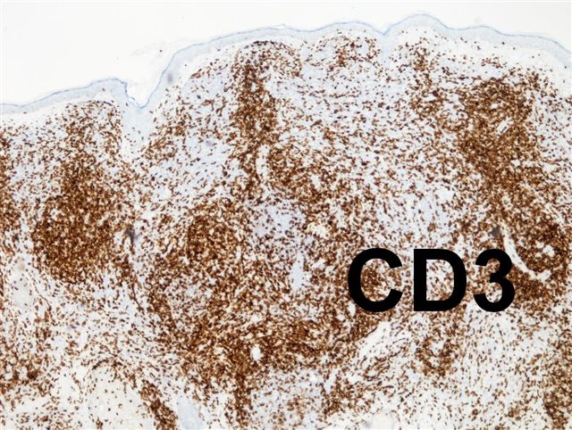
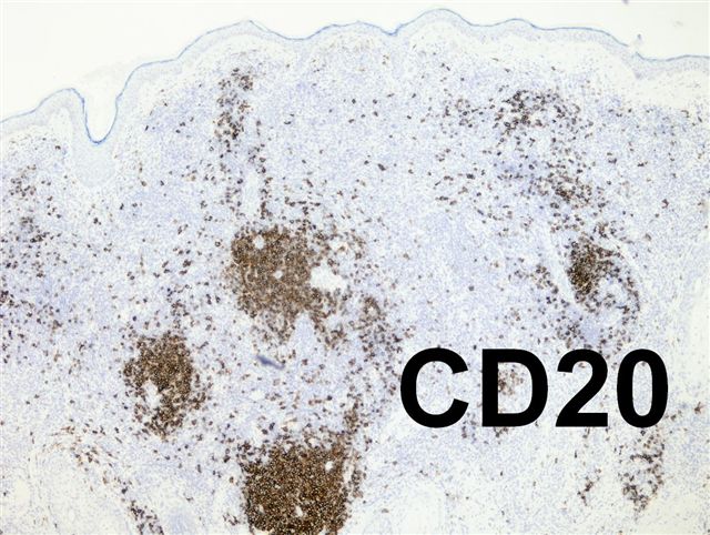
Presence of tumor-infiltrating lymphocytes may indicate better prognosis (J Clin Oncol 2012;30:2678)
For patients with localized melanoma, most important prognostic factors are tumor thickness and ulceration
For patients with nodal metastases, most important prognostic factors are number of metastatic nodes, microscopic versus macroscopic tumor and presence or absence of ulceration of primary melanoma
Note: there is excellent agreement between pathologists in assessing tumor thickness, ulcerative state and tumor mitotic rate
(Am J Surg Pathol 2003;27:1571)
Case reports
=========================================================================
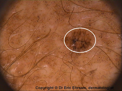
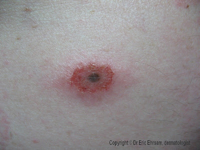
Black dots represent pigmented cells at dermal-epidermal junction and within epidermis in heavily pigmented columns; brown dots are similar to black dots, but with less pigment; blue dots are due to melanophages surrounding superficial vascular plexus; depigmentation is due to intense fibrosis of papillary dermis; radial streaming and pseudopods are due to cells in nests or centrifugal linear extensions (Am J Dermatopathol 2006;28:13); blue-whitish veil is associated with melanoma (Am J Dermatopathol 2001;23:463)
Amelanotic / hypomelanotic lesions: blue-white veil, scarlike depigmentation, multiple blue-gray dots, irregularly shaped depigmentation, irregular brown dots/globules, 5-6 colors and predominant central vessels are suggestive (Arch Dermatol 2008;144:1120)
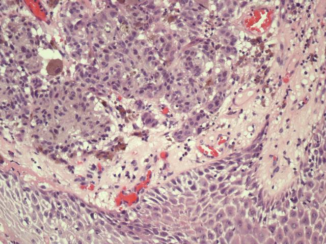
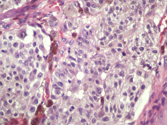
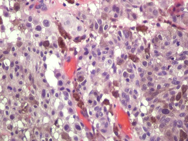
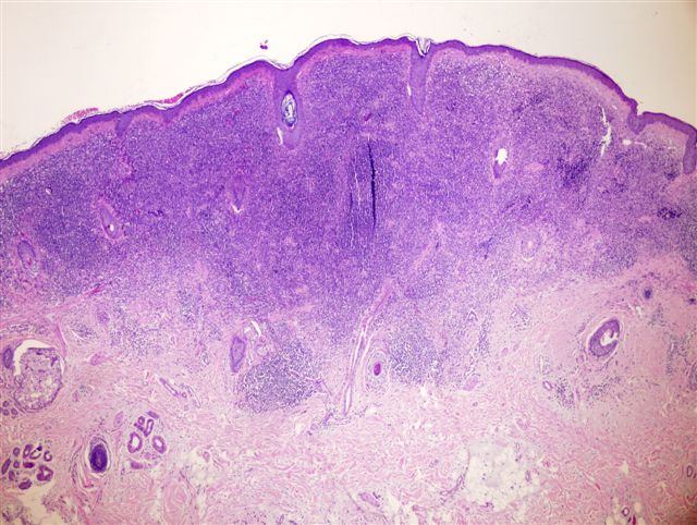
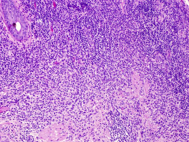
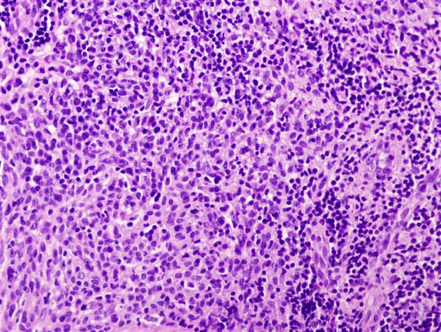
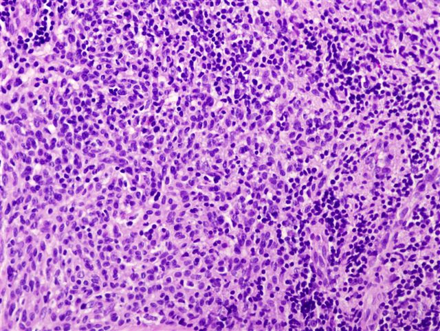
Features of melanoma in situ + dermal involvement of atypical melanocytes with cytologic atypia and no maturation
Classic features are junctional activity with obscured dermoepidermal junction and pagetoid spread individually and in clusters throughout epidermis
Prominent melanin pigmentation, invasion of surrounding tissue
Large cells with abundant eosinophilic and finely granular cytoplasm
Nuclear pseudoinclusions, folds or grooves
Marked atypia with pleomorphic nuclei with large eosinophilic nucleoli
Frequent mitotic figures
4 major subtypes: (described separately)
Acral lentiginous
Lentigo maligna
Nodular
Superficial spreading
Lack of a junctional component suggests a metastases, although epidermal component may have regressed or not been sampled, or melanoma may have developed from an intradermal nevus
Consumption of epidermis: usually (86%) present; thinning of epidermis with attenuation of basal and suprabasal layers and loss of rete ridges in areas of direct contact with neoplastic melanocytes; variable clefts separating epidermis and dermis, edema and telangiectasias (Am J Surg Pathol 2004;28:1621); is associated with increased Breslow depth and ulceration (Am J Dermatopathol 2007;29:527)
Lymphatic invasion identified in 5% on H&E but 33% using D2-40/podoplanin and S100 (Hum Pathol 2008;39:901)
Angiotropism is suggestive of epidermotropic metastatic disease versus recurrent disease (Am J Dermatopathol 2006;28:429)
Rarely paradoxical maturation occurs, but still have areas of cells with abundant cytoplasm and large nuclei, more mitotic figures, more confluence, high Ki-67 rate (Am J Surg Pathol 2000;24:1600)
Metastases may have spindle cells resembling malignant peripheral nerve sheath tumor
(Diagn Cytopathol 2008;36:754)
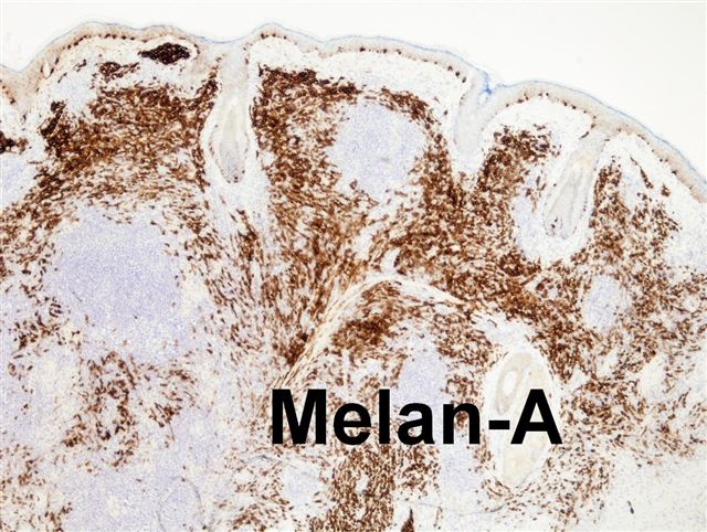
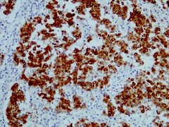
Positive stains
========================================================================= Distinguish melanocytes from non-melanocytes, but not malignant cells from benign cells:
S100: nuclear and cytoplasmic staining, 90%+ sensitive but not specific (although usually negative in tumors considered in the differential)
HMB45: cytoplasmic and weak nuclear staining (Mod Pathol 2008;21:1121),
less sensitive but more specific than S100; negative in desmoplastic melanoma
MelanA/Mart1: sensitive, but also stains steroid-producing cells in ovary, testis, adrenal cortex
Tyrosinase: sensitive, but also stains peripheral nerve sheath and neuroendocrine tumors
Microphthalmia transcription factor (MITF): sensitive, but also stains dermatofibroma and smooth muscle tumors; negative in spindle cell / desmoplastic melanoma
Main altered pathways include RAS-RAF-MEK-ERK, p16(INK4A)-CDK4-RB and ARF-p53 (APMIS 2007;115:1161)
20% of melanoma prone families have point mutation in CDKN2A locus at 9p21 which encodes p16(INK4a) and p14(ARF) (Br J Cancer 2008;99:364)
10% of cases may be familial due to CMM1 gene at 1p36
Microsatellite instability seen in pediatric melanoma (43%), adult melanoma (30%), nevi (9%, Am J Dermatopathol 2005;27:279)
Most melanomas have multiple chromosomal gains and losses, features that can be used to differentiate them from nevi, which do not have chromosomal alterations
Nevi: particularly Spitz nevi-desmoplastic type, halo nevi, activated and dysplastic nevi, vulval nevi and recurrent nevi after incomplete excision; features relatively specific for melanoma include absence of maturation, suprabasal melanocytes; also atypia, size >6 mm, mitotic figures, dermal lymphocytes and asymmetry, necrosis, asymmetrical melanin and melanin in deep cells (Melanoma Res 2008;18:253)
Pigmented basal cell carcinoma
Pigmented seborrheic keratosis
Helpful features of melanoma that differentiate from benign lesions (from Rosai):
Asymmetry
Atypia
Band like chronic inflammatory infiltrate in dermis
Lack of maturation of dermal tumor cells
Lateral extension of individual melanocytes
Melanocytes with clear cytoplasm and finely dispersed chromatin, individual melanocyte necrosis (compared to eosinophilic hyaline bodies in Spitz nevi)
Mitotic figures in melanocytes (particularly atypical ones)
Pleomorphism of tumor cells
Poor circumscription of intraepidermal component
Presence of chromosomal gains or losses
Transepidermal migration of melanocytes
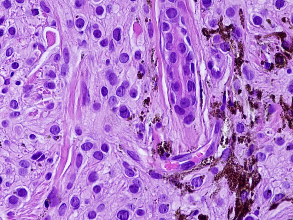
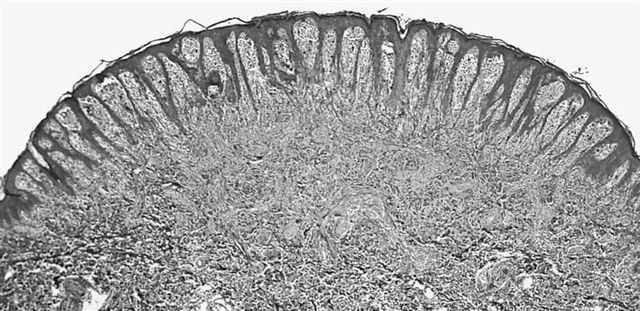
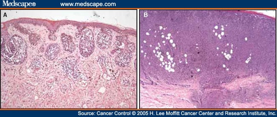
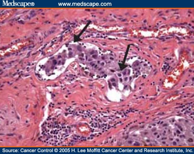
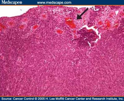
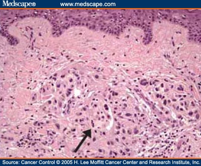
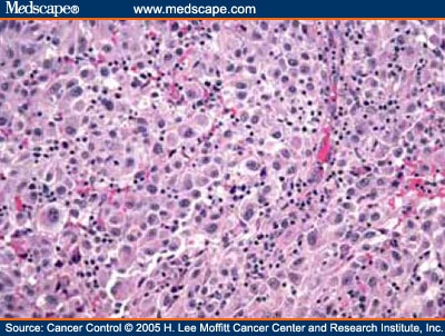
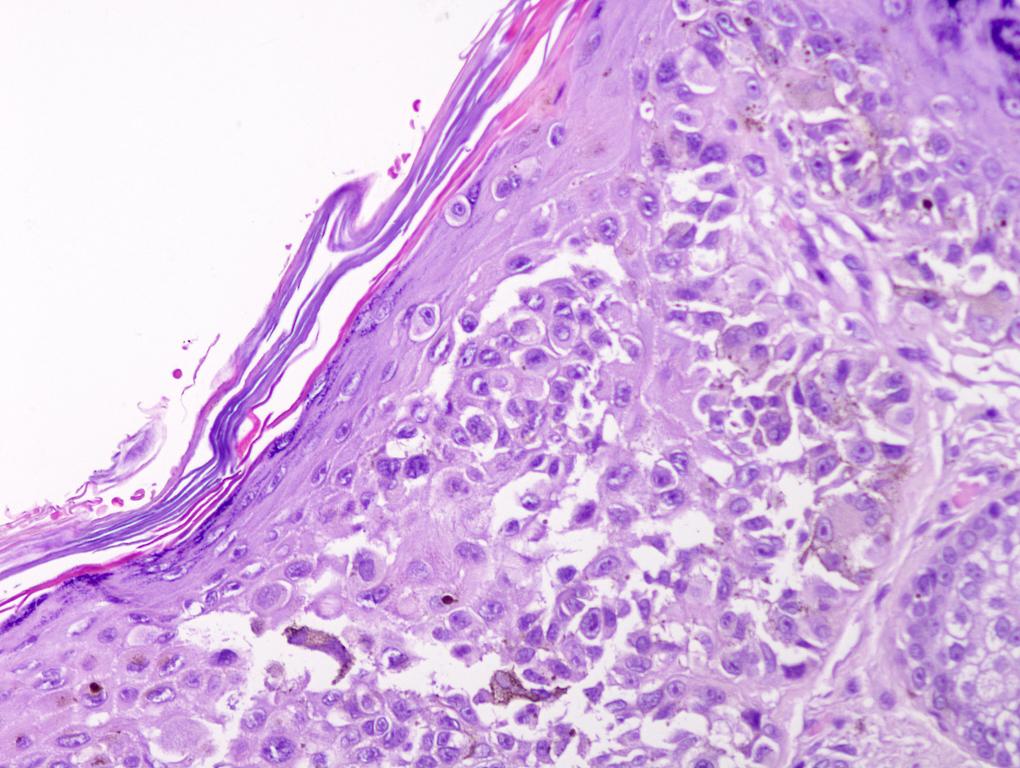
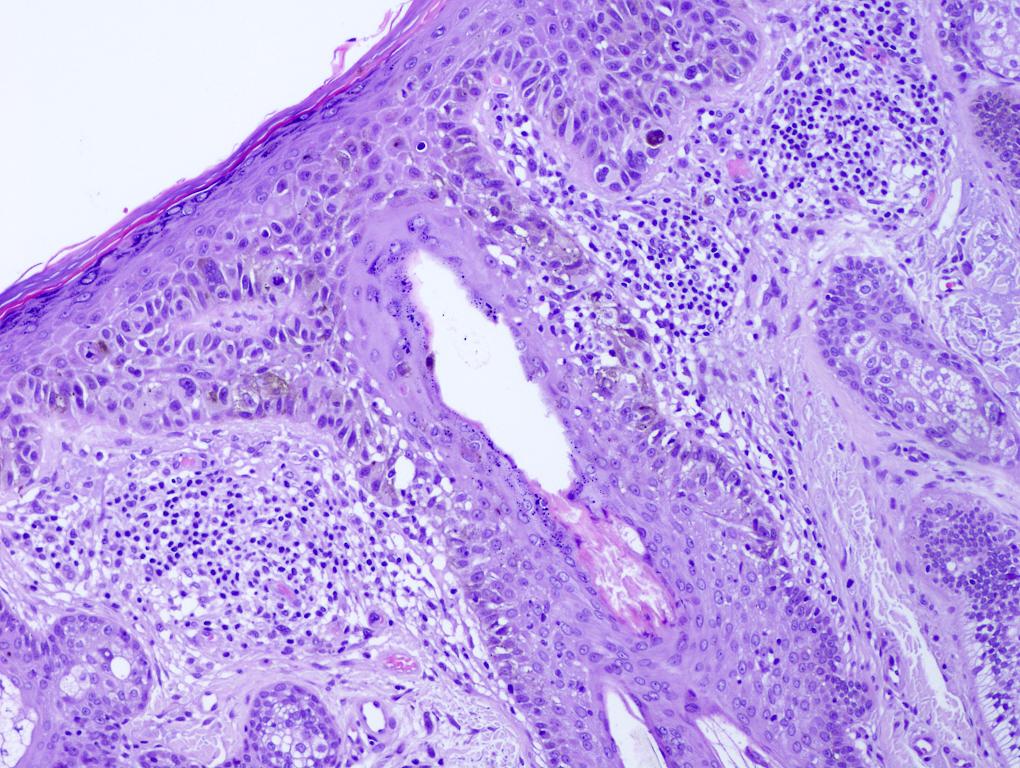
Melanoma Acral lentiginous melanoma (ALM) Reviewer: Christopher Hale, M.D. (see Reviewers page) Revised: 5 June 2013, last major update May 2013 Copyright: (c) 2002-2013, PathologyOutlines.com, Inc.
General
=========================================================================
Acral: relating to or affecting the glabrous (non-hair bearing) or volar skin of the soles, palms and digits as well as the nail apparatus
Note: all melanomas of acral sites do NOT have histology of acral lentiginous melanoma (Br J Dermatol 2012;166:727)
Usually palms and soles, subungual, mucocutaneous oral and nasal cavity or anus
Clinical features
=========================================================================
Often advanced at diagnosis because thickened, hyperkeratotic epidermis overlies and hides primary lesion; often initially misdiagnosed (J Am Acad Dermatol 2003;48:183)
May evolve slowly over years; mean 1 year to diagnosis in foot / ankle lesions (J Foot Ankle Res 2008;1:11)
In situ cases show longitudinal pigmented streak in nail plates, black pigmentation on proximal or lateral nail fold, irregular border or variegated pigmentation on sole or thumb (Am J Dermatopathol 2004;26:285)
Invasive cases show densely pigmented macules with irregular borders; mean 3 mm, usually ulcerated (74%, Cancer Causes Control 2009;20:115)
Prominent acanthosis of epidermis with elongated rete ridges
Pagetoid spread
Proliferation of melanocytes downward along eccrine ducts
Melanocytes may display prominent dendritic proceses
Invasive component often composed of spindle cells, but epithelioid, small cells and pleomorphic cells are occasionally noted
Intraepidermal lentiginous component is similar to lentigo maligna, but intraepidermal melanocytes are bizarre, epidermis is markedly hyperplastic and papillary dermis is widened and inflamed
Consumption of epidermis present (attenuation of basal / suprabasal layers with rete ridge loss, J Cutan Pathol 2012;39:577)
Early lesions may show proliferation of solitary melanocytes in crista profunda intermedia, the epidermal rete ridge underlying the ridge of the skin marking (Am J Dermatopathol 2006;28:21)
Nail lesions show confluent stretches of solitary melanocytes, multinucleation, lichenoid inflammatory reaction and florid pagetoid spread (Am J Surg Pathol 2008;32:835)
Melanoma Desmoplastic melanoma Reviewer: Christopher Hale, M.D. (see Reviewers page) Revised: 5 June 2013, last major update May 2013 Copyright: (c) 2002-2013, PathologyOutlines.com, Inc.
General
=========================================================================
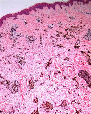
Rare variant of spindle cell melanoma seen in older adults in sun-exposed skin
A type of nodular (vertical growth) melanoma with scanty spindle cells, prominent desmoplastic stroma and often minimal atypia
Features of regression (white scar like areas, peppering); also multiple colors, linear irregular vessels or milky-red areas (Br J Dermatol 2008;159:360)
Poorly circumscribed; focal fascicular pattern of scanty spindle cells with prominent desmoplastic stroma
Tumor cells may have minimal atypia
Solar elastosis (82%), amelanotic (71%), deep invasion, perineural infiltration (35%), lymphoid aggregates at periphery (37%); may grow in peripheral nerve sheath pattern
Clean background; aggregates of pleomorphic spindle cells mixed with fibrous stroma and single cells
Fine, wispy and delicate cytoplasm at nuclear poles, nuclei are elongated and plump with irregular contours, deep grooves and folds and dark coarse chromatin with variably prominent nucleoli (Cytojournal 2007;4:18)
Compared to other melanomas, is less cellular and less often has intranuclear cytoplasmic inclusions and mitotic figures (Am J Clin Pathol 2008;130:715)
HMB45 and MelanA (usually, but may have focal staining of epithelial cells in junctional component or superficial dermis, Am J Dermatopathol 2004;26:452)
Reviewer: Christopher Hale, M.D. (see Reviewers page) Revised: 7 June 2013, last major update May 2013 Copyright: (c) 2002-2013, PathologyOutlines.com, Inc.
General
=========================================================================
Deep seated follicular structure with malignant melanocytes extending downward along follicular epithelium and permeating parts of follicle, as well as adjacent dermis
Reviewer: Christopher Hale, M.D. (see Reviewers page) Revised: 8 June 2013, last major update May 2013 Copyright: (c) 2002-2013, PathologyOutlines.com, Inc.
General
=========================================================================
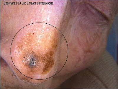
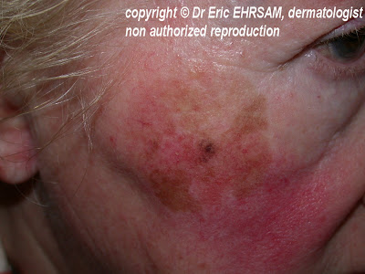
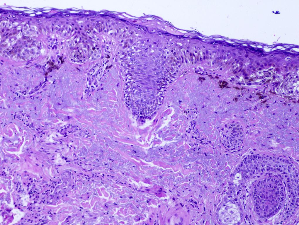
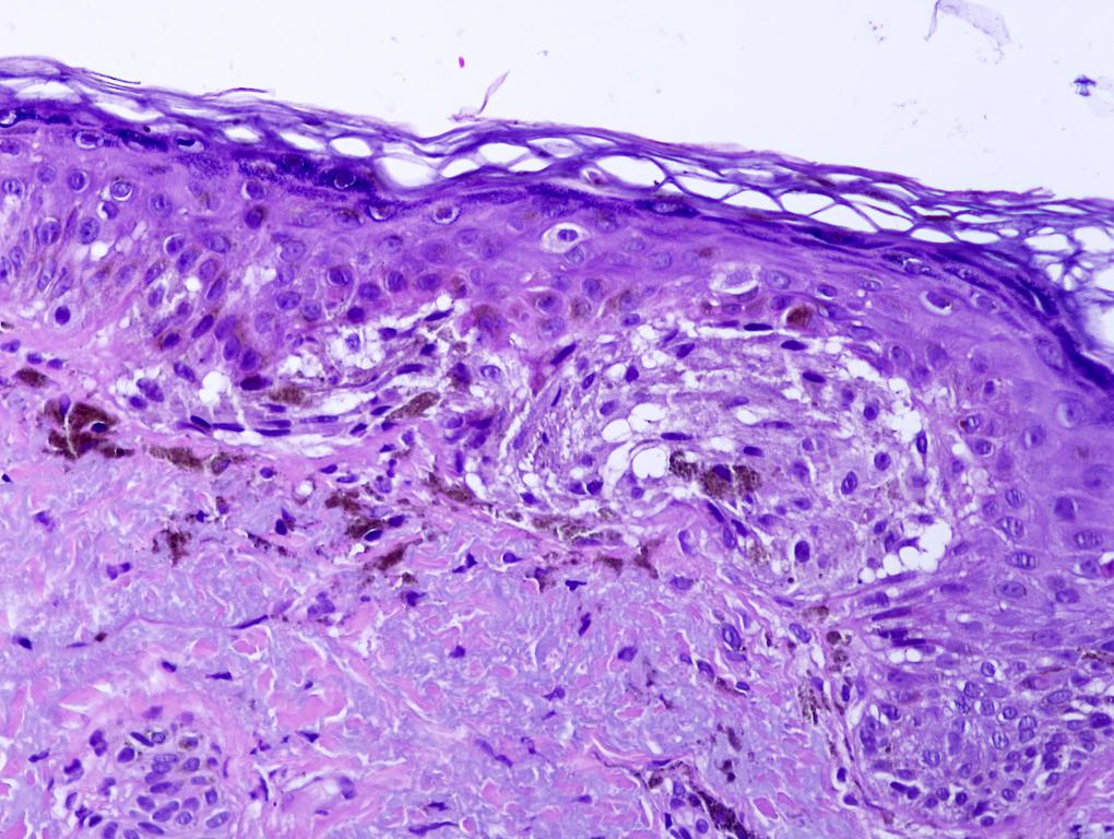
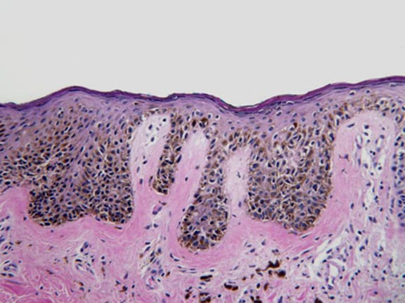
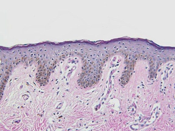
Atypical melanocytes in basal layer, individually and in nests (theques), associated with sun damage (eMedicine)
Also called melanoma arising in Hutchinson’s freckle, actinic melanosis, melanoma on sun damaged skin and solar melanoma
Note: lentigo maligna (a subtype of melanoma in situ), by definition, does NOT infiltrate into dermis, but lentigo maligna melanoma has at least single cell infiltration into papillary dermis
Clinical features
=========================================================================
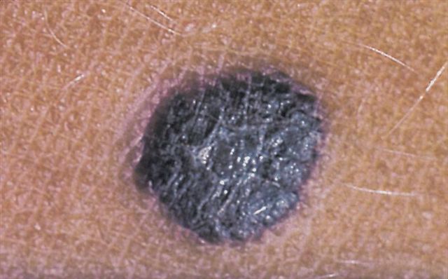
Flat, tan to black with irregular hyperpigmentation, > 2 cm
5-15% of (invasive) melanoma; increasing prevalence, particularly among men age 65+ years (J Invest Dermatol 2005;125:685)
Slow growing lesion of sun exposed skin of elderly whites, often cheek; partial regression is common
Similar behavior to other melanoma subtypes when depth of invasion is considered, but unusual to die of disease
Possible origin from progenitor stem cell (strongly CD133+ and CD34+) in outer root sheath of mid-lower hair follicles (Dermatol Ther 2008;21 Suppl 1:S1)
Case reports
=========================================================================
Staged excision for peripheral margin control using permanent sections is required because frozen sections are unreliable (Plast Reconstr Surg 2007;120:1249)
Reviewer: Christopher Hale, M.D. (see Reviewers page) Revised: 7 June 2013, last major update May 2013 Copyright: (c) 2002-2013, PathologyOutlines.com, Inc.
General
=========================================================================
Rare subset of slowly progressing, radial growth phase melanoma with lentiginous proliferation of moderately atypical epithelioid melanocytes, focal pagetoid spread and nesting; not associated with architectural changes at dermoepidermal junction (Arch Pathol Lab Med 2011;135:337)
Broad lentiginous proliferation of melanocytes at dermoepidermal junction, both as single cells and as small nests with areas of confluent growth, extending to edges of biopsy
Prominent atypia of melanocytes and invasion may be evident only at excision
Preservation of retiform epidermis
Similar to other histologic subtypes with a lentiginous junctional proliferation (lentigo maligna, acral lentiginous and mucosal lentiginous)
Melanoma Minimal deviation melanoma Reviewer: Christopher Hale, M.D. (see Reviewers page) Revised: 8 June 2013, last major update May 2013 Copyright: (c) 2002-2013, PathologyOutlines.com, Inc.
General
=========================================================================
Introduced to embrace the concept that a subset of invasive melanomas is characterized by lesser cytologic atypia and a better prognosis than conventional melanomas of the same thickness
Cells are more atypical than nevi, but less atypical than classic melanoma
Expansive nodule in vertical growth phase that fills papillary dermis and may extend into reticular dermis (Mod Pathol 2006;19 Suppl 2:S41)
Usually uniform cells with mild to moderate atypia that resemble nevus cells, but are moderately enlarged with irregular chromatin and increased N/C ratios
Growth displaces surrounding structures and remnants of residual benign nevus are often present
Equivalent to at least a level III melanoma due to extent of dermal invasion
May have perineural invasion and mitotic figures, but usually does not invade subcutaneous fat; no necrosis, no maturation
Nevoid melanoma: more mitotic figures, usually no residual nevus
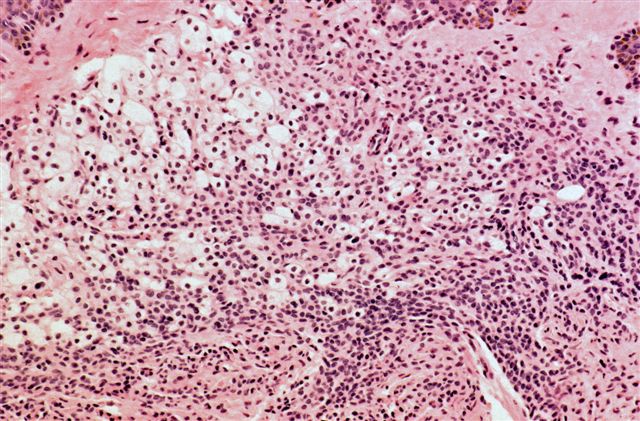
Melanoma Nevoid melanoma
Reviewer: Christopher Hale, M.D. (see Reviewers page) Revised: 8 June 2013, last major update May 2013 Copyright: (c) 2002-2013, PathologyOutlines.com, Inc.
General
=========================================================================
Resembles ordinary compound or dermal nevus at low power, with symmetrical dome-shaped or verrucous and papillomatous features, sharp lateral demarcation, inconspicuous junctional component and no pagetoid growth
High power shows relatively bland and monomorphic cells resembling classic nevus or epithelioid cells in Spitz nevus
Focal sheetlike growth pattern, nucleoli in tumor cells at base of lesion
Multiple dermal mitoses with atypical mitoses
Subtle pleomorphism and impaired maturation with depth
Melanoma arising in dermal nevus: residual nevus present, often tumor extension into deep reticular dermis and fat
Metastatic melanoma
Minimal deviation melanoma: at most moderate atypia, at least Level III due to dermal invasion, remnants of existing nevi usually present and usually few mitotic figures
Reviewer: Christopher Hale, M.D. (see Reviewers page) Revised: 8 June 2013, last major update May 2013 Copyright: (c) 2002-2013, PathologyOutlines.com, Inc.
General
=========================================================================
Aggressive subtype of melanoma with early vertical growth phase
Affects all body surfaces, but usually legs and trunk
Clinical features
=========================================================================
Screening methods have had little impact on this subtype (Cancer 2008;113:3341); may have less of an association with sun exposure than superficial spreading subtype (Melanoma Res 2012;22:460)
Non-specific global dermoscopic patterns of globules, blue-white veil, atypical vessels and structureless areas (Arch Dermatol 2008;144:1311)
Important features: peripheral black dots / globules, multiple brown dots, irregular black dots / globules, blue-white veil, homogeneous blue pigmentation, 5 to 6 colors and black color (JAMA Dermatol 2013;3:1)
Reviewer: Christopher Hale, M.D. (see Reviewers page) Revised: 8 June 2013, last major update May 2013 Copyright: (c) 2002-2013, PathologyOutlines.com, Inc.
General
=========================================================================
Extremities are most common site, although numerous sites are affected
Clinical features
=========================================================================
Mimics melanocytic neoplasms in gray horses and laboratory animals, although appears to have different molecular origin than equine melanomas (Am J Surg Pathol 2004;28:31)
Includes lesions previously described as epithelioid blue nevus of Carney complex (Am J Surg Pathol 2004;28:31)
Does not appear to be related to sun exposure
Nodal metastases in 46%, but death from disease is rare
Resembles combined nevus
Case reports
=========================================================================
Associated with loss of protein kinase A regulatory subunit type 1alpha (R1alpha), coded by the PRKAR1A gene, which is lost in both sporadic cases and patients with Carney complex (Am J Surg Pathol 2007;31:1764)
Nodular melanosis: pigmented cells are actually pigment laden macrophages
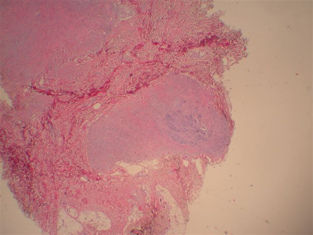
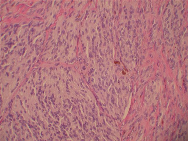
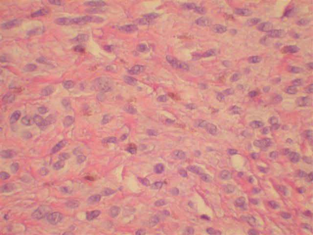
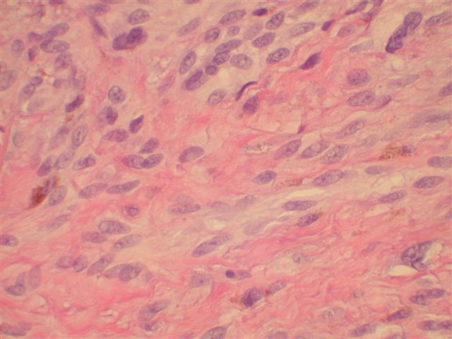
Melanoma Primary dermal melanoma
Reviewer: Christopher Hale, M.D. (see Reviewers page) Revised: 8 June 2013, last major update May 2013 Copyright: (c) 2002-2013, PathologyOutlines.com, Inc.
General
=========================================================================
Lack of atypical pigmented network and radial streaks; papillomatous projections with light brown and dark brown blotchy areas (Am J Dermatopathol 2009;31:574)
Reviewer: Christopher Hale, M.D. (see Reviewers page) Revised: 8 June 2013, last major update May 2013 Copyright: (c) 2002-2013, PathologyOutlines.com, Inc.
General
=========================================================================
Clinical features
=========================================================================
Often delay in diagnosis because lesion is attributed to trauma; most (73%) cases are AJCC stage II/III, acral lentiginous subtype (66%) and Clark level IV/V (79%, Am J Surg Pathol 2007;31:1902)
Sentinel node metastases in 24%
Case reports
=========================================================================
Reviewer: Christopher Hale, M.D. (see Reviewers page) Revised: 8 June 2013, last major update May 2013 Copyright: (c) 2002-2013, PathologyOutlines.com, Inc.
General
=========================================================================
Traditional considered most common type of melanoma (50-75%), but lentigo maligna may actually be more common in patients with extensive sun exposure (J Am Acad Dermatol 2008;58:1013)
Recently diagnosed tumors are thinner with less ulceration than in the past (Cancer 2008;113:3341)
Risk factors:
Extensive sun exposure during childhood, family history of melanoma, large numbers of benign nevi and dysplastic nevi
Recommended to evaluate vertical growth phase as prognostic factor for thin (< 0.76 mm) tumors (Am J Surg Pathol 2003;27:717)
Multi-component pattern, asymmetry and multiple colors
Also atypical reticular pattern (irregular holes and thick lines) with a sharp demarcation, blue-white veil, irregular linear vessels, central ulceration and irregularly distributed dots
Classified based on radial growth component (has nothing to do with level of dermal invasion)
Non-invasive areas have asymmetry and poor circumscription, irregular acanthosis, irregular lentiginous and nested proliferation, uniform atypical melanocytes with nests and pagetoid cells
Also transdermal migration, nuclear pleomorphism, dusty pigmentation, apoptosis of individual melanocytes and pigmented parakeratosis
Invasive component generally of epithelioid subtype
Miscellaneous Breslow’s system for tumor thickness
Reviewer: Christopher Hale, M.D. (see Reviewers page) Revised: 7 June 2013, last major update May 2013 Copyright: (c) 2002-2013, PathologyOutlines.com, Inc.
General
=========================================================================
Measure tumor thickness with ocular micrometer at right angles to surface of adjacent normal skin from (a) top of granular layer of overlying epidermis OR from ulcer base over deepest point of invasion to (b) deepest invasive tumor cells
Traditional categories are 0 to 0.76 mm, 0.76 to 1.49 mm, 1.50 mm to 3.99 mm and greater than 4.00 mm
2009 AJCC Staging System employs following thresholds for thickness: T1: ≤ 1.00 mm thickness, T2: 1.01 to 2.00 mm thick, T3: 2.01 to 4.00 mm thick, T4: more than 4.00 mm (J Clin Oncol 2009;27:6199, table)
10 year survival: 92% for melanoma < 1.00 mm thick; 80% if 1.01 to 2.00 mm thick; 63% if 2.01 to 4.00 mm thick; 50% if > 4.00 mm thick (J Clin Oncol 2009;27:6199)
5 year survival in non-ulcerated tumors is 97% for Breslow thickness of 0 to 1.0 mm, 91% for 1.01 to 2.0 mm, 79% for 2.01 to 4.0 mm and 71% for > 4.0 mm (AJCC 7th Edition, 2010)
Reviewer: Christopher Hale, M.D. (see Reviewers page) Revised: 7 June 2013, last major update May 2013 Copyright: (c) 2002-2013, PathologyOutlines.com, Inc.
General
=========================================================================
I: not penetrating basement membrane (in situ)
II: in papillary dermis (difficult to differentiate II versus III, SEER)
III: filling and expanding the papillary dermis and stopping at the interphase between the papillary and reticular dermis
Less reproducible than Breslow’s system (J Pathol 1991;163:245); also not always uniform due to variation in depth of skin layers
8 year disease free survival is 96.3% with level II, 76.1% with level III, 60.7% with level IV and 38.5% with level V (J Natl Cancer Inst 1989;81:1893)
Must recognize that deep collection of malignant melanocytes may be attached to a pilosebaceous unit
Miscellaneous Melanoma - features to report
Reviewer: Christopher Hale, M.D. (see Reviewers page) Revised: 7 June 2013, last major update May 2013 Copyright: (c) 2002-2013, PathologyOutlines.com, Inc.
Features to report =========================================================================
Specimen type / procedure
If biopsy, specify shave, punch or incisional; excision or re-excision
If lymphadenectomy, specify if sentinel node(s) or regional nodes
Tumor location
Histologic subtype
Margin of excision (distance from tumor) - lateral and deep
Angiolymphatic invasion (if present)
Mandatory features used for pathologic staging:
Maximum tumor thickness (Breslow)
Tumor ulceration
Laterality
Tumor size
Macroscopic satellite nodule(s) (required for excision specimens only)
Macroscopic pigmentation
Histologic type
Anatomic level: I (melanoma in situ), II (melanoma present in, but does not fill and expand, papillary dermis), III (melanoma fills and expands papillary dermis), IV (melanoma invades reticular dermis) and V (melanoma invades subcutaneum)
Ulceration
Features used in some protocols for treatment choice:
Tumor depth (Clark’s levels)
Margin of excision (distance from tumor) - lateral and deep
Mitotic index: less or greater than 1/mm2
Microsatellitosis
Lymph-vascular invasion
Perineural invasion
Tumor-infiltrating lymphocytes
Tumor regression
Growth phase: radial, vertical or indeterminate
Lymph nodes
Number of nodes examined, number with melanoma (if available):
Synoptic reports are more complete than non-synoptic reports; recommendations include free text option, tailoring to each institution and regular updates (Histopathology 2008;52:130), example - Michigan Cancer Consortium
Reviewer: Christopher Hale, M.D. (see Reviewers page) Revised: 8 June 2013, last major update May 2013 Copyright: (c) 2002-2013, PathologyOutlines.com, Inc.
General
=========================================================================
Sentinel lymph node is defined as first extracutaneous target of lymphogenous tumor cell spread and potential source of subsequent lymph node metastases and distant metastases (eMedicine)
Interval sentinel nodes: nodes receiving direct lymphatic drainage from primary site but lying between tumor and a recognized node field (Ann Surg Oncol 2011;18:3292)
Lymphatic mapping and sentinel lymph node biopsy are widely used for staging
Predictors of positive non-sentinel nodes in sentinel node positive patients (occurs in 20-35%): amount of tumor in sentinel node, Breslow thickness of primary melanoma and density of dendritic WBCs in sentinel node paracortex (Mod Pathol 2004;17:747); also perinodal intralymphatic tumor (Ann Surg Oncol 2008;15:1723), depth of invasion in sentinel node (Ann Surg Oncol 2008;15:1202)
4 sequential sections of both halves of each sentinel node examined - first and fourth sections stained with H&E, second section stained for S100, and third section for HMB45 (Semin Diagn Pathol 2008;25:100)
RT-PCR: prognostic significance of melanocytic mRNA in histologically negative nodes is controversial (significant-APMIS 2008;116:199, not significant-Mod Pathol 2007;20:427)
RT-PCR detects significant sentinel node metastases that are missed by histopathology, but overestimates number of patients with clinically significant metastases (Melanoma Res 2003;13:313)
FISH detects chromosomal aberrations in 83% of nodal metastatic melanoma vs. 6% of nodal nevi (Am J Surg Pathol 2010;34:231)
Reviewer: Christopher Hale, M.D. (see Reviewers page) Revised: 8 June 2013, last major update May 2013 Copyright: (c) 2002-2013, PathologyOutlines.com, Inc.
Primary difference between clinical and pathologic stage grouping is whether regional lymph nodes are staged clinically (including radiologic exam) or pathologically (after excision)
NX: regional lymph nodes cannot be assessed (e.g. previously removed for another reason)
N0: no regional lymph node metastasis
N1: metastasis in one lymph node
N1a: micrometastasis
N1b: macrometastasis
N2: metastasis in 2-3 regional nodes
N2a: micrometastasis
N2b: macrometastasis
N2c: satellite or in-transit metastasis without nodal metastasis
N3: metastasis in four or more regional nodes, or matted metastatic nodes, or in-transit metastasis or satellite(s) with metastasis in regional node(s)
Micrometastases are diagnosed after sentinel lymph node biopsy and completion lymphadenectomy (if performed)
Macrometastases are defined as clinically detectable nodal metastases confirmed by therapeutic lymphadenectomy or when nodal metastasis exhibits gross extracapsular extension
Satellite metastases: defined arbitrarily as intralymphatic metastases occurring within 2 cm of the primary melanoma
In-transit metastases: defined arbitrarily as intralymphatic metastases occurring more than 2 cm from the primary melanoma, but before the first echelon of regional lymph nodes
Note: HMB45 or MelanA positive isolated cells in sentinel nodes appear to have no prognostic significance, at least short term (Am J Surg Pathol 2007;31:1175)
Pathologic staging includes microstaging of the primary melanoma and pathologic information about the regional lymph nodes after partial or complete lymphadenectomy
Pathologic Stage 0 or Stage IA patients are the exception; they do not require pathologic evaluation of their lymph nodes
End of Skin-melanocytic tumor > Superpage
This information is intended for physicians and related personnel, who understand that medical information is often imperfect, and must be interpreted in the context of a patient's clinical data using reasonable medical judgment. This website should not be used as a substitute for the advice of a licensed physician.
All information on this website is protected by copyright of PathologyOutlines.com, Inc. Information from third parties may also be protected by copyright. Please contact us at copyrightPathOut@gmail.com
with any questions (click here for other
contact information).