

Skin melanocytic tumor
Lentigines, melanotic macules and melanocytic hyperplasia
Lentigo
Author: Christopher S. Hale, M.D.

Last author update: 1 April 2013
Last staff update: 10 March 2023
Copyright: 2002-2024, PathologyOutlines.com, Inc.
PubMed Search: Solar lentigo
Table of Contents
Definition / general | Terminology | Epidemiology | Clinical features | Treatment | Dermoscopy | Clinical images | Microscopic (histologic) description | Differential diagnosisCite this page: Hale CS. Lentigo. PathologyOutlines.com website. https://www.pathologyoutlines.com/topic/skintumormelanocyticsolarlentigo.html. Accessed April 19th, 2024.
Definition / general
- Benign melanocytic proliferation due to sun exposure (Br J Dermatol 2007;156:1214, eMedicine)
- Multiple lesions, often poorly circumscribed
Terminology
- Also called solar lentigines, age spots
Epidemiology
- Sun exposed skin of elderly (90% of whites age 60+); also truck drivers on sun exposed face (J Dermatol 2008;35:146)
- Multiple large solar lentigos on upper back and shoulders suggest prior severe sunburn, a risk factor for melanoma (Dermatology 2007;214:25)
- PUVA (psoralen + ultraviolet A) treatment for psoriasis cause numerous solar lentigines with atypia; similar findings after severe radiation exposure (Arch Dermatol 1997;133:209)
Clinical features
- Macular hyperpigmentation
- Often > 1 cm
- "Ink spot lentigo" variant: small, darkly pigmented, stellate
Treatment
- Laser (J Am Acad Dermatol 2006;54(5 Suppl 2):S262)
- Intense pulsed light (Dermatol Surg 2007;33:449)
- Cryotherapy
- Trichloroacetic acid (J Eur Acad Dermatol Venereol 2008;22:316)
Dermoscopy
- Sharply demarcated border and fingerprint like structures
Clinical images
Images hosted on other servers:
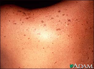
Back lesions


Pre and post laser treatment
Microscopic (histologic) description
- Elongation of rete ridges and increased pigmentation at tips of retes; pigmentation may be irregular
- Also solar elastosis, telangiectasia, variable chronic inflammatory infiltrate in dermis
Differential diagnosis
- Actinic keratosis
- Lentigo maligna melanoma: clusters of MelanA+ cells at dermoepidermal junction, versus scattered for solar lentigo (J Cutan Pathol 2008;35:931)
- Macular seborrheic keratosis: thicker lesion, horn cysts / pseudocytes and on continuum with solar lentigo
