

Skin melanocytic tumor
Spitz neoplasms
Reed nevus
Author: Christopher S. Hale, M.D.

Last author update: 1 February 2013
Last staff update: 12 November 2024 (update in progress)
Copyright: 2002-2024, PathologyOutlines.com, Inc.
PubMed Search: Pigmented spindle cell nevus

Table of Contents
Definition / general | Terminology | Epidemiology | Sites | Clinical features | Case reports | Treatment | Dermoscopy | Clinical images | Microscopic (histologic) description | Microscopic (histologic) images | Positive stains | Molecular / cytogenetics description | Videos | Differential diagnosisCite this page: Hale CS. Reed nevus. PathologyOutlines.com website. https://www.pathologyoutlines.com/topic/skintumormelanocyticpigmentedspindlecellnevus.html. Accessed December 27th, 2024.
Definition / general
- First described by Reed in 1975
- Heavily pigmented nevus, often recent onset, widely considered a Spitz nevus variant (J Am Acad Dermatol 1993;28:565)
- Clinically and histologically simulates melanoma
Terminology
- Also called Reed's nevus
Epidemiology
- Young adults, commonly women (Am J Surg Pathol 1984;8:645)
- Median age 25 (Dermatol Online J 2004;10:5)
Sites
- Proximal extremities or trunk (Am J Surg Pathol 1984;8:645)
Clinical features
- < 1 cm, solitary, deeply pigmented and well circumscribed maculopapule
- Clinically resembles melanoma
Case reports
- 23 year old woman with pigmented lesion since childhood on lower leg (Australas J Dermatol 2011;52:104)
Treatment
- Conservative but complete excision
- Does not recur
Dermoscopy
- 2 patterns reported:
- Brown to blue pigmentation with peripheral rim of large brown globules (globular pattern)
- Dark diffused pigmentation and pseudopods regularly distributed at periphery in stellate or radiate pattern (starbust pattern) (Dermatol Online J 2004;10:5)
Clinical images
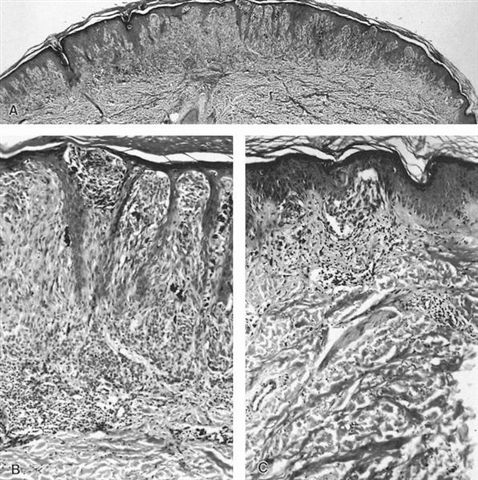
AFIP images

Small and
symmetrical
lesion differs
from Spitz nevus
Images hosted on other servers:

Rim of brown globules around lesion

Pseudopods arranged regularly

Small multicolored lesion

Slightly raised dark lesion
Microscopic (histologic) description
- Some similarity with Spitz nevi
- Symmetric with cytologic maturation
- Nests and fascicles of spindled melanocytes along dermoepidermal junction and within dermal papillae
- May be junctional or compound
- Expansive, not infiltrative growth pattern
- Extends no deeper than reticular dermis
- Nevus cells typically contain abundant melanin pigment, may be associated with melanophages
- Nuclei are monotonous, resemble normal keratinocytes and may have small nucleoli
- Often has architectural or cytologic atypia (Hum Pathol 1991;22:52)
- Variable lymphocytic infiltrate at base of lesion
- Variable transepidermal elimination of junctional nests
- No / rare mitotic figures
- Note: hypopigmented variant is similar but without abundant melanin (J Cutan Pathol 2008;35 Suppl 1:87)
Microscopic (histologic) images
AFIP images
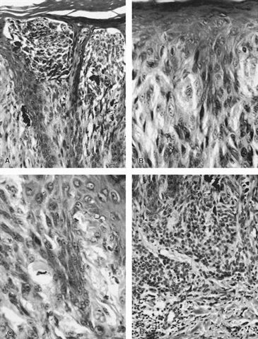
Various images
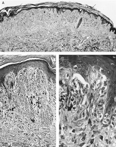
Images hosted on other servers:


Symmetric pigmented tumors (figs 6 - 9)


Symmetric pigmented tumor

Pigmented spindle cell tumor
Positive stains
- HMB45 highly expressed in intraepidermal component of pigmented spindle cell nevus (PSCN) and spindle cell melanoma but dermal component negative in PSCN, irregularly positive in spindle cell melanoma (Am J Surg Pathol 2011;35:1733)
- Other melanocytic markers (S100, MelanA)
Molecular / cytogenetics description
- FISH targeting 6p25 (RREB1), 11q13 (CCND1), 6q23 (MYB) and centromere 6 may differentiate from melanoma (Am J Surg Pathol 2011;35:1733, Adv Anat Pathol 2011;18:229)
Videos
Pigmented spindle cell nevus
Differential diagnosis
- Spindle cell melanoma
- Spitz nevus
- Superficial spreading melanoma (Dermatol Online J 2004;10:5)

