

Skin nontumor
Pigmentary disorders
Vitiligo
Last author update: 1 August 2011
Last staff update: 18 November 2020
Copyright: 2002-2024, PathologyOutlines.com, Inc.
PubMed Search: Vitiligo [title] skin


Table of Contents
Definition / general | Epidemiology | Sites | Clinical features | Treatment | Clinical images | Microscopic (histologic) description | Electron microscopy description | Differential diagnosisCite this page: Hamodat M, Hale CS. Vitiligo. PathologyOutlines.com website. https://www.pathologyoutlines.com/topic/skinnontumorvitiligo.html. Accessed April 26th, 2024.
Definition / general
Epidemiology
- Affects 1% of world’s population; more noticeable in dark skinned individuals
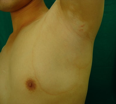
- Usually hands / wrists, axilla, perioral, periorbital, anogenital skin
Sites
- Focal: only a few areas
- Segmented: one side of the body only
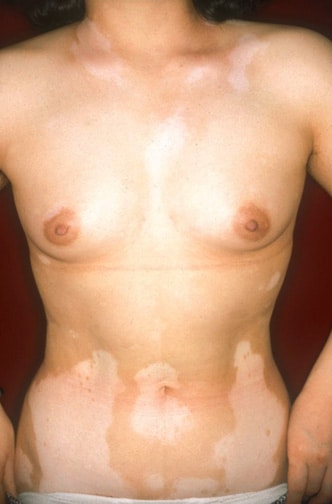
- Generalized: most common, both sides of body
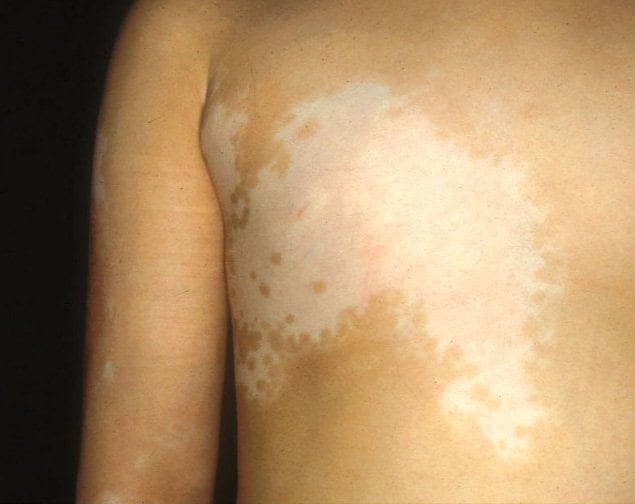
- Trichrome: patient has three shades of skin color
Clinical features
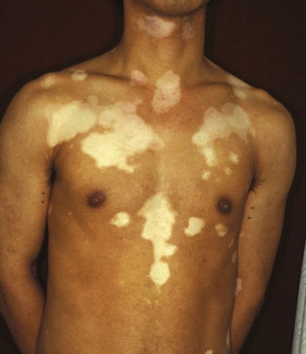
- Asymptomatic, flat, well-demarcated zones of pigment loss
- Autoimmune disorder associated with pernicious anemia, Addison’s disease, Hashimoto’s thyroiditis
- Perilesional skin up to 5 cm from vitiligo spot is still lighter than normal (Photodermatol Photoimmunol Photomed 2008;24:314)
- Associated with polymorphisms in COX2 gene (J Dermatol Sci 2009;53:176), mutations of autoimmune regulator gene (Br J Dermatol 2008;159:591)
- May cause severe psychological distress
- May worsen with local trauma (cuts, scrapes, burns, Koebner phenomenon)
- Decreased risk for melanoma and nonmelanoma skin cancer (Br J Dermatol 2013;168:162)
Treatment
- Laser skin ablation; phototherapy; 5 FU (Photodermatol Photoimmunol Photomed 2008;24:322)
- Topical steroids or immunomodulators (J Dermatol 2008;35:503)
- Hydroxyacetone (Dermatol Online J 2008;14:23)
Clinical images
Images hosted on other servers:

Various images
Marginal inflammatory
Segmental
Nonsegmental

Hands

Autologous epidermal graft using suction blister
Microscopic (histologic) description
- Difficult to diagnose by histology; decreased melanocytes (use S100 or MelanA and control biopsy from adjacent normal skin)(Am J Dermatopathol 2008;30:112)
- At advancing border, melanocytes may be increased in size with an increased number of dendrites; occasionally lymphocytes are present in this region, particularly if an inflammatory border is present; epidermotropic lymphocytes may form small Pautrier-like collections in the basal layer, with an associated perivascular infiltrate of mononuclear cells involving the superficial plexus and some superficial edema
- Focal spongiosis may be present in marginal areas
- Degenerative changes have also been reported in nerves and sweat glands
- Langerhans cells are usually increased
- Melanocytes are always reduced more in vitiligo than they are in nevus depigmentosus
Electron microscopy description
- No melanocytes
- Keratinocyte apoptosis (Ann Dermatol 2012;24:115)
Differential diagnosis
- Leukoderma: chemical, melanoma related, scleroderma related; acquired condition with localized loss of skin pigmentation associated with inflammatory skin conditions, burns, intralesional steroid injections, postdermabrasion (Wikipedia)
- Oculocutaneous albinism: melanocytes present, but no melanin due to defect in tyrosinase enzyme or melanogenesis
