
Skin nontumor
Infectious disorders
Scrub typhus
Last author update: 1 September 2010
Last staff update: 16 November 2020
Copyright: 2002-2025, PathologyOutlines.com, Inc.
PubMed Search: Scrub typhus [title]


Table of Contents
Definition / general | Terminology | Epidemiology | Etiology | Clinical features | Treatment | Clinical images | Microscopic (histologic) description | Positive stains | Molecular / cytogenetics description | Additional referencesCite this page: Hamodat M. Scrub typhus. PathologyOutlines.com website. https://www.pathologyoutlines.com/topic/skinnontumorscrubtyphus.html. Accessed April 3rd, 2025.
Definition / general
- Chigger-borne zoonosis of tropical Asia and western Pacific islands, caused by Orientia (formerly Rickettsia) tsutsugamushi
Terminology
- Formerly called Rickettsia tsutsugamushi but now distinguished from Rickettsiaceae family by differences in cell wall
- Similar in presentation to other forms of typhus, but caused by agent in a different genus (Wikipedia: Scrub Typhus [Accessed 28 August 2018])
- "Scrub" refers to the type of vegetation (i.e., terrain between woods and clearings) that harbors the vector but name is not entirely correct because endemic areas can be sandy and semiarid (eMedicine: Scrub Typhus [Accessed 28 August 2018])
Epidemiology
- Infected large number of soldiers in World War II
- Today, a frequent cause of febrile illness leading to hospital admissions in indigenous populations in eastern Asia, the southwestern Pacific (Korea to Australia) and from Japan to India and Pakistan
Etiology
- Caused by Orientia (formerly Rickettsia) tsutsugamushi, an obligate intracellular gram-negative bacteria
- Maintained in nature by trombiculid mites (Wikipedia: Trombiculidae [Accessed 28 August 2018]), transmitted to humans by larval mite (chigger) during feeding
- Orientia is grouped with the Rickettsial infections (Centers for Disease Control: Travelers' Health [Accessed 28 August 2018]) but distinguished from Rickettsiaceae family by differences in cell wall (Int J Syst Bacteriol 1995;45:589)
Clinical features
- Travelers with imported disease often become sick before or within a few days of return from an endemic region; unlikely diagnosis if illness begins > 18 days after return (Curr Infect Dis Rep 2009;11:66)
Treatment
- Doxycycline (eMedicine: Scrub Typhus [Accessed 28 August 2018])
- Death rates up to 30% without treatment; death is now rare with treatment
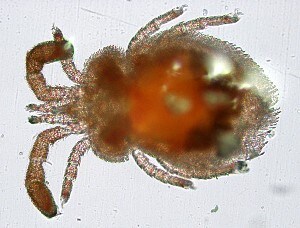
Clinical images
Images hosted on other servers:

Adult trombiculid mite
Chigger

Eschar
Microscopic (histologic) description
- Lymphocytic vasculitis of arterioles (not postcapillary venules) causes cutaneous findings
- Variable thrombosis of vessels
- Microorganisms are detected in endothelium of skin and other organs using fluorescein-labelled antiserum; also present in macrophages as fine granules on high power
- Eschar exhibits necrotic ulceration, due to coagulative necrosis of of the epidermis and underlying dermis
- Often only few neutrophils in the inflammatory infiltrate
Positive stains
- Immunohistochemical staining of Orientia tsutsugamushi, particularly in eschars, is confirmatory (Am J Clin Pathol 2008;130:543)
Molecular / cytogenetics description
- PCR is useful for confirmation
Additional references
