

Skin nontumor
General
Histology
Author: Mariantonieta Tirado, M.D.
Editorial Board Member: Robert E. LeBlanc, M.D.
Editor-in-Chief: Debra L. Zynger, M.D.

Last author update: 29 September 2020
Last staff update: 19 July 2021
Copyright: 2002-2024, PathologyOutlines.com, Inc.
PubMed Search: Skin[TIAB] histology[TI] review[PT]

Table of Contents
Definition / general | Essential features | Terminology | Physiology | Diagrams / tables | Microscopic (histologic) description | Microscopic (histologic) images | Positive stains | Negative stains | Electron microscopy description | Electron microscopy images | Additional references | Board review style question #1 | Board review style answer #1 | Board review style question #2 | Board review style answer #2 | Board review style question #3 | Board review style answer #3Cite this page: Tirado M. Histology. PathologyOutlines.com website. https://www.pathologyoutlines.com/topic/skinnontumorgeneral.html. Accessed December 22nd, 2024.
Definition / general
- Membrane covering the exterior of the body composed of epidermis and dermis
Essential features
- Skin is the largest organ of the body with a weight of approximately 5 kg and an area close to 2 m²
- General functions of the skin:
- Protection (against UV light, dehydration, microbial invasion, mechanical injury, chemical and thermal stresses)
- Retains water and excretes drugs and waste
- Thermoregulation through blood vessels and eccrine glands
- Metabolic: vitamin D production
- Sensorial: contains peripheral endings of sensory nerves
Terminology
- Also called integument
Physiology
- Epidermis
- Outer stratified squamous epithelium composed of epidermal and intraepidermal adnexal (acrotrichial and acrosyringeal) keratinocytes
- Main purpose is cornification
- Keratinocytes are connected by desmosomes, adherens junctions, tight junctions and gap junctions
- Basal layer
- 10% are stem cells
- Produces keratin 5
- Squamous layer
- Desmosomes develop and composition of keratin intermediate filaments change (↑ intracellular calcium)
- Produces keratins 1 and 10
- Langerhans cells and Merkel cells present
- Granular layer
- Profilaggrin which forms filaggrin: facilitates filament aggregation
- Loricrin and involucrin: contribute to the formation of the insoluble cell envelope
- Cells begin to lose nuclei
- Production of the cell envelope
- Located beneath the cell membrane
- Composed of crosslinked proteins dependent on transglutaminase
- Odland bodies
- Lipid rich lamellated granules (mainly ceramide) secreted into intercellular spaces
- Contribute to permeability barrier
- Cornified layer
- Formed because of keratinocytic maturation (cells flatten as they ascend to the surface)
- Synthesis of lamellar granules and proteins
- Cells lose their nuclei, cytoplasmic organelles, metabolic activity and eventually desquamate
- Dermal epidermal junction: components bind the epidermis to the dermis
- 4 major structural regions proceeding from the epidermis to the dermis
- Basal cell plasma membrane
- Lamina lucida: low electron density region, contains laminins (major laminin 332)
- Lamina densa: electron dense region; its major component is collagen IV
- Sublamina densa: upper papillary dermis contains loops of type VII collagen (anchoring fibrils)
- Attachment structures
- Hemidesmosomes: plaque proteins extend from basal plasma membrane of the keratinocyte to the lamina lucida (associated to the anchoring filaments)
- Anchoring filaments: thread-like structures that cross the lamina lucida
- Anchoring fibrils: extend from the lower lamina densa to the upper reticular dermis
- Total epidermal renewal time, approximately 2 months
- Cells take 26 - 42 days to transit from the basal layer to the granular layer
- 14 days for keratin layer to be shed
- 4 major structural regions proceeding from the epidermis to the dermis
- Melanocytes
- Neural crest derived dendritic cells that synthesize melanin
- Melanin absorbs ultraviolet (UV) and protects from damaging UV Induced mutations
- Melanin (produced from tyrosine) is transferred in melanosomes (lysosome type organelle), through melanocytic dendritic processes into adjacent keratinocytes and hair shafts
- 2 types of melanin are produced: pheomelanin (yellow-red) and eumelanin (black-brown)
- Racial skin color is due to amount of melanin in keratinocytes, not number of melanocytes
- Dermis
- Mainly supports the epidermis and plays varied other roles due to the presence of vessels, nerves and adnexa
- Blood vessels provide nutrients and help regulate temperature
- Sucquet-Hoyer canals
- Specialized acral arteriovenous anastomoses
- Allow blood shunting from arterioles to venules, bypassing capillaries
- Surrounded by modified smooth muscle glomus cells (latter function as sphincters)
- Primary function is of thermoregulation
- Lymphatics
- Play a role in tumor spread and removal of debris (fluid, cells and macromolecules)
- Represent the primary route for Langerhans cells to reach regional lymph nodes
- Nerves
- Somatic sensory nerves mediate pain, itch temperature and touch
- Autonomic motor nerves mediate vascular tone, pilomotor response and sweating
- Afferent nerves consist of myelinated and nonmyelinated free nerve endings
- 2 types of sensory receptors are present
- Specialized (encapsulated): Meissner corpuscles have tactile function and Pacinian corpuscles detect pressure
- Unspecialized: sensory non-encapsulated nerves linked to Merkel cells
- Subcutis
- Plays a role in cushioning, insulation, endocrine function and energy stores
- 2 types of fat:
- White
- Brown: mostly present in infants and children, rich in mitochondria and produces heat
- Adnexa
- Skin associated structures each with specific functions including hair follicles, sebaceous glands, eccrine sweat glands and apocrine glands
- Folliculosebaceous apocrine units: functional complex of hair follicle, sebaceous gland, erector pili muscle and (depending on site) apocrine gland
- Hair follicle
- Functions include temperature regulation, protection of other structures and tactile sensory input
- Types of hair
- Lanugo: present during late gestation and first month of life
- Vellus: fine hair (face, trunk and extremities)
- Terminal: coarse (scalp, eyebrows, eyelashes)
- Cycle of hair
- Anagen: growing phase (lasts 2 - 7 years)
- Catagen: involuting phase (lasts 2 - 3 weeks, 1 - 2% of total hair)
- Telogen: resting phase (lasts 100 days, 10 - 20% of total hair)
- Sebaceous glands
- Main function is protection by production and release of sebum
- Secretion (governed by androgens) plays a role in waterproofing, control of epidermal water loss and inhibition of fungal and bacterial growth
- Glands are holocrine (secretion depends on degeneration of acini with release of cells and lipid)
- Sebaceous secretions (sebum) carry to the surface a mixture of fat, corneocytes and normal flora (yeasts, bacteria and mites)
- Flora includes Propionobacterium acnes, Staphylococcus epidermidis and Demodex brevis
- Apocrine sweat glands
- Concentrated in axilla, groin, perineum, face, periumbilical, external auditory meatus, eyelid and areola
- Function in humans is unknown; in other mammals they produce scent and play a role in sexual attraction
- Odorless secretion is initially released and then modified by superficial bacteria producing body odor
- Eccrine sweat glands
- Regulate body temperature
- Present almost everywhere in the skin except oral lips, clitoris, labia minora and external auditory canal
- Eccrine duct possesses a conduit and metabolic (modifies secretion and reabsorbs water) function
- Possess merocrine secretion
- Nail unit
- Protects tissues of the distal fingertip from injuries and enhances delicate movements by counterpressure
- Langerhans cells
- Bone marrow derived dendritic cells, described in 1868 by Paul Langerhans, a German pathologist, physiologist and biologist (Am J Dermatopathol 1985;7:347)
- Function as intraepidermal macrophages phagocytosing antigens and then migrate to regional lymph nodes where they present antigens to T cells
- Merkel cells
- Part of the affector limb mechanoreceptors related with touch sensation
- Described by in 1875 by Friedrich Merkel a Germany anatomist and histopathologist (Am J Dermatopathol 1982;4:521)
- Located in the basal epidermis and concentrated in tactile areas of hairy skin, glabrous skin, lips, eccrine sweat glands and anal canal
- Their close relation with nerve fibers represents a Merkel cell neurite complex
- Heavily granulated cells containing keratin filaments and neuropeptides (Anat Rec A Discov Mol Cell Evol Biol 2003;271:225)
Diagrams / tables
Contributed by Mariantonieta Tirado, M.D.
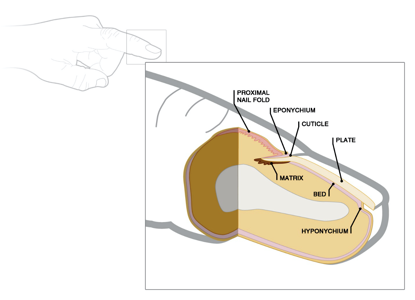
Normal nail anatomy
Microscopic (histologic) description
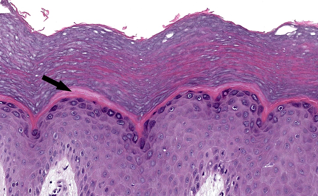
- Epidermis
- Composed of 4 layers
- Basal cell layer (stratum basale)
- Prickle cell layer (stratum spinosum or Malpighian layer)
- Granular cell layer (stratum granulosum)
- Corneocyte layer (stratum corneum, horny layer)
- Basal layer
- Proliferating cell population with cuboidal shape, larger nuclei, conspicuous nucleoli and basophilic cytoplasm
- Few mitotic figures may be present
- Melanocytes surrounded by clear halo are present
- Toker cells are clear cells present in the basal and suprabasal layers of the nipple epidermis of both males and females
- Squamous layer
- Several layers of larger eosinophilic polygonal cells with oval nuclei and conspicuous nucleoli
- Cells attached to each other by spine-like processes (intercellular bridges)
- Granular layer: 3 layers of flattened, diamond shaped cells with keratohyaline granules
- Cornified layer: composed of flat, eosinophilic corneocytes without nuclei
- Stratum lucidum
- Homogenous eosinophilic zone
- Present only in soles and palms, between granular and cornified layer
- Composed of 4 layers
- Melanocytes
- Appear as clear cells (truly an artifact of fixation, secondary to shrinkage of the cytoplasm), with dendritic cytoplasm and a smaller and more basophilic nucleus than that of a basal keratinocyte
- Ratio of melanocytes to basal cells ranges from approximately 1:4 on the cheek to 1:10 on the limbs
- Dermis and subcutis
- Divided into superficial papillary dermis and deeper reticular dermis
- Papillary dermis: thin collagen fibers, located beneath the epidermis and around adnexa
- Reticular dermis: thicker, extends from the base of the papillary dermis to the surface of the subcutis
- Varies in thickness depending on anatomic location (eyelid: 0.5 mm; back: 5 mm)
- Consists of connective tissue composed of collagen, elastic fibers and ground substance of mucopolysaccharides and mucoproteins
- Harbors:
- Scattered cells (fibrocytes, dendrocytes, histiocytes, mast cells, Langerhans cells and rare lymphocytes)
- Adnexa
- Smooth muscle
- Nerves
- Vessels: small arteries, arterioles and lymphatics
- Small arteries, arterioles, venules, lymphatics and nerves conform a network of 2 connected plexuses parallel to the surface
- Superficial plexus: located in the upper reticular dermis, supplies the papillary dermis with a capillary loop system
- Deep plexus: located in the lower reticular dermis
- Sensory receptors
- Meissner corpuscles: ellipsoid lamellated structures, localized in the papillary dermis of lips, palms and soles
- Pacinian corpuscles: ovoid structure with concentric lamellae, localized in the deep dermis and subcutis of genitalia, lips, palms and soles
- Subcutis
- Contains lobules of mature adipose tissue divided by thin connective tissue septa
- Composed of adipocytes with a single globule of lipid that compresses the nucleus to the periphery
- Divided into superficial papillary dermis and deeper reticular dermis
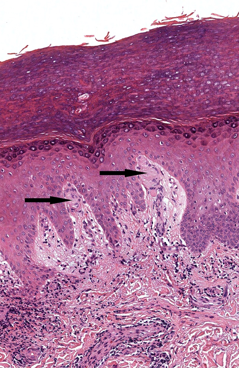
- Hair follicles
- Segments of the hair follicle in longitudinal sections
- Upper segment: stationary
- Infundibulum: from ostium of the follicle to the opening of the sebaceous duct; shape of a funnel with similar layers as the epidermis, with granular layer
- Isthmus: from the opening of the sebaceous duct to the attachment of the arrector pili muscle at the hair bulge; contains a basal layer, spinous layer, absence of granular layer and an eosinophilic cornified layer
- Lower segment: transient
- Stem: from base of the isthmus to Adamson fringe (latter area between anucleated cells of the stem and nucleated cells of the bulb)
- Bulb: contains matrix cells with large pale nucleus and prominent nucleoli and melanocytes that surround the dermal papillae
- Upper segment: stationary
- Layers of a terminal anagen hair follicle of the suprabulbar area in horizontal sections, from the center to the periphery
- Hair shaft (medulla, cortex and cuticle)
- Inner root sheath
- Cuticular layer of the inner root sheath: 1 cell thick
- Huxley layer: 2 cells thick with abundant eosinophilic trichohyalin granules
- Henle layer: 1 cell thick, bright eosinophilic trichohyalin granules
- Outer root sheath: composed of clear keratinocytes and keratohyaline granules
- Vitreous and external fibrous layer (perifollicular connective tissue sheath)
- Hair often contains Demodex folliculorum mites, clumps of Staphylococcus epidermidis or Pityrosporum yeasts
- Segments of the hair follicle in longitudinal sections
- Sebaceous glands
- Lobulated structures mostly connected to hair follicles
- Distributed all over the skin with exception of palms, soles and dorsum of the feet
- Have outer cuboidal or flattened, basophilic germinative cells that differentiate, move inward and accumulate intracytoplasmic lipid droplets, causing multivacuolation and indentations of nuclei
- Excretory ducts are lined by keratinizing squamous epithelium
- Apocrine sweat glands
- Empty into the follicle above the sebaceous duct
- Includes 2 components
- Secretory
- Located in the deep dermis or subcutis
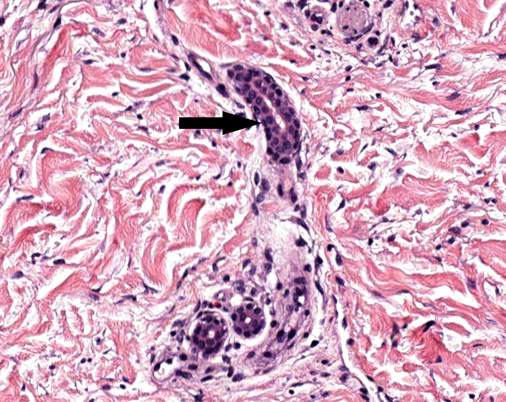
- Possesses an outer layer of myoepithelial cells and an inner layer of cuboidal to columnar eosinophilic cells
- Shows luminal "decapitation" secretion
- Ductal
- Connects with the pilosebaceous follicle
- Composed by a double layer of cuboidal cells
- Histologically indistinguishable from eccrine ducts
- Secretory
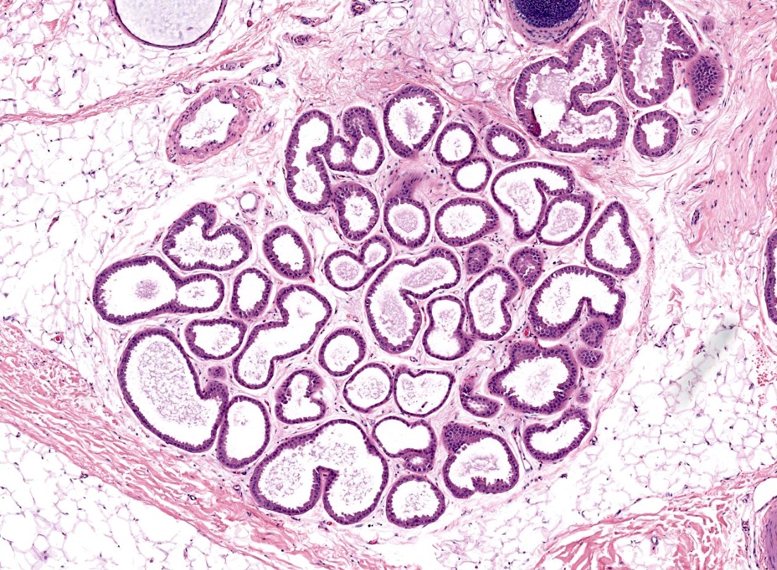
- Eccrine sweat glands
- Includes 2 components
- Secretory
- Located deep in the dermis or subcutis
- Has an outer layer of myoepithelial cells
- Also possesses an inner layer of large clear pyramidal cells (secrete water) and smaller darker cells (secrete glycoproteins, mostly line the luminal surface)
- Ductal
- Opens directly into the epidermis
- Composed of a double layer of basophilic cuboidal cells
- Luminal surface is lined by an eosinophilic cuticle
- Divided in 4 subunits: coiled secretory unit, coiled dermal duct, straight dermal duct, coiled intraepidermal duct (acrosyringium)
- Secretory
- Includes 2 components
- Nail unit
- Comprised of the nail plate and surrounding tissues
- Located in the dorsal aspect of the distal phalanx of fingers and toes
- Anatomic structures include
- Proximal nail fold: layer that extends superficially with the skin and deeply with the nail matrix
- Eponychium (cuticle): cornified layer of the nail fold located between the nail plate and matrix
- Nail matrix: produces the superficial and ventral portions of the nail plate
- Lunula: white crescent shaped area representing the junction between the matrix and the bed
- Nail plate: consists of corneocytes and is attached to the nail bed
- Nail bed: epithelium lying over a vascularized dermis that provides support to the nail plate
- Hyponychium: intermediate epithelium between the junction of the distal ventral edge of the free nail and the fingertip skin
- Lateral nail folds: lateral overhanging skin folds that guide the growth of the nail plate
- Langerhans cells
- Dendritic cells with reniform nucleus scattered in the superficial epidermal spinous layer into the granular layer and in the dermis, difficult to see on H&E
- Merkel cells
- More common in outer root sheath of hair follicles and tactile hair discs
- Not identified with H&E but with immunohistochemistry and electron microscopy
Microscopic (histologic) images
Contributed by Mariantonieta Tirado, M.D.

Layers of the epidermis
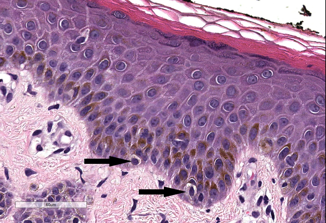
Melanocytes
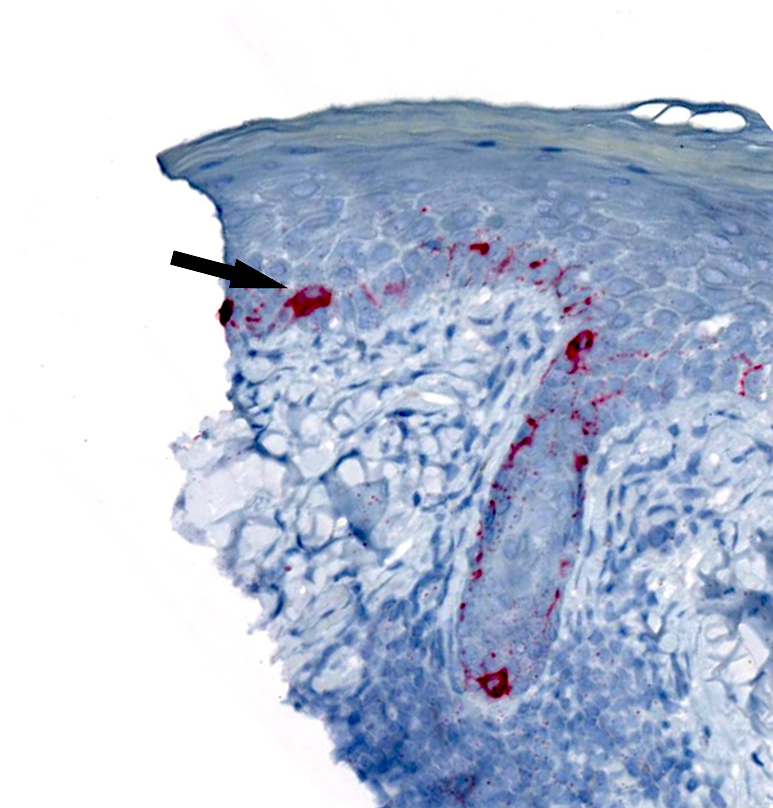
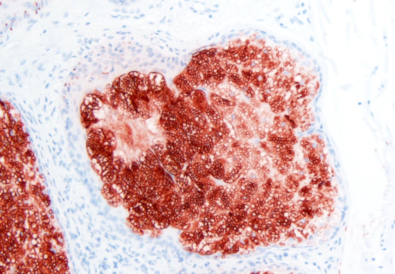
MelanA+ melanocytes
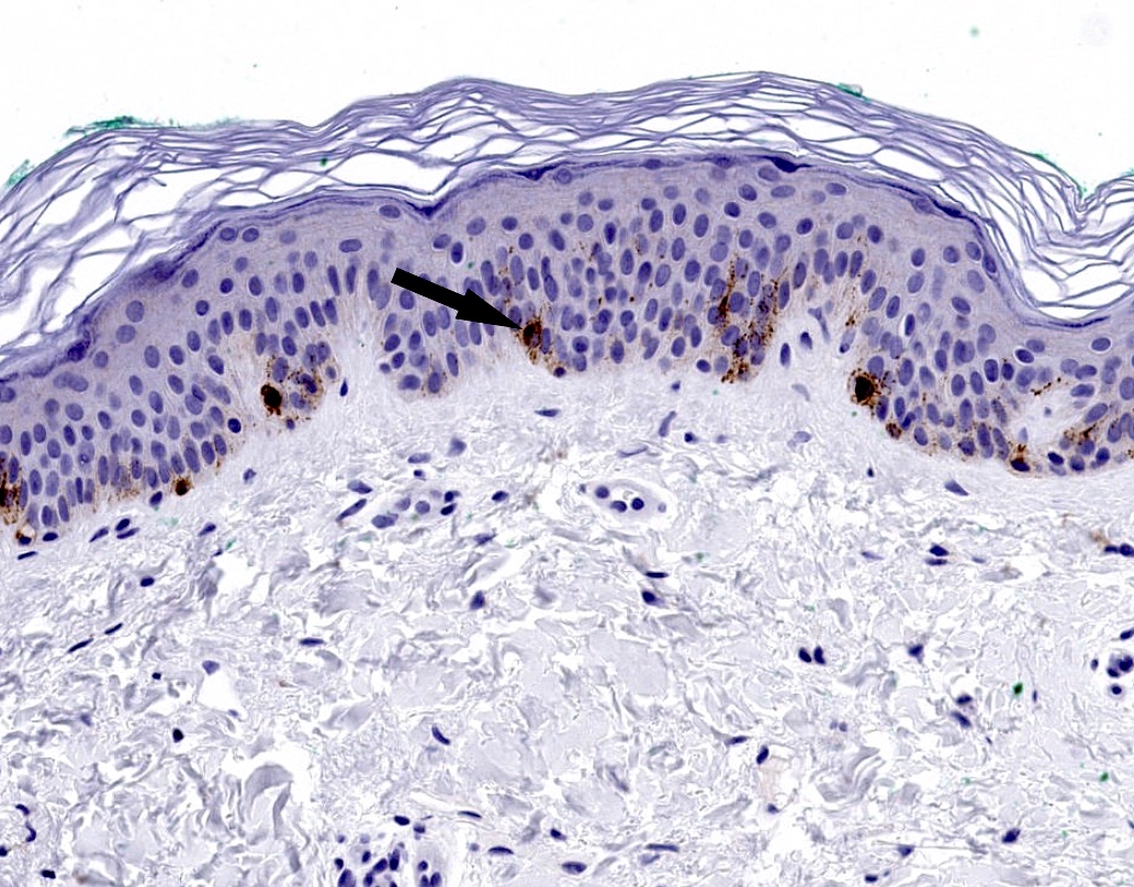
HMB45+ melanocytes
Stratum lucidum
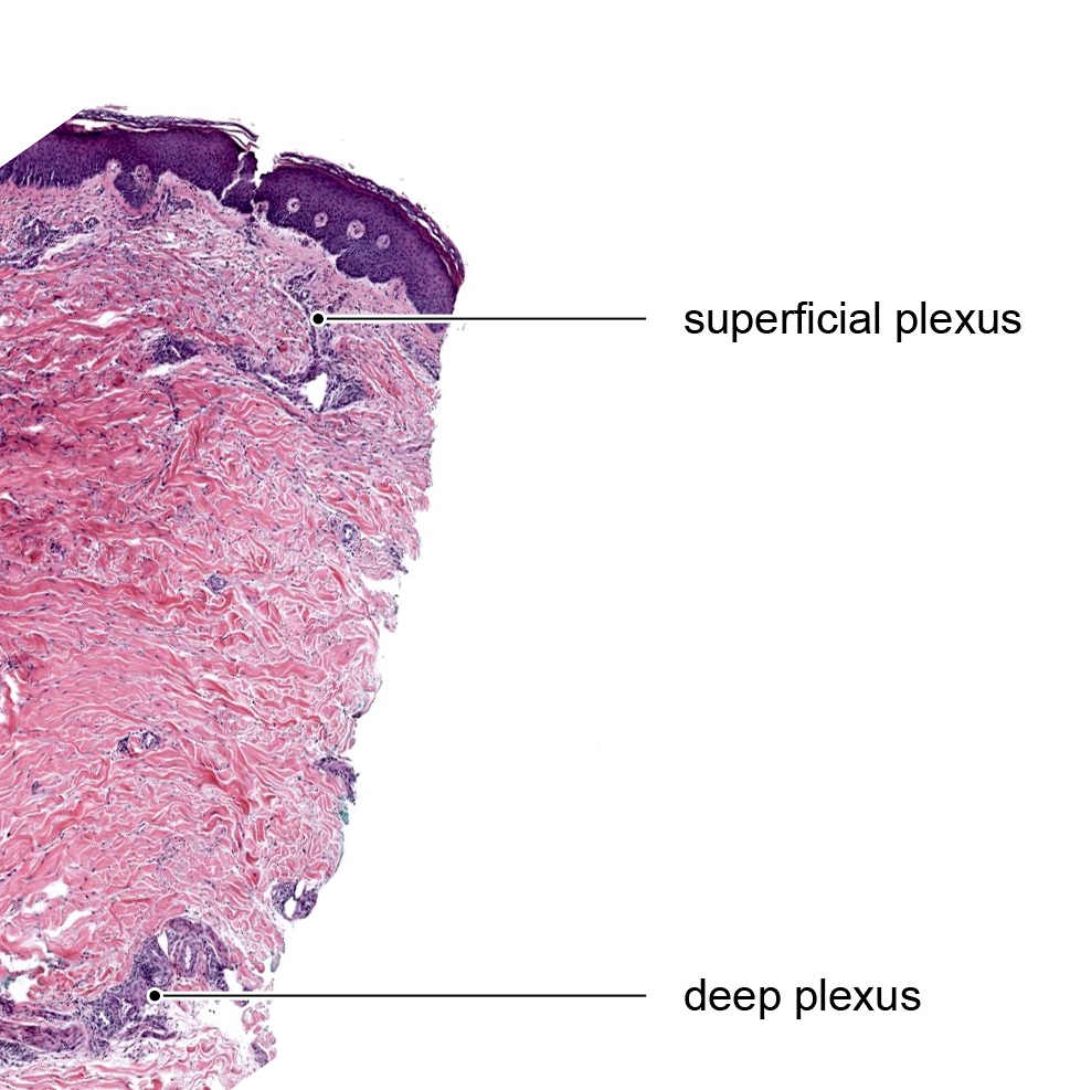
Superficial and deep neurovascular plexuses

Pacinian corpuscle
Meissner corpuscles
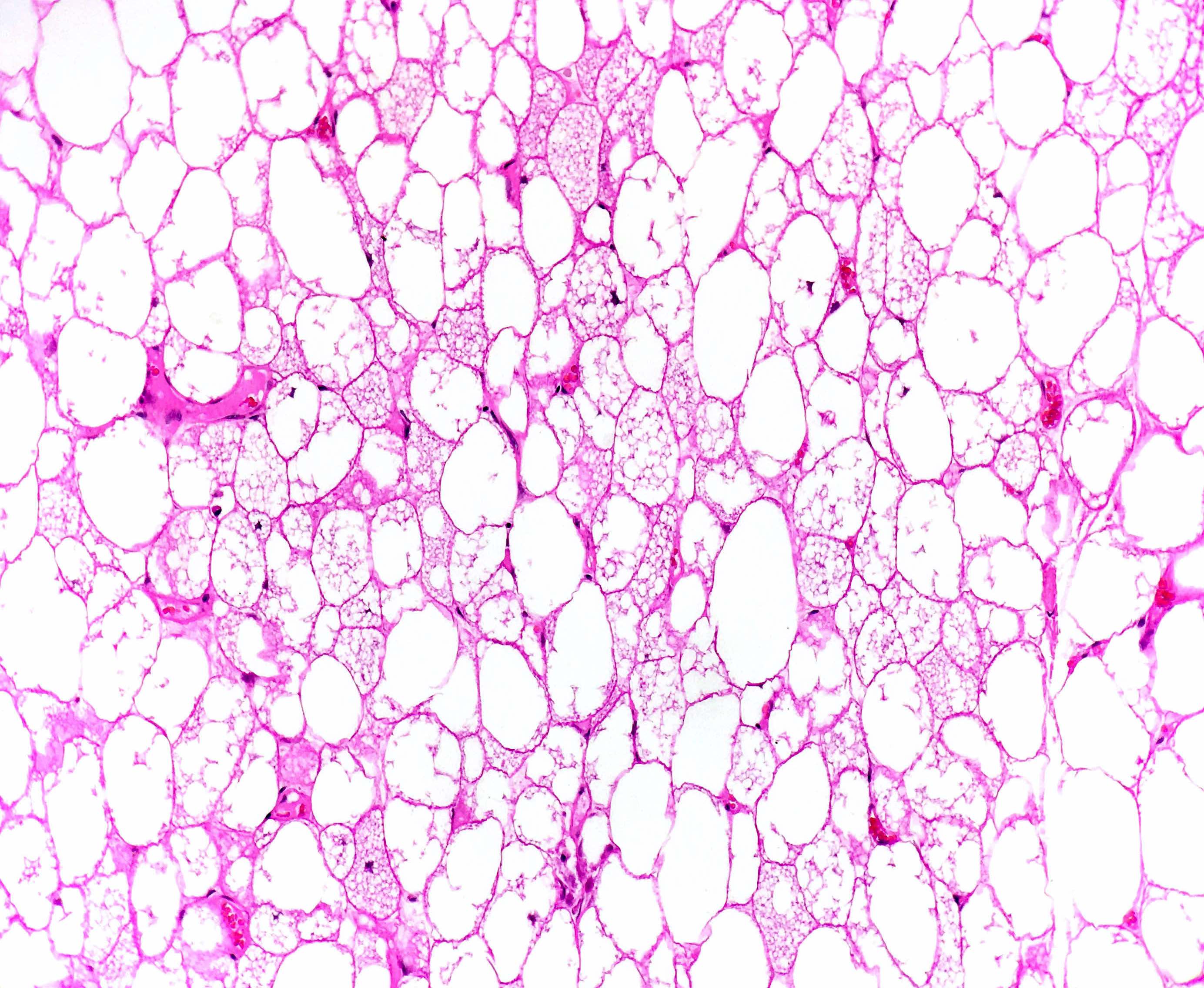
Brown fat
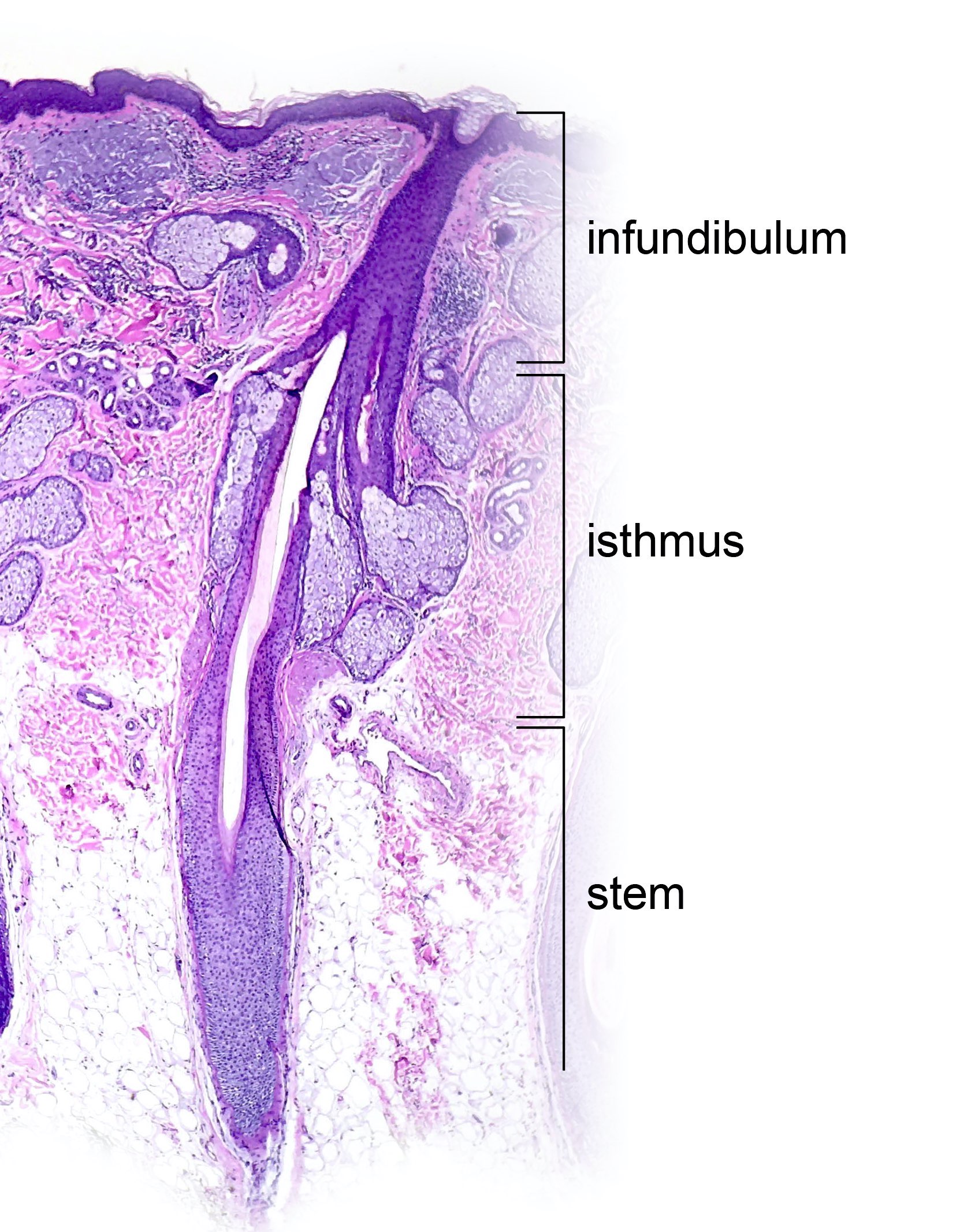
Segments of hair follicle
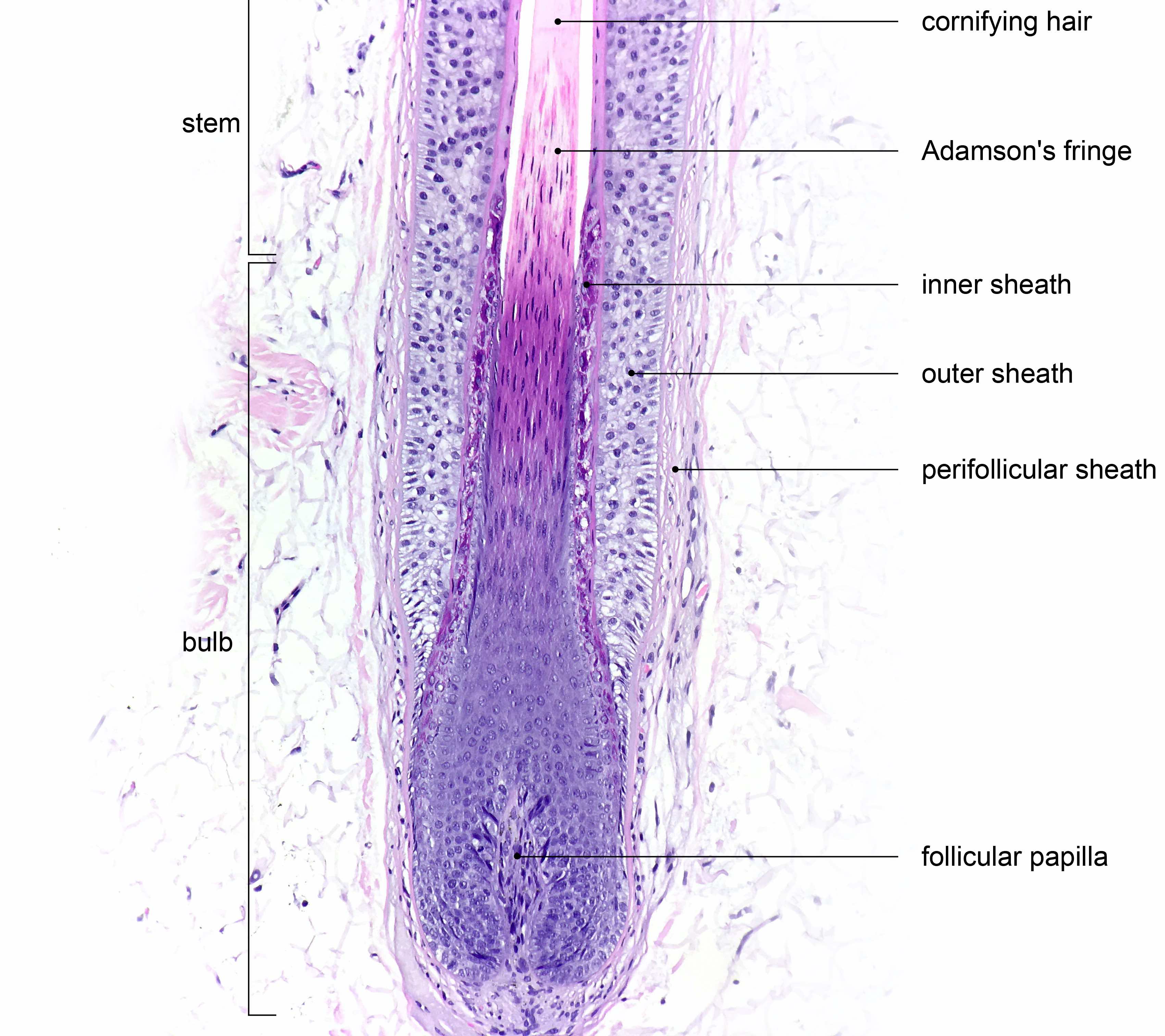
Layers of terminal anagen hair follicle
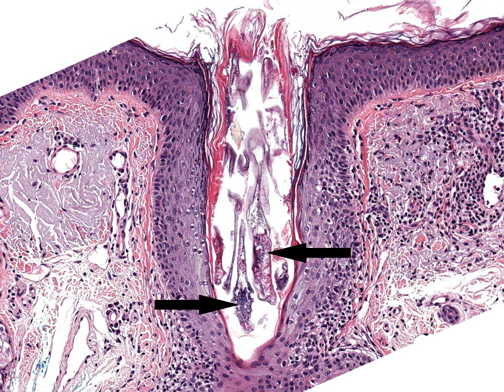
Demodex folliculorum mites
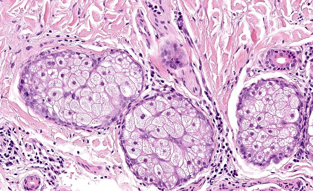
Sebaceous gland
Adipophilin+ sebaceous gland

Eccrine gland, myoepithelial cell
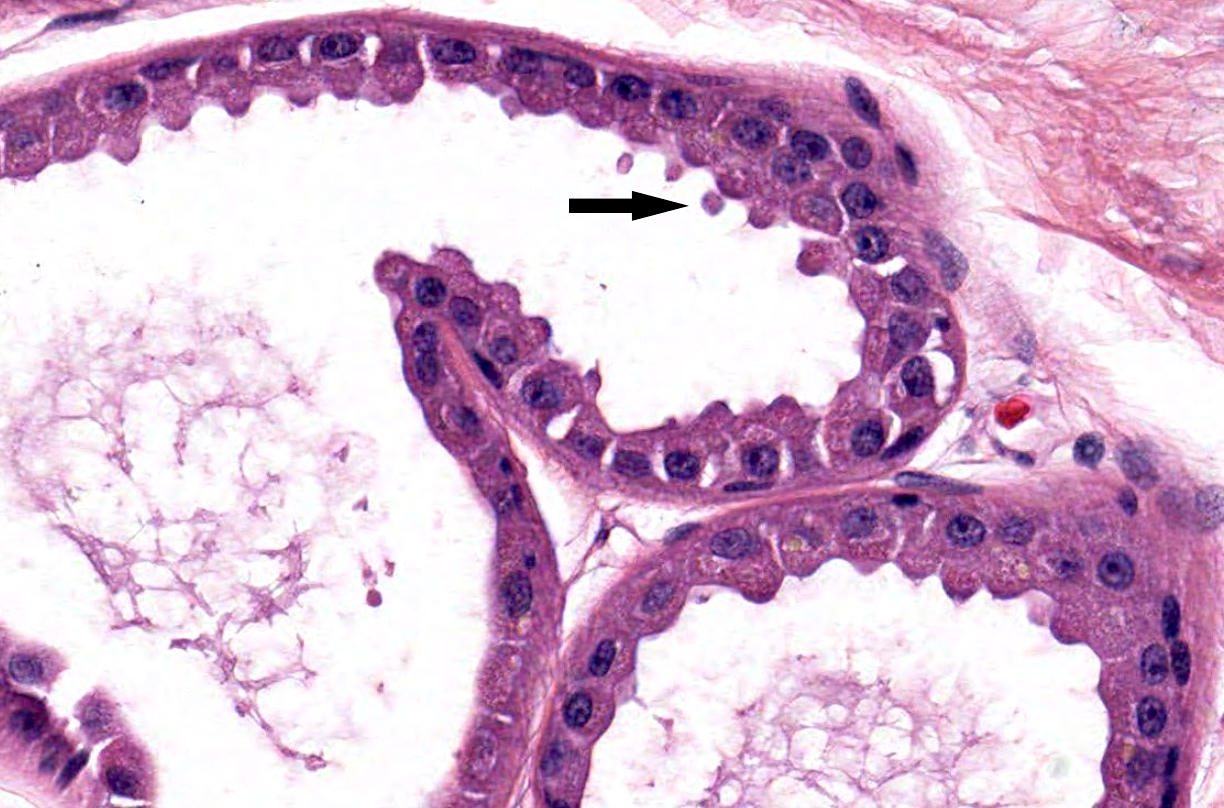
Eccrine duct
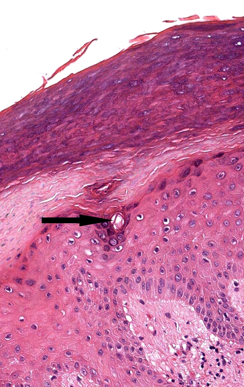
Intraepidermal portion of eccrine duct (acrosyringium)
Apocrine gland
Apocrine gland with decapitation secretion
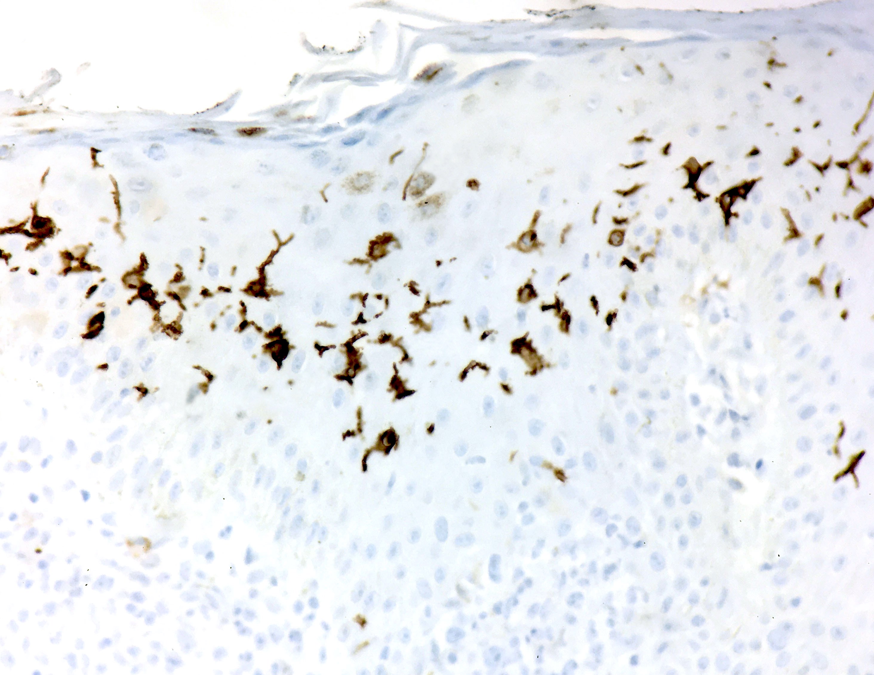
CD1a+ Langerhans cells
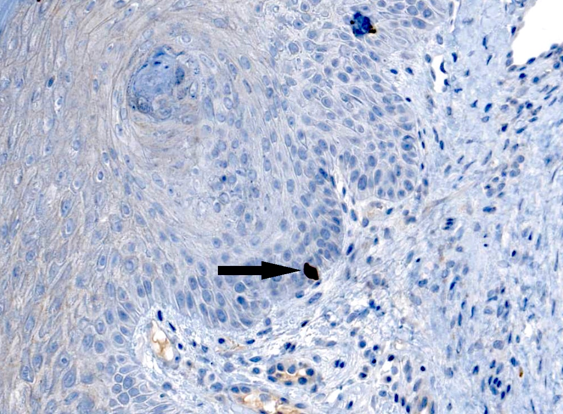
CK20+ Merkel cell
Positive stains
- Basal keratinocytes: CK5 / CK14
- Toker cells: CK7, AE1, CAM5.2, EMA, HER2, ER and PR (Am J Dermatopathol 1995;17:487, Hum Pathol 2008;39:1295)
- Melanocytes: Fontana-Masson, tyrosinase, S100, SOX10, MelanA / MART1, MITF, HMB45, CD117, p75 (Dermatoendocrinol 2011;3:32)
- Dermal dendrocytes: factor XIIIa and CD34
- Adipocytes and nerves: S100
- Vascular
- Arteries, arterioles and veins: alpha smooth muscle actin, CD31 (endothelial) and CD34 (endothelial)
- Lymphatics: D2-40
- Hair follicle: outer root sheath
- Sebaceous glands
- EMA
- Adipophilin (membranous staining of intracytoplasmic lipid droplets) (Mod Pathol 2010;23:567)
- Factor XIIIa (AC‐1A1) (J Cutan Pathol 2016;43:649)
- Eccrine and apocrine glands
- Myoepithelial cells: actin, calponin, caldesmon, S100, SOX10 (J Cutan Pathol 2014;41:353)
- Langerhans cells: S100, CD1a, CD207 (Langerin)
- Merkel cells: CK20 (dot pattern), CK8, CK8/18, neuron specific enolase, neurofilament, synaptophysin
Negative stains
- Toker cells
- Mucicarmine and PAS (Cancer 1970;25:601)
- p53 or CD138 (Hum Pathol 2008;39:1295)
Electron microscopy description
- Melanosomes: spherical membrane bound particle with periodic longitudinal concentric lamellae
- Birbeck granules (rod shaped structure with zipper-like striations, often with bulbous end)
Electron microscopy images
Images hosted on other servers:


Birbeck granules
Additional references
Board review style question #1
What is this structure and what does it make?
- Apocrine gland produces odorless secretion
- Eccrine gland produces merocrine secretion
- Hair follicle produces hair shafts
- Sebaceous gland produces sebum
Board review style answer #1
Board review style question #2
Which skin cell type is CK20+?
- Keratinocyte
- Langerhans cell
- Melanocyte
- Merkel cell
Board review style answer #2
Board review style question #3
Which immunostain is expected to be expressed by this cell?
- CD1a
- CK5/6
- CK20
- MelanA
Board review style answer #3

