
Skin nontumor
Vesiculobullous and acantholytic reaction patterns
Epidermolysis bullosa
Copyright: 2002-2025, PathologyOutlines.com, Inc.
PubMed Search: Epidermolysis bullosa (EB)


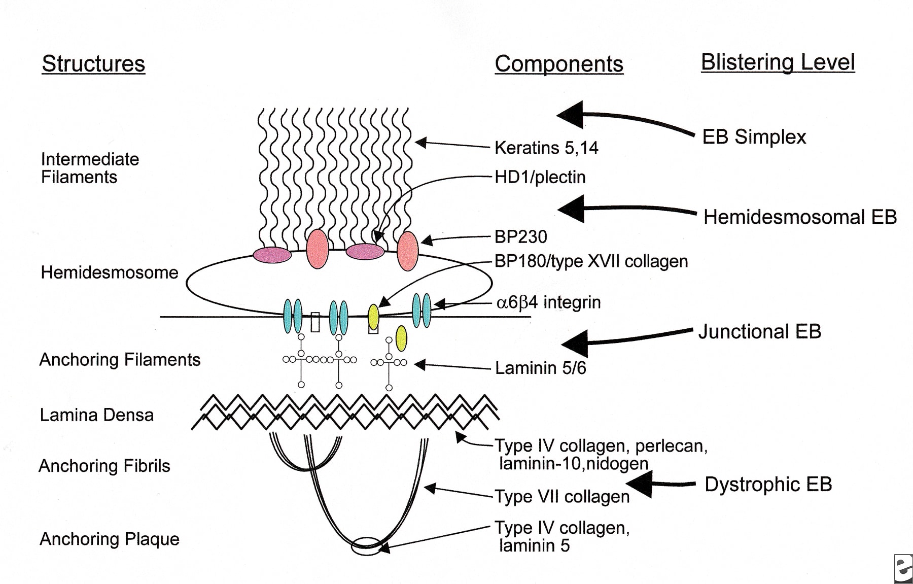
- Rare genetic blistering disorder (incidence of 8 - 19 per million) with cleavage in dermis, lower epidermis or at dermoepidermal junction
- Classified as EB simplex, junctional EB, dystrophic EB and Kindler syndrome, based on level of tissue separation within the cutaneous basement membrane zone (Orphanet J Rare Dis 2010;5:12)
- Also classified and subclassified based on clinical, electron microscopic, immunohistochemical and genotypic features (J Am Acad Dermatol 2008;58:931, Orphanet J Rare Dis 2010; 5: 12)
- EB simplex: intraepidermal tissue separation; degeneration of basal cell layer causes clinical bullae; usually due to dominant mutations in keratin 14 or keratin 5 (J Clin Invest 2009;119:1784)
- Junctional: blisters within lamina lucida, skin appears normal
- Dystrophic: blisters are sub-lamina densa; dominant or recessive
- Kindler syndrome: blisters at multiple levels (intra-lamina lucida and sub-lamina densa)
- Defects in keratin K5 / K14 filament network architecture cause basal keratinocytes to become fragile and account for their trauma-induced rupture
Images hosted on other servers:

Normal epidermis and components
Level of disruption in EB subtypes
- Blisters form shortly after birth due to pressure, rubbing or trauma
- Blisters cause scarring or milia on dorsum of hands, elbows and knees and oromucosal lesions
- EB Simplex-Dowling Meara: associated with marked morbidity; may cause death during early infancy; exhibits intact vesicles or small blisters in a grouped or arcuate (curved) configuration
- Junctional EB: characterized by enamel hypoplasia, with pitting of some or all of the tooth surfaces
- Junctional EB-Herlitz subtype: severe symptoms; presents at birth and involves all skin surfaces with exuberant granulation tissue of skin and possibly upper airway
- Recessive dystrophic EB: subtypes are severe generalized (formerly Hallopeau-Siemens), non-Hallopeau-Siemens and inverse; each arises at birth; severe generalized is "one of the most devastating multiorgan genetically transmitted diseases of mankind"; has generalized blistering at birth, progressive and often mutilating scarring of skin, corneal blisters or scarring, profound growth retardation, multifactorial anemia, failure to thrive, esophageal strictures and debilitating hand and foot deformities
- Diagnosis: usually requires focused lab testing, such as transmission EM (gold standard), immunofluorescence antigen mapping or immunohistochemical staining with EB specific monoclonal antibodies
Images hosted on other servers:

Typical noninflammatory blister

EB simplex blistered foot of infant

EB simplex, generalized, with Dowling-Meara variant

Junctional EB: marked pitting of enamel

Junctional EB-Herlitz
subtype: exuberant
granulation tissue
on child's neck

Dominant dystrophic
EB-milia arising within
erythematous patch

Generalized dominant dystrophic EB

Generalized dominant
dystrophic EB:
hypertrophic scarring

Generalized dominant
dystrophic EB: dystrophy
of all 20 nails


Recessive dystrophic EB


Severe generalized recessive dystrophic EB

Congenital absence of
skin in neonate with Bart
syndrome (not specific
for any EB subtype)
- Subepidermal blister with variable inflammation
- Superficial dermis is fibrotic (type IV collagen positive)
Images hosted on other servers:

EB simplex: intact stratum corneum and upper epidermis, with vesicle formation in the lower epidermis at the basal layer caused by degeneration of individual epidermal cells

Dystrophic EB: pretibial lesion
- Linear C3 and IgG deposits along epidermal basement membrane
- Diagnostically useful differences in antigenic staining in selected EB subtypes
Images hosted on other servers:

EB simplex: epidermal basal keratinocytes
