

Skin nontumor
Lichenoid and interface reaction patterns
Dermatomyositis
Last author update: 1 July 2011
Last staff update: 21 September 2023
Copyright: 2002-2024, PathologyOutlines.com, Inc.
PubMed Search: Dermatomyositis skin
See Also: Muscle & peripheral nerve nontumor - Dermatomyositis

Table of Contents
Definition / general | Clinical features | Treatment | Clinical images | Microscopic (histologic) description | Microscopic (histologic) images | Positive stains | Negative stains | Additional referencesCite this page: Hamodat M. Dermatomyositis. PathologyOutlines.com website. https://www.pathologyoutlines.com/topic/skinnontumordermatomyositis.html. Accessed April 25th, 2024.
Definition / general
- See also Muscle chapter
- Autoimmune inflammatory disease of skeletal muscle and skin, usually affects women
- Symmetric proximal muscle weakness and skin lesions
- Affects face, dorsal hands and feet, particularly knuckles
- 20% of cases lack muscle involvement (Arch Dermatol 2010;146:26)
- 15% have coexisting adenocarcinoma of stomach, breast, ovary, lung or colon, with remission of dermatomyositis following tumor resection; high risk of nasopharyngeal carcinoma in Asian patients (Ann Acad Med Singapore 2010;39:843)
- Increased risk of thyroid disease, particularly hypothyroidism, especially in patients with interstitial lung disease
- Polymyositis: similar muscle changes without skin changes
Clinical features
- Poorly demarcated, scaly, erythematous patches
- Also heliotrope erythema of upper eyelids and extensor joint surfaces
Treatment
- Steroids, immunosuppressants (Arthritis Care Res (Hoboken) 2010;62:219), tumor resection (if present)
Clinical images
Images hosted on other servers:

Purple plaques on knees

Heliotrope rash and Gottron papules

Various images
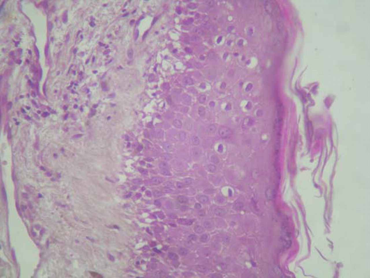
Microscopic (histologic) description
- Chronic nonspecific dermatitis or interface dermatitis resembling systemic lupus erythematosus
- Often atrophic epidermis with prominent vacuolar interface change
- Sparse perivascular lymphocytic infiltrate with markedly increased dermal mucin
- Muscles show myositis with myofiber necrosis, fragmentation and phagocytosis; late myofiber atrophy, fibrosis and fatty change
Microscopic (histologic) images
Contributed by Hillary Rose Elwood, M.D.
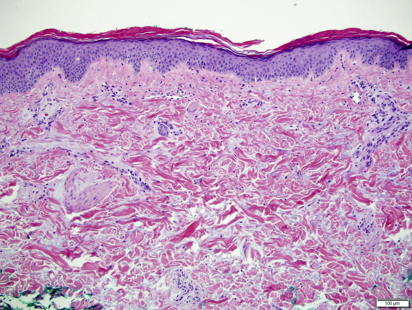
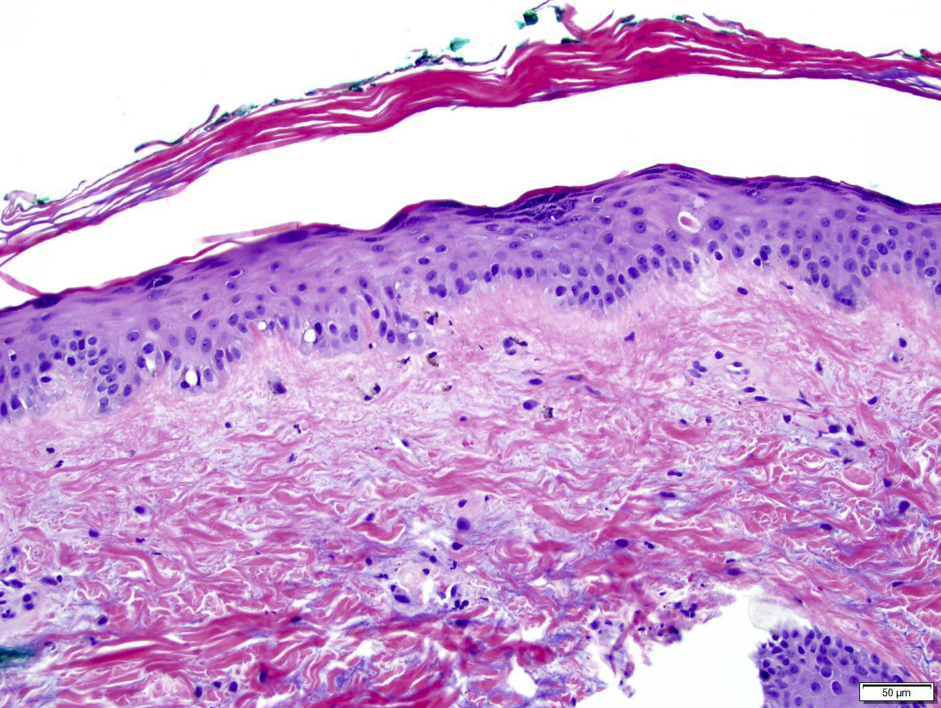
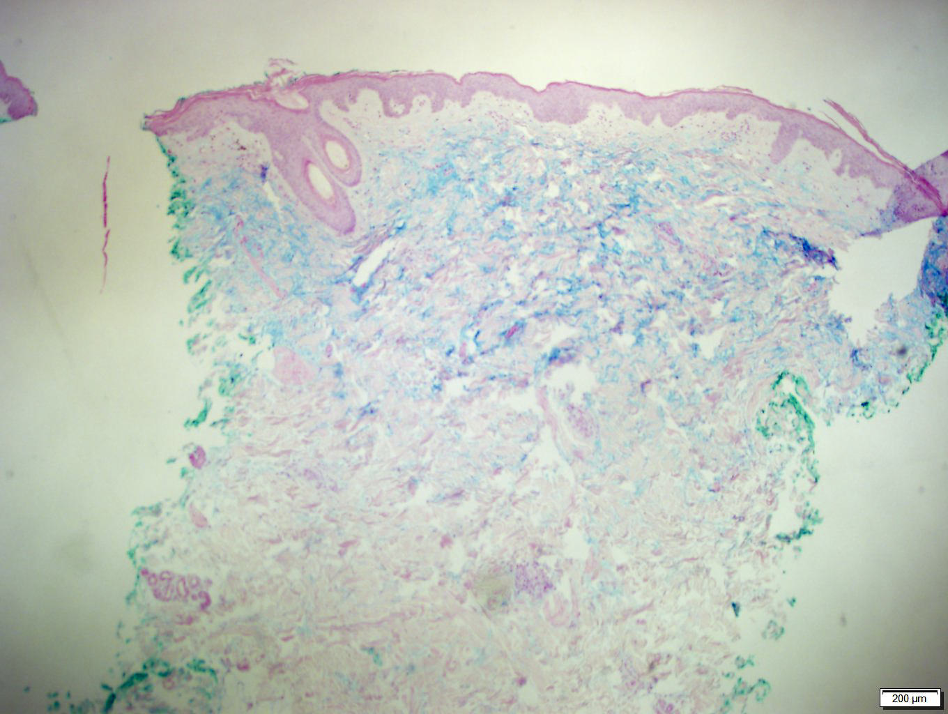
Subtle vacuolar interface change and increased dermal mucin highlighted by Alcian blue stain
Images hosted on other servers:

Erythrodermic dermatomyositis
Interface dermatitis
with vacuolar
alteration at dermal
epidermal junction
Positive stains
- C5-9 (by immunofluorescence)
Negative stains
- IgG, IgA and IgM by immunofluorescence
Additional references
