

Skin nontumor
Vesiculobullous and acantholytic reaction patterns
Keratosis follicularis (Darier disease)
Authors: Alicia McNish, M.B.B.S., Michael Fitz-Henley, M.B.B.S., D.M., Jonathan D. Ho, M.B.B.S., D.Sc.
Resident / Fellow Advisory Board: Caroline I.M. Underwood, M.D.


Last author update: 29 July 2021
Last staff update: 15 February 2024
Copyright: 2002-2024, PathologyOutlines.com, Inc.
PubMed Search: Darier disease skin pathology
Table of Contents
Definition / general | Essential features | Terminology | ICD coding | Epidemiology | Sites | Pathophysiology | Etiology | Clinical features | Diagnosis | Prognostic factors | Case reports | Treatment | Clinical images | Microscopic (histologic) description | Microscopic (histologic) images | Virtual slides | Positive stains | Molecular / cytogenetics description | Sample pathology report | Differential diagnosis | Additional references | Board review style question #1 | Board review style answer #1 | Board review style question #2 | Board review style answer #2 | Board review style question #3 | Board review style answer #3Cite this page: McNish A, Fitz-Henley M, Ho JD. Keratosis follicularis (Darier disease). PathologyOutlines.com website. https://www.pathologyoutlines.com/topic/skinnontumordariersdisease.html. Accessed April 26th, 2024.
Definition / general
- Autosomal dominant genodermatosis
- Clinical: greasy keratotic papules in a seborrheic distribution, nail changes and mucosal findings
- Histologic: acantholytic dyskeratosis
Essential features
- Autosomal dominant genodermatosis due to mutations in ATP2A2 gene
- Clinical features: family history, greasy hyperkeratotic papules in a seborrheic / flexural / acral distribution, mucosal lesions and nail changes
- Histologic features: acantholytic dyskeratosis with corp rond and grain formation
- Clinical correlation is crucial to distinguish from mimickers with identical histopathologic features
Terminology
- Darier-White disease, keratosis follicularis and dyskeratosis follicularis
ICD coding
Epidemiology
- Worldwide distribution (Br J Dermatol 2002;146:107)
- Prevalence varies regionally between 1:30,000 and 1:100,000 (Br J Dermatol 2002;146:107, Br J Dermatol 1992;127:126)
- M = F
Sites
- Seborrheic distribution: central chest, back, marginal scalp and face
- Dorsal hands
- Palms and soles
- Oral cavity
- Nails
- Intertriginous skin
Pathophysiology
- Autosomal dominant mutations in ATP2A2 gene (Hum Mutat 2017;38:343)
- ATP2A2 encodes the sarco / endoplasmic reticulum Ca2+ ATPase isoform 2 (SERCA2) (Hum Mol Genet 1999;8:1611)
- Abnormal SERCA2 results in endoplasmic reticulum stress and impaired formation of keratinocyte adhesion proteins (J Invest Dermatol 2014;134:1961)
- Resultant lack of epidermal integrity with acantholysis and impaired keratinization (Biochim Biophys Acta 2011;1813:1111)
Etiology
- Autosomal dominant mutations in ATP2A2 gene (Hum Mutat 2017;38:343)
Clinical features
- Onset 6 - 20 years (J Am Acad Dermatol 1992;27:40)
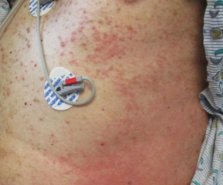
- Keratotic, red-brown papules on the trunk, face, neck and intertriginous areas; often malodorous
- Hypopigmented macules may predominate in darker skin types (JAAD Case Rep 2018;4:262)
- Flat topped acral papules and palmoplantar pits are common
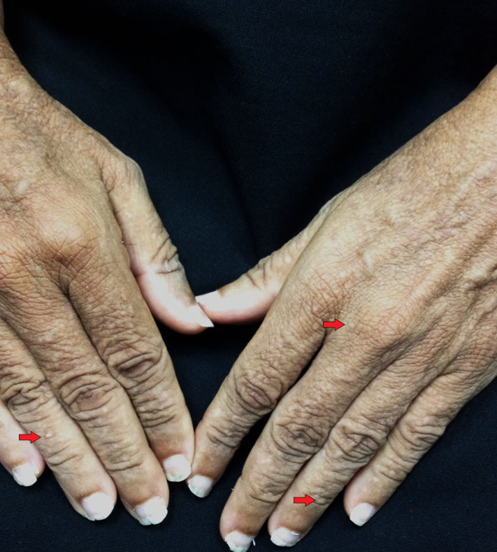
- Nail changes: longitudinal erythro / leukonychia, subungual hyperkeratosis and V shaped nicking of the free edge of the nail (J Am Acad Dermatol 1992;27:40)
- White papules involving the hard palate, buccal mucosa or tongue
- Mucosal disease may be associated with salivary gland swelling (Br Dent J 1991;171:133)
- Less common presentations include acral hemorrhagic lesions, segmental lesions and comedonal variants (Actas Dermosifiliogr 2017;108:e49, Clin Case Rep 2019;7:1362, J Dermatol 2019;46:e211)
- Associated with neuropsychiatric conditions, particularly mood disorders (Br J Dermatol 2010 Sep;163:515)
Diagnosis
- Family history
- Mucocutaneous findings
- Skin biopsy
- PCR DNA amplification to detect ATP2A2 mutations (Mol Med Rep 2015;12:1845)
Prognostic factors
- Chronic, with intermittent exacerbations and no tendency for resolution (J Dermatol 2016;43:275)
- Prone to secondary bacterial, viral and fungal colonization / infections (J Eur Acad Dermatol Venereol 2013;27:1405, Am J Dermatopathol 2017;39:370, Dermatol Ther 2020;33:e14500, Br J Dermatol 2015;172:837, Acta Derm Venereol 2017;97:139)
- Increased morbidity from associated mood disorders (Br J Dermatol 2010;163:515)
- Increased risk of squamous cell carcinoma within affected skin (Br J Dermatol 2008;159:1378, Clin Exp Dermatol 2009;34:e1015)
Case reports
- 28 year old woman, 50 year old man and 60 year old woman presenting with acral hemorrhagic lesions (Actas Dermosifiliogr 2017;108:e49)
- 31 year old woman with asymptomatic acneiform papules on the face (Ann Dermatol 2011;23:S398)
- 48 year old woman with worsening of Darier disease after interferon alpha treatment (Case Rep Dermatol 2016;8:218)
- Man in his 50s with leonine face and keratotic papules all over his body, a condition present since childhood (Case of the Month #517)
- 67 year old woman with segmental Darier disease treated with doxycycline (Dermatol Online J 2018;24:13030)
Treatment
- Mild disease: topical agents, including retinoids, vitamin D analogues and calcineurin inhibitors (Pediatr Dermatol 2011;28:197, J Dermatol 2010;37:718, Eur J Dermatol 2011;21:301)
- Systemic retinoids (Indian J Dermatol Venereol Leprol 2021;87:14)
- Laser ablation (Arch Dermatol 1999;135:423)
- Botulinum toxin injections (Orphanet J Rare Dis 2021;16:93)
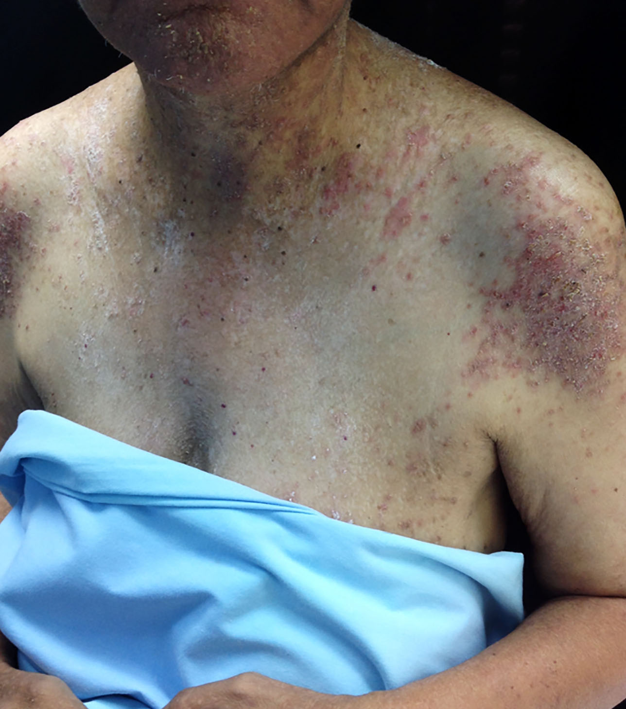
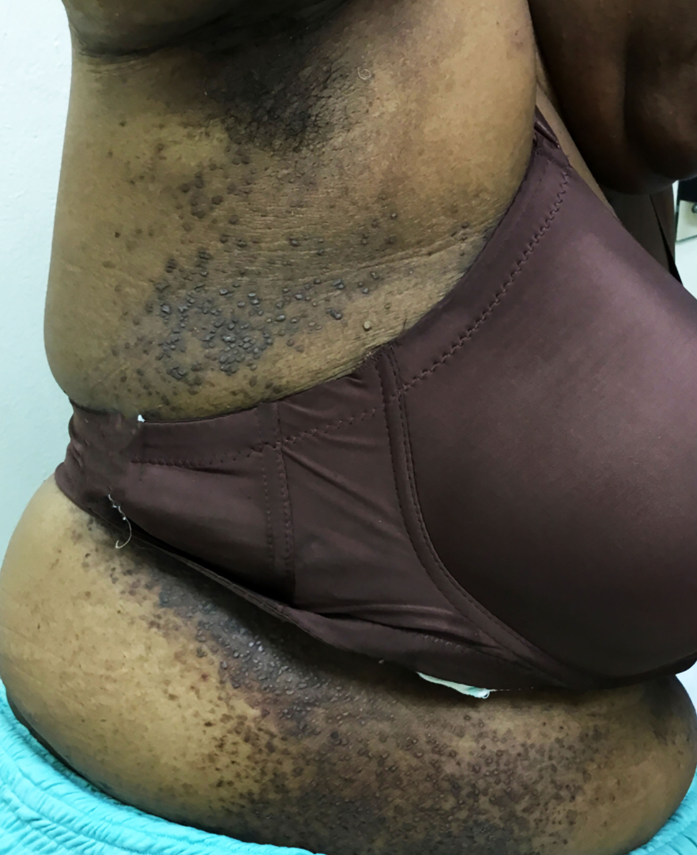
Clinical images
Contributed by Michael Fitz-Henley, M.B.B.S., D.M. and Viktoryia Kazlouskaya, M.D. (Case #517)
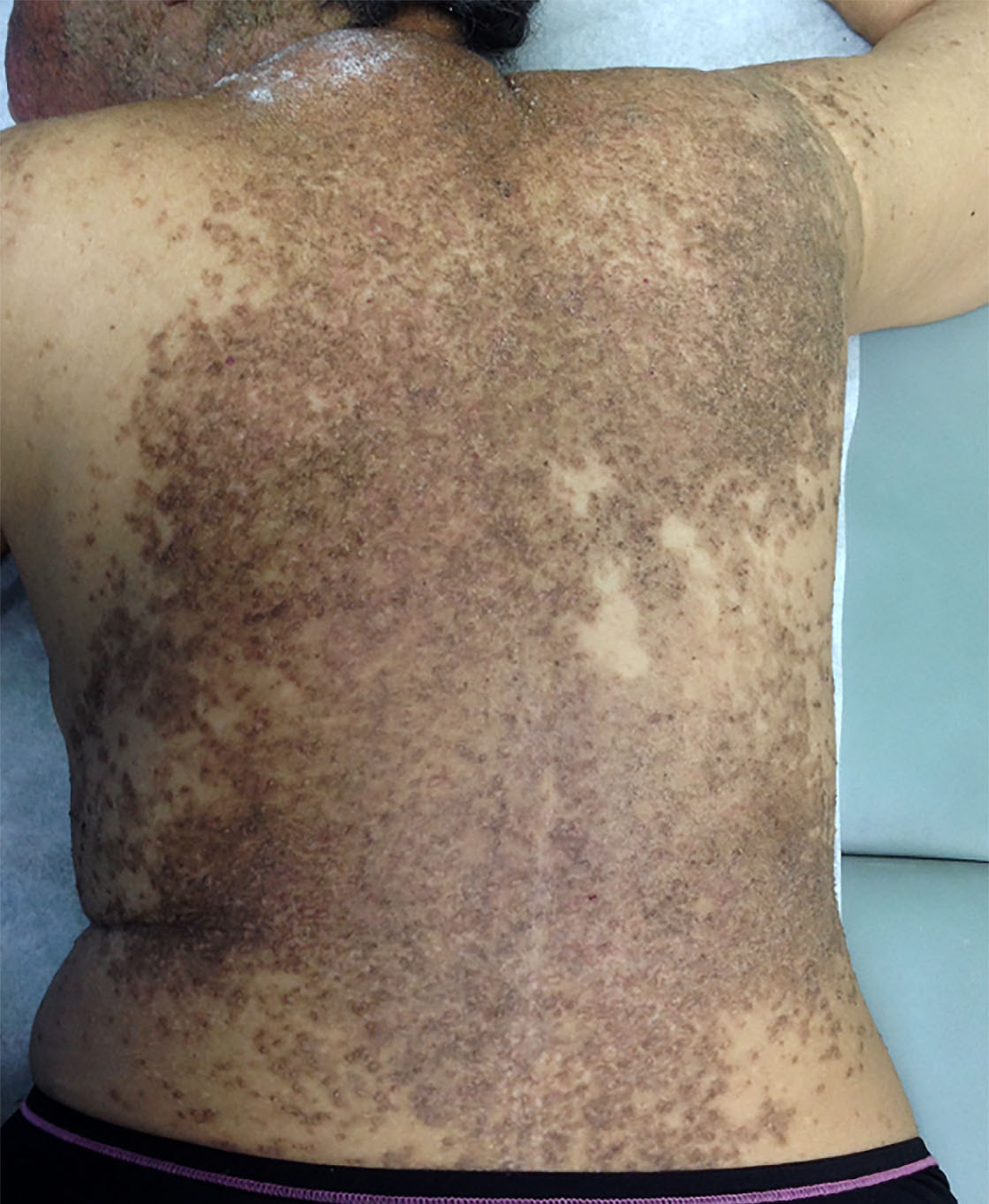
Darier disease
Flexural Darier disease
Subtle flat topped acral papules

Nail changes in Darier disease
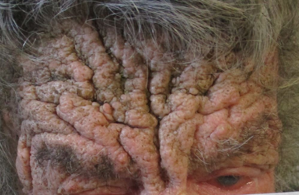
Leonine facies
Keratotic papules
Microscopic (histologic) description
- Parakeratosis
- Variable epidermal thickness
- Acantholysis with characteristic dyskeratosis forming corp ronds and grains
- Corp rond: rounded keratinocyte in superficial spiny and granular layer with basophilic / pyknotic nucleus, perinuclear halo and often a rim of eosinophilic cytoplasm (J Dermatol 2017;44:232)
- Grain: elongated keratinocyte in the stratum corneum with small basophilic nuclei and intensely pink cytoplasm; appears as plump parakeratosis; may form tiers (J Dermatol 2017;44:232)
- Corp rond and grain type dyskeratosis is classical but not specific for Darier disease (see Differential diagnosis)
- Suprabasal acantholysis and clefting with retained single layer of basal keratinocytes overlying dermal papillae which appear to project into the acantholytic cavity (villi) (J Dermatol 2016;43:275)
- Frank bullae may occur in cases with extensive acantholysis and large clefts (Arch Dermatol 1982;118:278)
- Superimposed fungal, bacterial and herpetic infections may be seen (J Eur Acad Dermatol Venereol 2013;27:1405, Am J Dermatopathol 2017;39:370, Dermatol Ther 2020;33:e14500, Br J Dermatol 2015;172:837, Acta Derm Venereol 2017;97:139)
- Variable mild perivascular inflammatory cell infiltrate
- Variants: extensive pseudoepitheliomatous hyperplasia, comedonal lesions with prominent villus formation and hemorrhagic bullous lesions (Am J Dermatopathol 2015;37:323, J Dermatol 2006;33:477, J Cutan Med Surg 2016;20:478)
- Flat topped acral papules demonstrate orthokeratosis (may be church spire type), hypergranulosis and papillomatous epidermal hyperplasia; acantholytic dyskeratosis is often subtle or absent
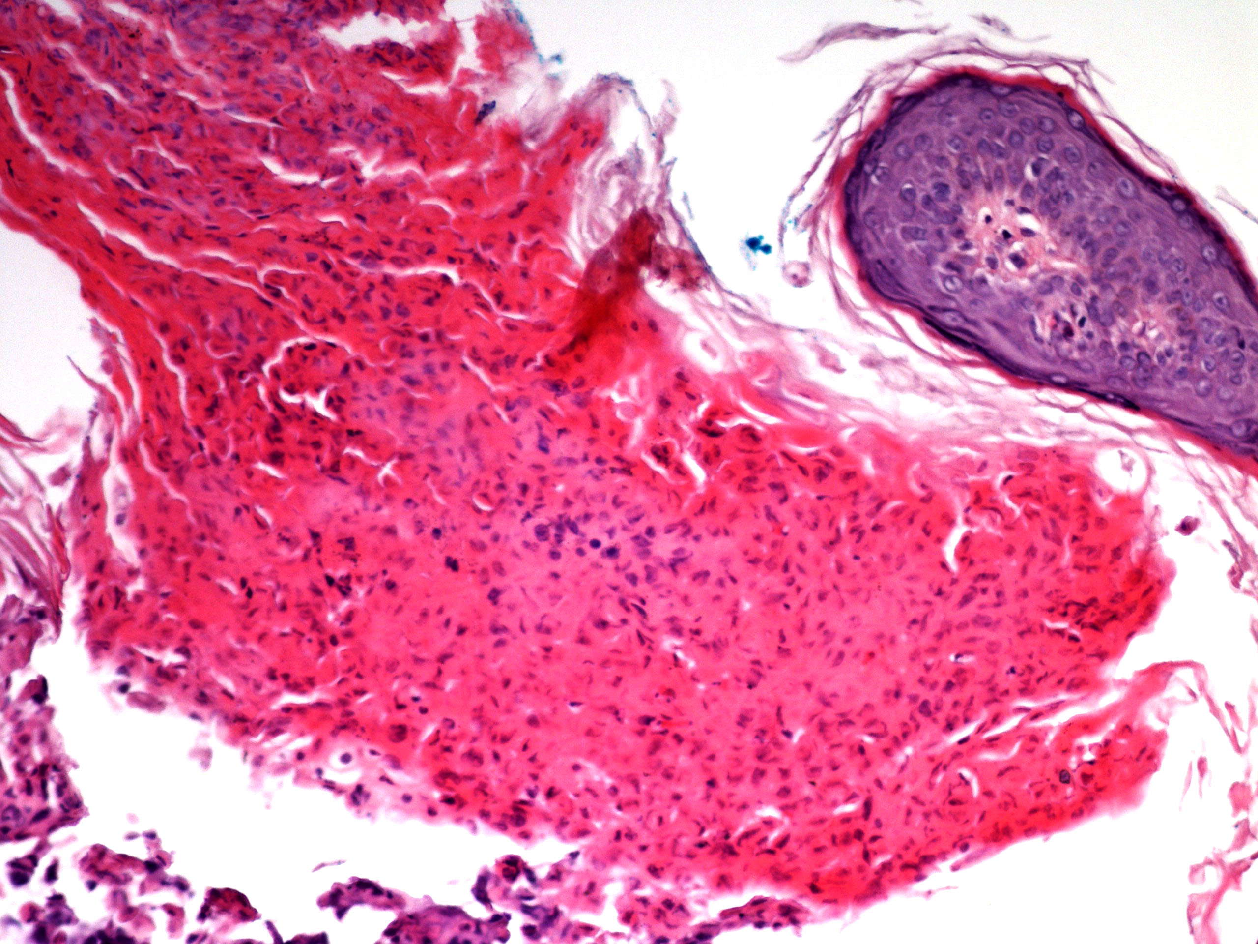
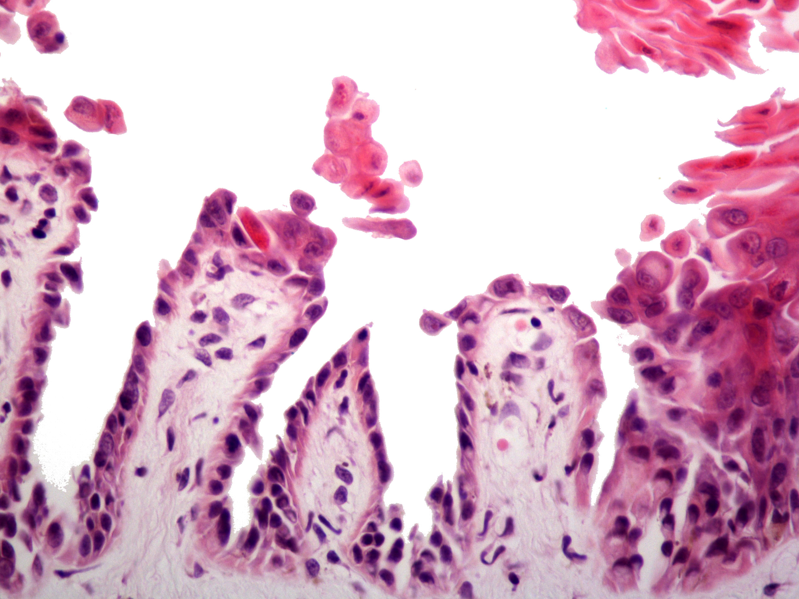
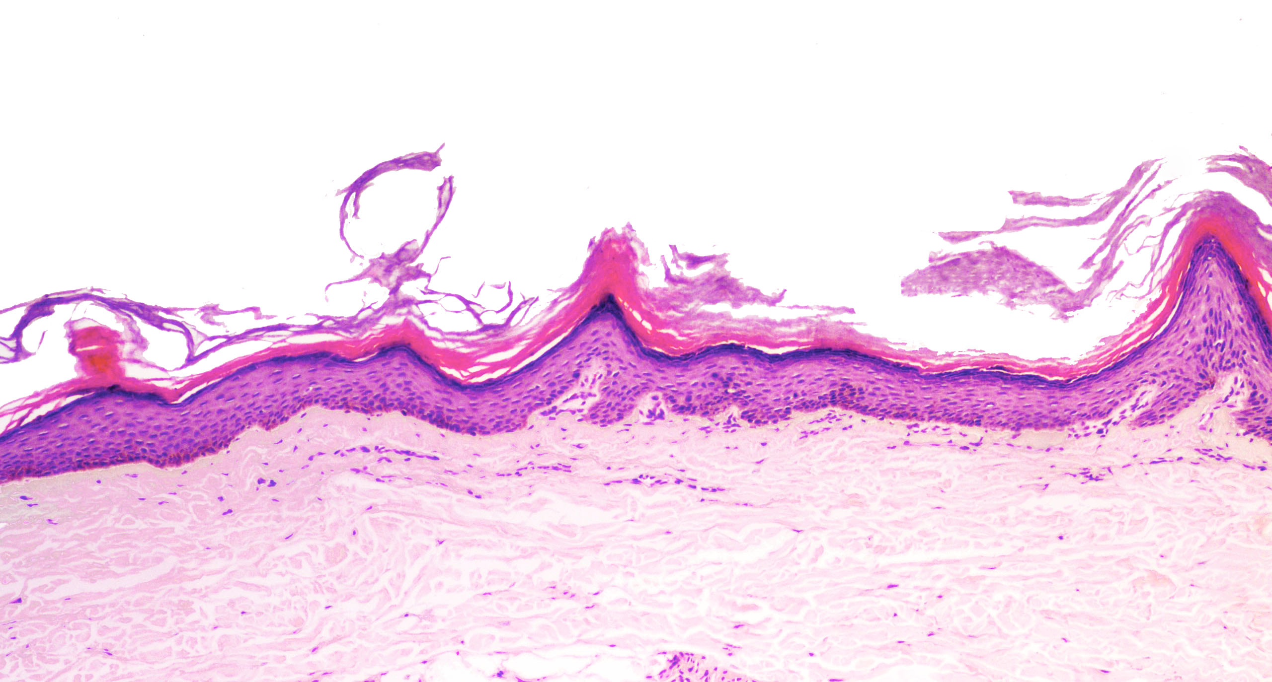
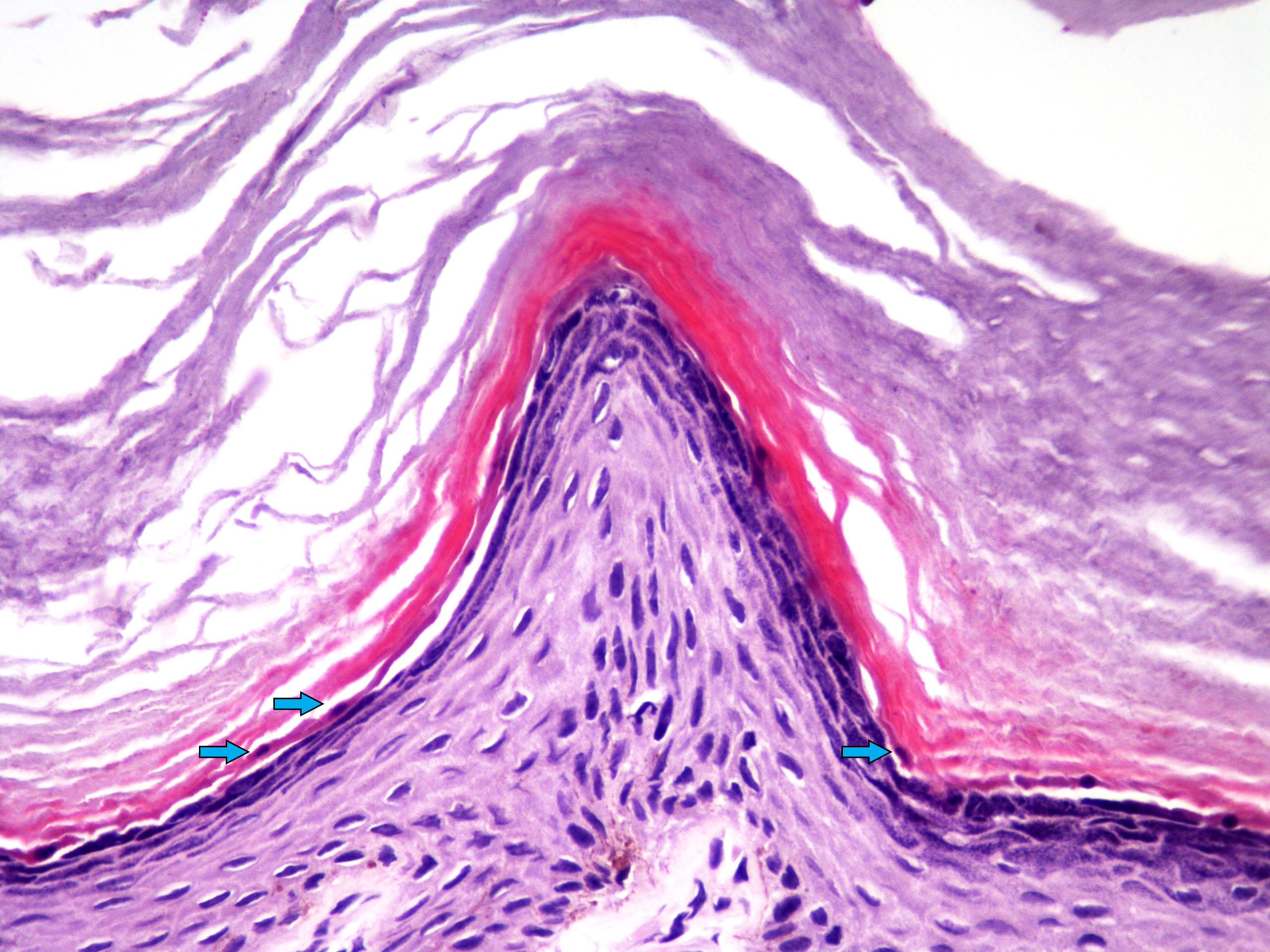
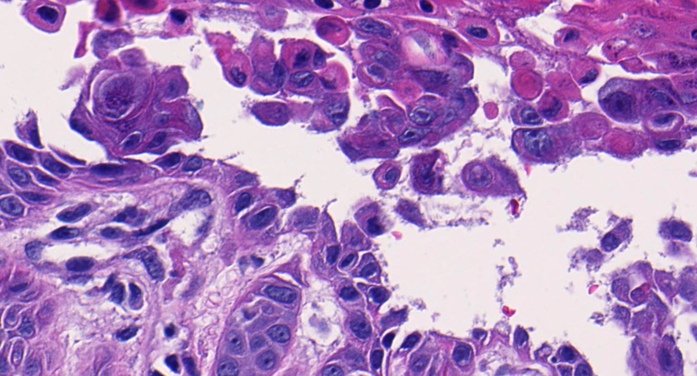
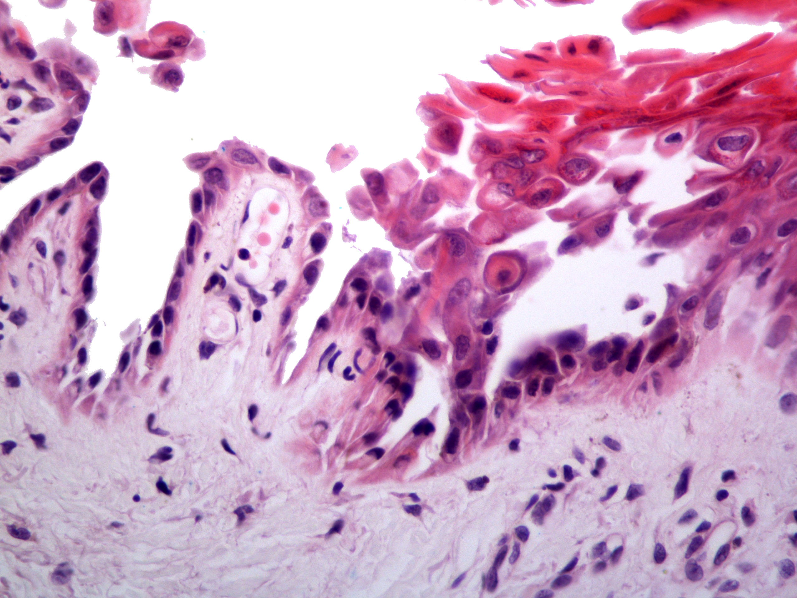
Microscopic (histologic) images
Contributed by Jonathan D. Ho, M.B.B.S., D.Sc. and Viktoryia Kazlouskaya, M.D. (Case #517)
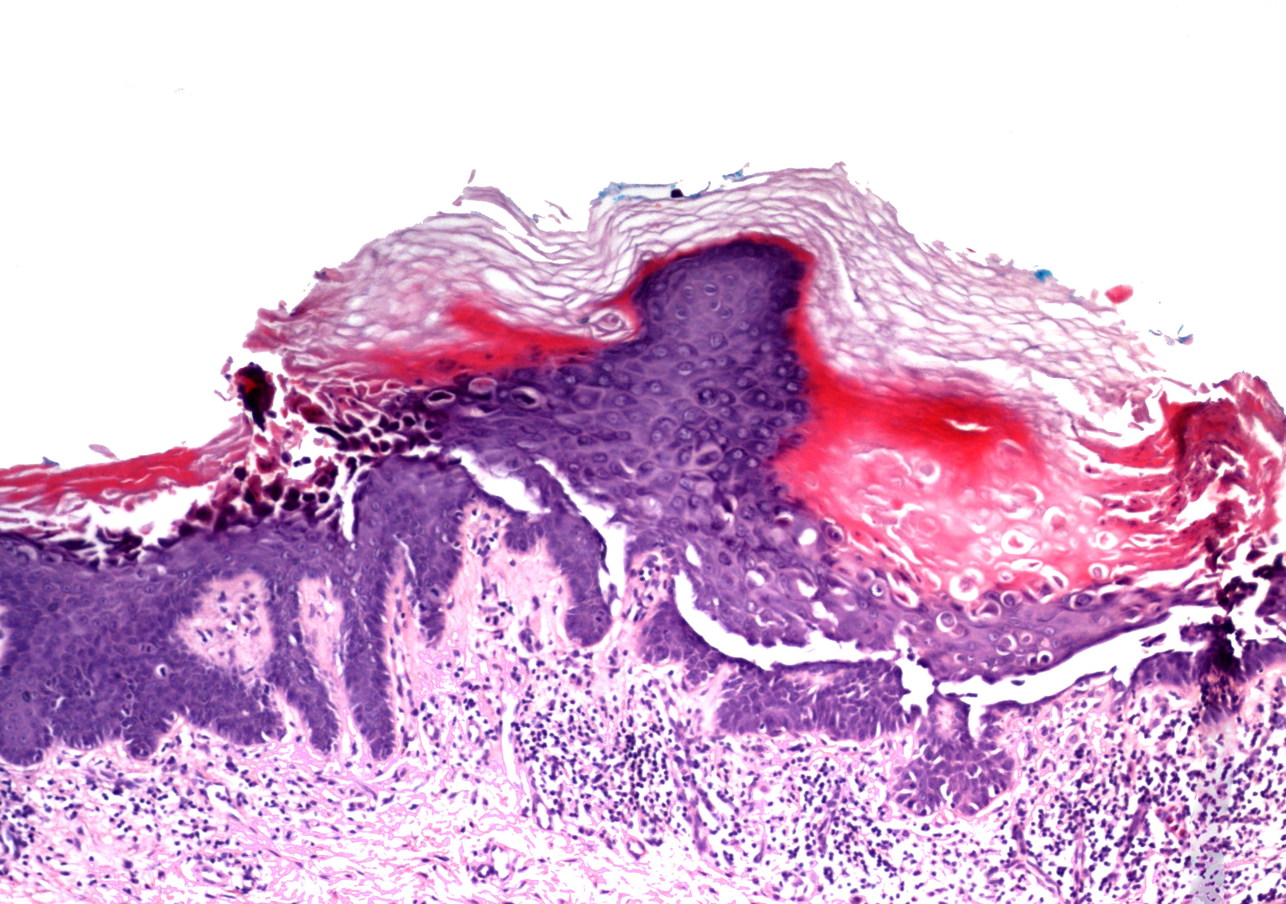
Hyperkeratosis and
intraepidermal
clefting
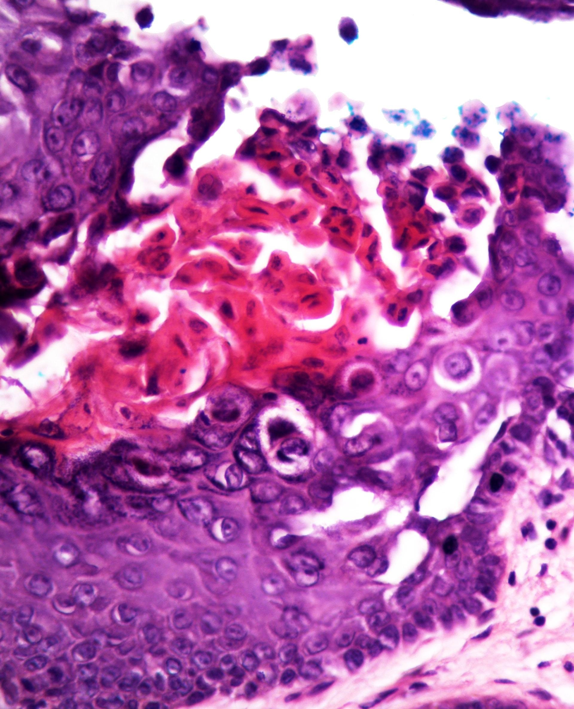
Acantholytic dyskeratosis

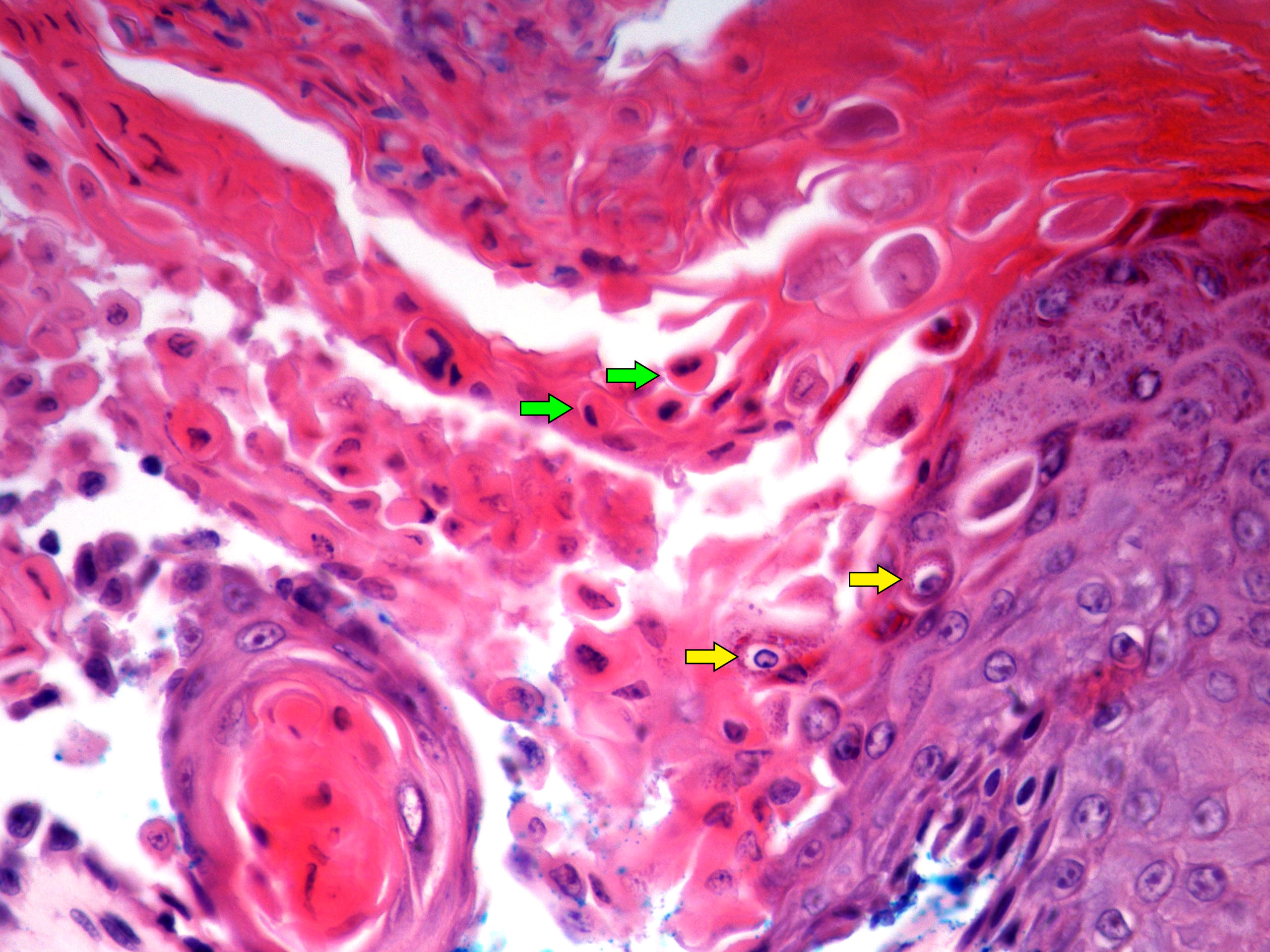
Corp ronds and grains
Grains forming tier
Villus formation
Acral papule

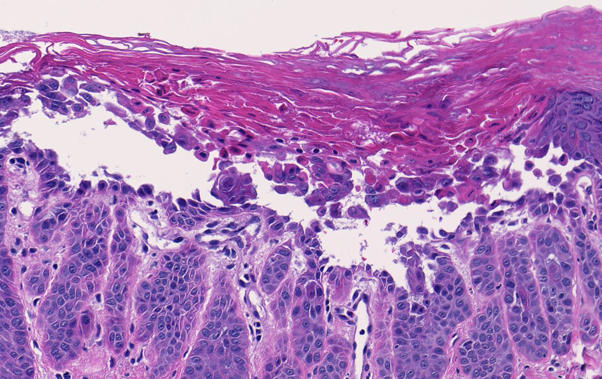
Mild perivascular superficial lymphocytic inflammation, intraepidermal acantholysis and dyskeratosis
Virtual slides
Images hosted on other servers:

Darier disease
Positive stains
- PAS stain - should be performed to rule out superimposed fungal infections but may not always be positive
Molecular / cytogenetics description
- > 270 unique ATP2A2 mutations described (missense / nonsense > deletion / splice site / insertion mutations) (PLoS One 2017;12:e0186356, Hum Mutat 2017;38:343)
Sample pathology report
- Skin of chest, punch biopsy:
- Darier disease (see comment)
- Comment: The specimen exhibits parakeratosis, epidermal hyperplasia, acantholytic dyskeratosis with prominent corp rond and grain formation, suprabasal clefting with formation of villi and a mild superficial perivascular lymphocytic infiltrate. Although similar acantholytic dyskeratosis may be seen in a number of entities, given the clinical history of greasy papules in a seborrheic distribution, a positive family history and persistence of lesions, the findings are most consistent with Darier disease.
Differential diagnosis
- Hailey-Hailey disease:
- Intraspinous to full thickness acantholysis
- Less prominent dyskeratosis
- Dilapidated brick wall appearance
- Acrokeratosis verruciformis of Hopf:
- Church spire hyperkeratosis, hypergranulosis and papillomatous epidermal hyperplasia
- Lesions may be identical to flat topped acral papules of Darier disease (J Dermatol 2016;43:275, J Invest Dermatol 2003;120:229)
- No acantholytic dyskeratosis seen
- Pemphigus vulgaris:
- Suprabasal and intraspinous acantholysis without corp rond and grain formation
- Intraepidermal, intercellular deposition of IgG/C3
- Transient acantholytic dermatosis (Grover disease):
- May have identical histopathologic features
- Distinction is easy based on clinical features (relapsing remitting pruritic papular eruption in middle aged to elderly males)
- Warty dyskeratoma:
- Identical Darier type acantholytic dyskeratosis but solitary lesion
- Tends to have distinct cup shaped epidermal invagination and very prominent villus formation (J Dermatol 2017;44:232)
- Acantholytic dyskeratotic acanthoma:
- Darier or pemphigus type acantholytic dyskeratosis but solitary papule or nodule; rarely erythronychia (J Cutan Pathol 2007;34:494)
- Regular acanthosis without a cup shaped invagination (Dermatol Pract Concept 2014;4:25)
- Acantholytic dermatosis of the genitocrural area:
- Darier or Hailey-Hailey type histopathologic appearance
- Limited to the genitocrural area
- No other clinical features of heritable acantholytic disease
- Focal acantholytic dyskeratosis:
- Incidental acantholytic dyskeratosis without a clinical correlate of an acantholytic condition (J Am Acad Dermatol 1998;38:243)
- May be seen in normal skin or adjacent to a variety of benign or malignant tumors (J Am Acad Dermatol 1998;38:243)
Additional references
Board review style question #1
A 35 year old man presents with a 10 year history of scaly papules on the face and chest. Biopsy reveals the findings shown in the image above. What is the most likely diagnosis?
- Darier disease
- Hailey-Hailey disease
- Herpes simplex virus infection
- Pemphigus vulgaris
- Seborrheic dermatitis
Board review style answer #1
A. Darier disease. The photomicrograph shows acantholysis with dyskeratosis (corp ronds and grains) as well as the formation of villi classically seen in Darier disease. While Hailey-Hailey disease may have acantholysis with dyskeratosis, prominent corp ronds and grains are lacking. Pemphigus has bland acantholysis and herpes simplex shows distinct viral cytopathic change. Seborrheic dermatitis is a spongiotic dermatitis.
Comment Here
Reference: Darier disease
Comment Here
Reference: Darier disease
Board review style question #2
Which of the following diseases may demonstrate histopathologic features identical to Darier disease?
- Grover disease
- Hailey-Hailey disease
- Inflammatory and linear verrucous epidermal nevus
- Pemphigus foliaceus
- Pemphigus vulgaris
Board review style answer #2
A. Grover disease. Grover disease has multiple histopathologic patterns including those with Darier type histology. All other options lack typical corp rond and grain formation.
Comment Here
Reference: Darier disease
Comment Here
Reference: Darier disease
Board review style question #3
Where is the abnormal protein located in Darier disease?
- Cytoplasm
- Endoplasmic reticulum
- Golgi apparatus
- Mitochondria
- Nucleus
Board review style answer #3
B. Endoplasmic reticulum. An abnormal SERCA2 protein is located in the endoplasmic reticulum and plays a role in the Ca2+ signaling pathway regulating cell to cell adhesion and differentiation of the epidermis (Nat Genet 1999;21:271).
Comment Here
Reference: Darier disease
Comment Here
Reference: Darier disease
