
Placenta
Gross / macroscopic variations and conditions
Umbilical cord
Knots
Author: Paul J. Kowalski, M.D.

Last author update: 1 September 2015
Last staff update: 28 October 2020
Copyright: 2002-2025, PathologyOutlines.com, Inc.
PubMed Search: Knots umbilical cord [title]


Table of Contents
Definition / general | Terminology | Epidemiology | Pathophysiology | Etiology | Clinical features | Radiology description | Prognostic factors | Case reports | Clinical images | Gross description | Gross images | Microscopic (histologic) descriptionCite this page: Kowalski PJ. Knots. PathologyOutlines.com website. https://www.pathologyoutlines.com/topic/placentaknots.html. Accessed April 1st, 2025.
Definition / general
- Looping or interweaving of the umbilical cord that occurs during intrauterine growth of a fetus
Terminology
- True knot is present when the umbilical cord loops upon itself and can be physically released / untied
- Pseudoknot (false knot) is merely a varicosity or redundancy of an umbilical vessel (usually the vein) within the cord substance and cannot be physically released in an intact cord
Epidemiology
- True knots occur in 1 per 2000 deliveries (0.5% of pregnancies)
- Pseudoknots are very common and can be seen in a majority of pregnancies (especially small pseudoknots)
Pathophysiology
- True knots probably develop early in pregnancy when intrauterine space is available for excessive fetal movement
- As the fetus enlarges, a true knot may tighten or tightening may occur at delivery when the umbilical cord undergoes traction
- Constriction or hematoma development may lead to fetal hypoxia, neurologic impairment or fetal demise
Etiology
- True knots are associated with conditions that allow for increased fetal movement including multigravidae, long cord length, male fetuses, monoamnionic twins and increased amnionic fluid
Clinical features
- Clinically significant circulatory compromise may often be associated with edema, congestion, constriction, hemorrhage or thrombosis
- True knots (especially those that are tight) are associated with an overall mortality rate of 10%
- Pseudoknots are currently viewed as having no clinical significance
Radiology description
- Ultrasound examination can identify true knots and Doppler blood flow indices can be used to assess for cord constriction
Prognostic factors
- Constriction of the umbilical cord at the knot, curling of the unknotted portion of cord and thrombosis in the cord are negative prognostic features
Case reports
- 34 year old woman with heterotopic pregnancy with a monoamniotic monochorionic twin gestational pregnancy (J Reprod Med 2013;58:77)
- Three dimensional ultrasound with color Doppler imaging of an umbilical cord true knot (Ultrasound Obstet Gynecol 2014;43:360)
- Multiple true knots together with single artery and four umbilical cord nuchal loops (Eur J Obstet Gynecol Reprod Biol 2013;168:117)
Clinical images
Images hosted on other servers:

True knot in umbilical cord
Gross description
- True knots demonstrate single or multiple / complex loops
- Tightness of the knot, constriction or grooving of the umbilical cord, loss of Wharton jelly, cord congestion or hemorrhage and the diameter of the cord both proximal and distal to knot should all be noted
- Mural thrombosis or complete obstruction of the umbilical vein or placental surface veins can be seen
- With chronicity, the unknotted portion of cord may tend to curl
- Pseudoknots show variable tortuosity of the cord and range from very small to several centimeters
Gross images
AFIP images
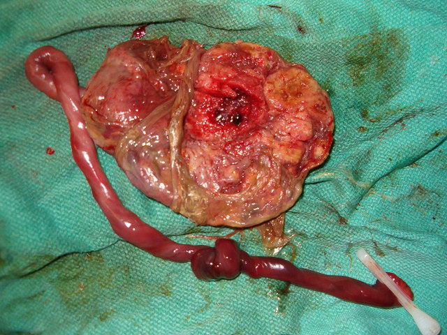
True knot with placental infarction
Images hosted on other servers:


True knot

False knot
Microscopic (histologic) description
- Changes are typically minimal unless evidence of significant hemorrhage or thrombosis is present
- Veins may be thickened or contain mural "cushions," which are myxoid appearing, often fusiform shaped regions of intramural fibrin
