

Penis & scrotum
General
Anatomy & histology-scrotum
Authors: Alcides Chaux, M.D., Antonio L. Cubilla, M.D.

Last author update: 1 May 2010
Last staff update: 26 October 2020
Copyright: 2002-2024, PathologyOutlines.com, Inc.
PubMed Search: Scrotum[TI] normal[TIAB]
Table of Contents
Definition / general | Embryology | Diagrams / tables | Anatomic layers | Drawings | Clinical images | Microscopic (histologic) imagesCite this page: Chaux A, Cubilla AL. Anatomy & histology-scrotum. PathologyOutlines.com website. https://www.pathologyoutlines.com/topic/penscrotumscrotumnormal.html. Accessed April 18th, 2024.
Definition / general
- Cutaneous fibromuscular sac containing testes, epididymis and distal spermatic cord
Embryology
- Derives from genital swellings or labioscrotal folds which enlarge and fuse in midline to form scrotal sac
- Formation is mediated by 5 alpha dihydrotestosterone
Diagrams / tables
AFIP images
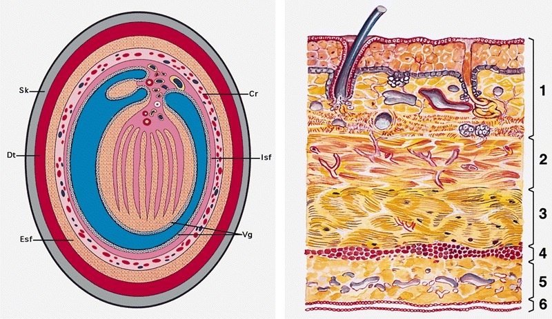
Cross section of the scrotal wall
Images hosted on other servers:


Embryologic development
Anatomic layers
- Skin: thin, corrugated and pigmented; includes keratinized squamous epithelium with skin adnexae, dermis and scattered adipocytes but no subcutaneous tissue; divided in half by a midline cutaneous raphe, which continues to inferior penile surface and along perineum to anus
- Dartos muscular layer: two coherent plexuses of smooth muscle cells; contracts in cold or during sexual stimulation
- External spermatic fascia (intercrural layer of Colles fascia): continuation of external oblique aponeurosis
- Cremasteric muscle (cremasteric layer of Colles fascia): bundles of skeletal muscle, continuation of internal oblique muscle
- Internal spermatic fascia (infundibuliform layer of Colles fascia): partitioned in the midline, continuation of transversalis fascia, attached to tunica vaginalis
- Parietal layer of tunica vaginalis
Drawings
Images hosted on other servers:


Various images

Inguinal ring
Clinical images
Images hosted on other servers:

Constricted human
scrotum (without
hair) with the raphe
clearly exposed
Microscopic (histologic) images
AFIP images

Muscle bundles of the
dartos are beneath
the keratinized
squamous epithelium
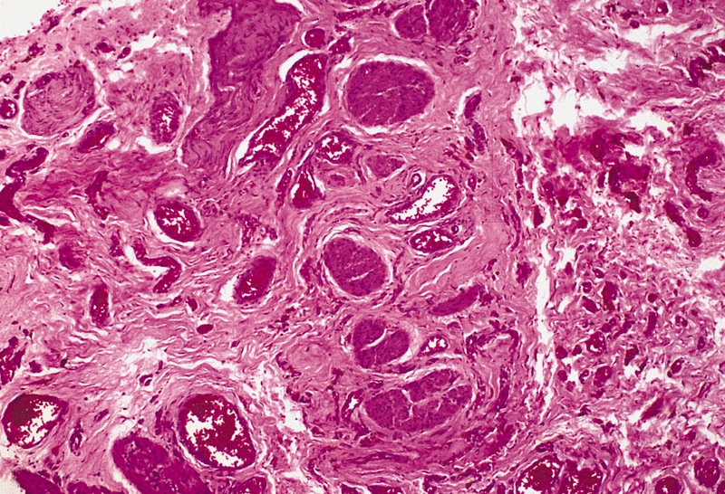
Thick bundles of
conspicuous smooth
muscle in the deep
reticular dermis
