Microbiology & infectious diseases
Parasites-gastrointestinal (not liver)
Enteromonas hominis
Author: Nat Pernick, M.D.

Last author update: 9 August 2019
Last staff update: 5 October 2020
Copyright: 2019-2025, PathologyOutlines.com, Inc.
PubMed Search: Enteromonas hominis

Table of Contents
Case reports | Microscopic (histologic) description | Microscopic (histologic) images | Differential diagnosisCite this page: Pernick N. Enteromonas hominis. PathologyOutlines.com website. https://www.pathologyoutlines.com/topic/parasitologyehominis.html. Accessed April 2nd, 2025.
Case reports
- Small trophozoites were seen on a trichrome stained stool specimen (Pritt: Creepy Dreadful Wonderful Parasites Blog - Case of the Week 552 [Accessed 9 August 2019])
Microscopic (histologic) description
- Small trophozoites, measuring only 5 - 7 micrometers in greatest dimension (Pritt: Creepy Dreadful Wonderful Parasites Blog - Answer to Case 552 [Accessed 9 August 2019])
Microscopic (histologic) images
Contributed by Bobbi Pritt, M.D., Blaine Mathison and Henry Bishop
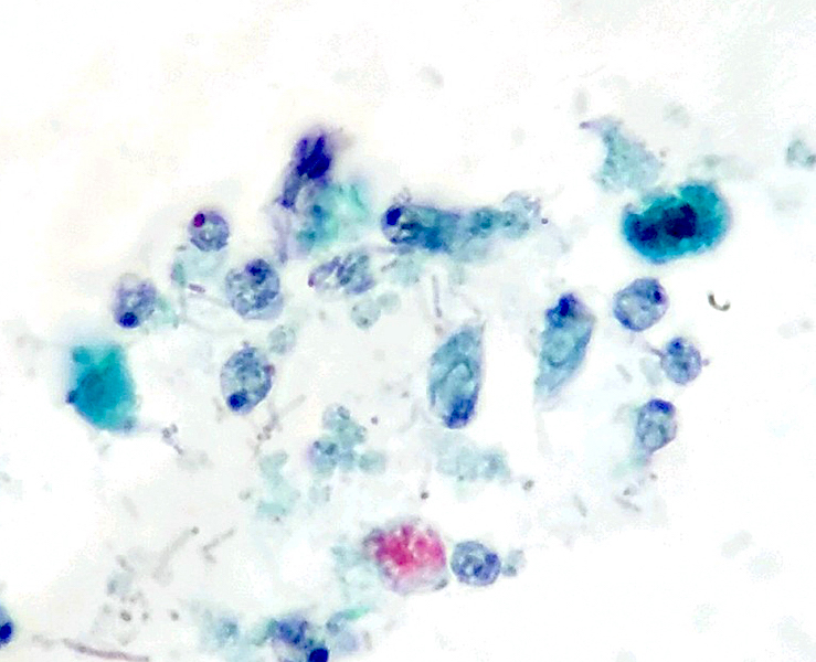
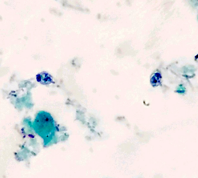

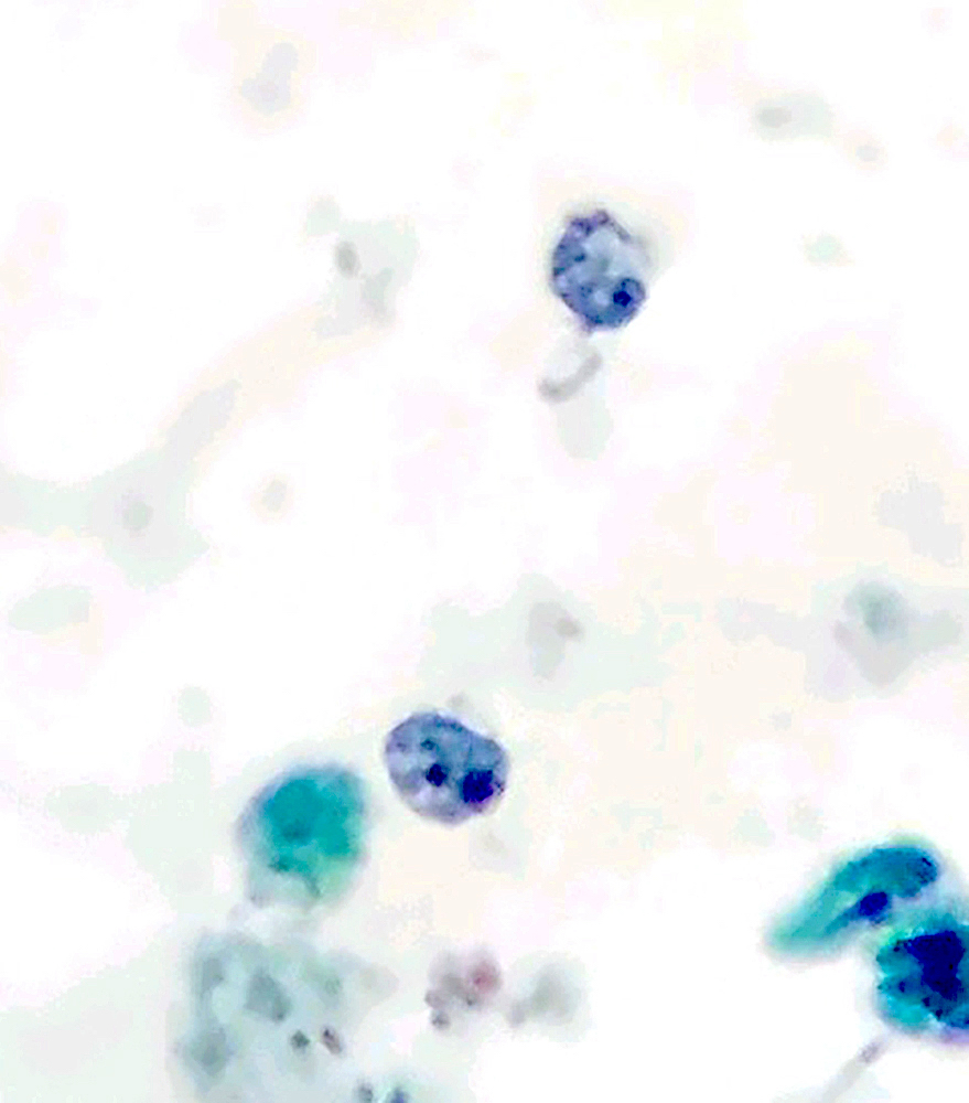
Trichrome stained stool specimen shows Enteromonas hominis trophozoites

Diagnostic features
Differential diagnosis
- Chilomastix mesnili:
- E. hominis trophozoites have smaller size, less elongated shape without a well defined terminal point, and larger karyosome (Pritt: Creepy Dreadful Wonderful Parasites Blog - Answer to Case 552 [Accessed 9 August 2019])