Table of Contents
Definition / general | Case reports | Gross description | Microscopic (histologic) description | Microscopic (histologic) imagesCite this page: Pernick, N. Antrochoanal polyps. PathologyOutlines.com website. https://www.pathologyoutlines.com/topic/nasalpolypantrochoanal.html. Accessed December 27th, 2024.
Definition / general
- 4 - 6% of nasal polyps
- Frequently occur in childhood
- 90% solitary
- Arise from wall of maxillary antrum, extending through large primary or secondary maxillary ostium into nasal cavity
- May pass into choanae or nasopharynx
Case reports
- 27 year old woman with right nasal polyp (Case of the Week #390)
Gross description
- Long narrow stalk with firm, fibrous body
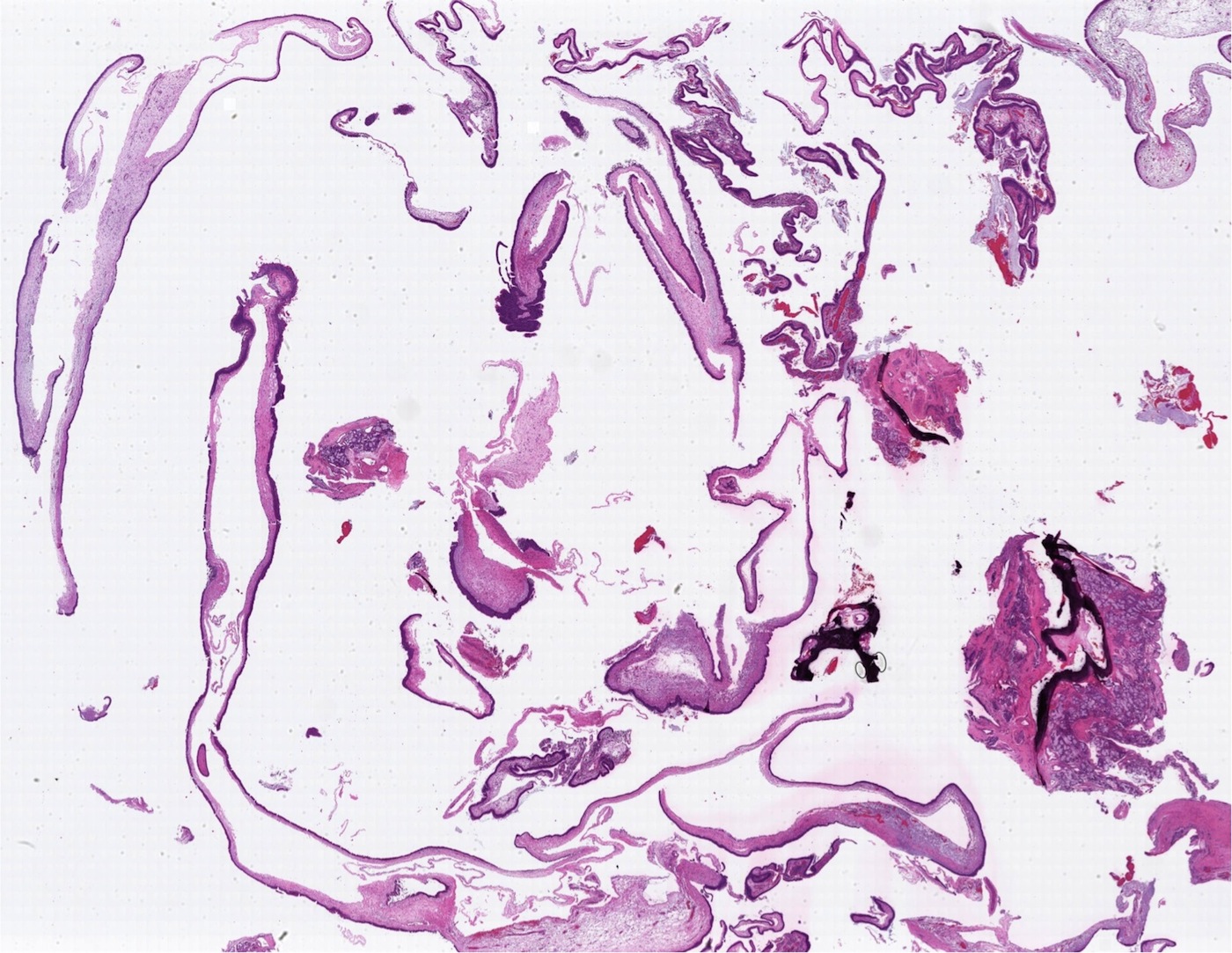
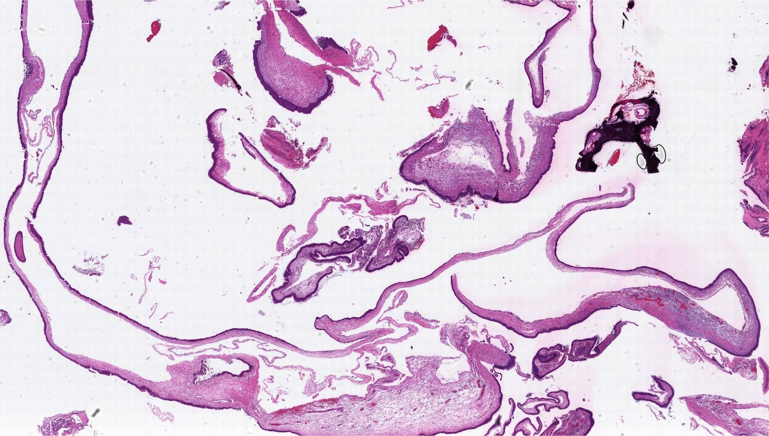
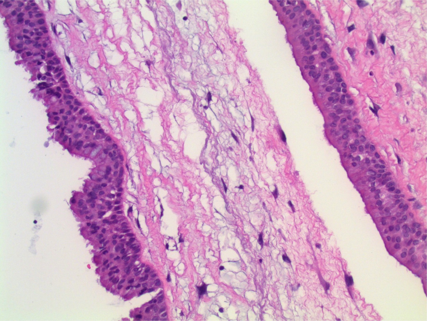
Microscopic (histologic) description
- Thin surface mucosa with no thickened basement membrane
- Stroma with stellate cells, less edema and fewer glands than inflammatory polyp
- May have prominent dilated vessels with thrombosis or infarct
- Prominent eosinophils in only 20%


