
Mandible & maxilla
Benign odontogenic tumors
Squamous odontogenic tumor
Author: Nat Pernick, M.D.

Last author update: 1 January 2004
Last staff update: 23 August 2022
Copyright: 2004-2024, PathologyOutlines.com, Inc.
PubMed Search: Squamous odontogenic tumor (mandible OR maxilla)
Table of Contents
Definition / general | Radiology description | Case reports | Treatment | Microscopic (histologic) description | Microscopic (histologic) images | Differential diagnosis | Additional referencesCite this page: Pernick N. Squamous odontogenic tumor. PathologyOutlines.com website. https://www.pathologyoutlines.com/topic/mandiblemaxillasquamousodon.html. Accessed April 18th, 2024.
Definition / general
- Uncommon
- Most common in 20s but occurs in all ages, 2/3 male
- Usually in anterior maxilla or posterior mandible in soft tissue or bone; 25% are multiple lesions
- Arises from rests of Malassez in periodontal ligament
- Low probability of recurrence, no malignant transformation reported
Radiology description
- Well circumscribed
- Semicircular radiolucency
- Sclerotic border
- Near teeth roots
Case reports
- 30 year old woman with an incidental finding during orthodontic treatment (Arch Pathol Lab Med 2001;125:297)
Treatment
- Excision with extraction of involved teeth or en bloc resection
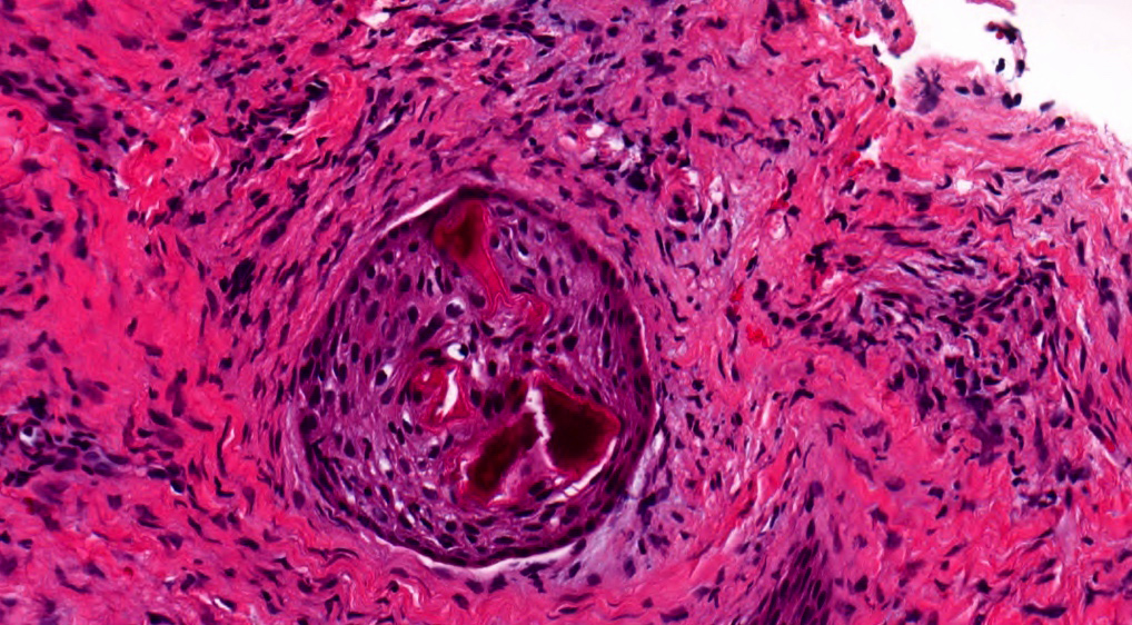
Microscopic (histologic) description
- Anastomosing islands of benign, stratified squamous epithelium within fibrous stroma, often well defined nests with clear cells
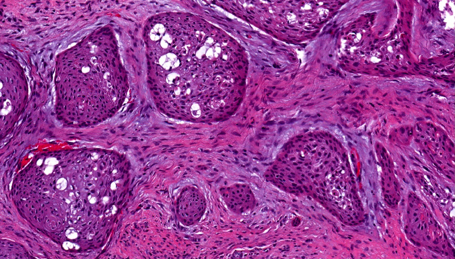
- May contain keratin or psammoma bodies
- Often epithelial vacuolization and microcysts
- No atypia, no mitotic figures, no inflammation, no peripheral palisading
Microscopic (histologic) images
Contributed by Kelly Magliocca, D.D.S., M.P.H.
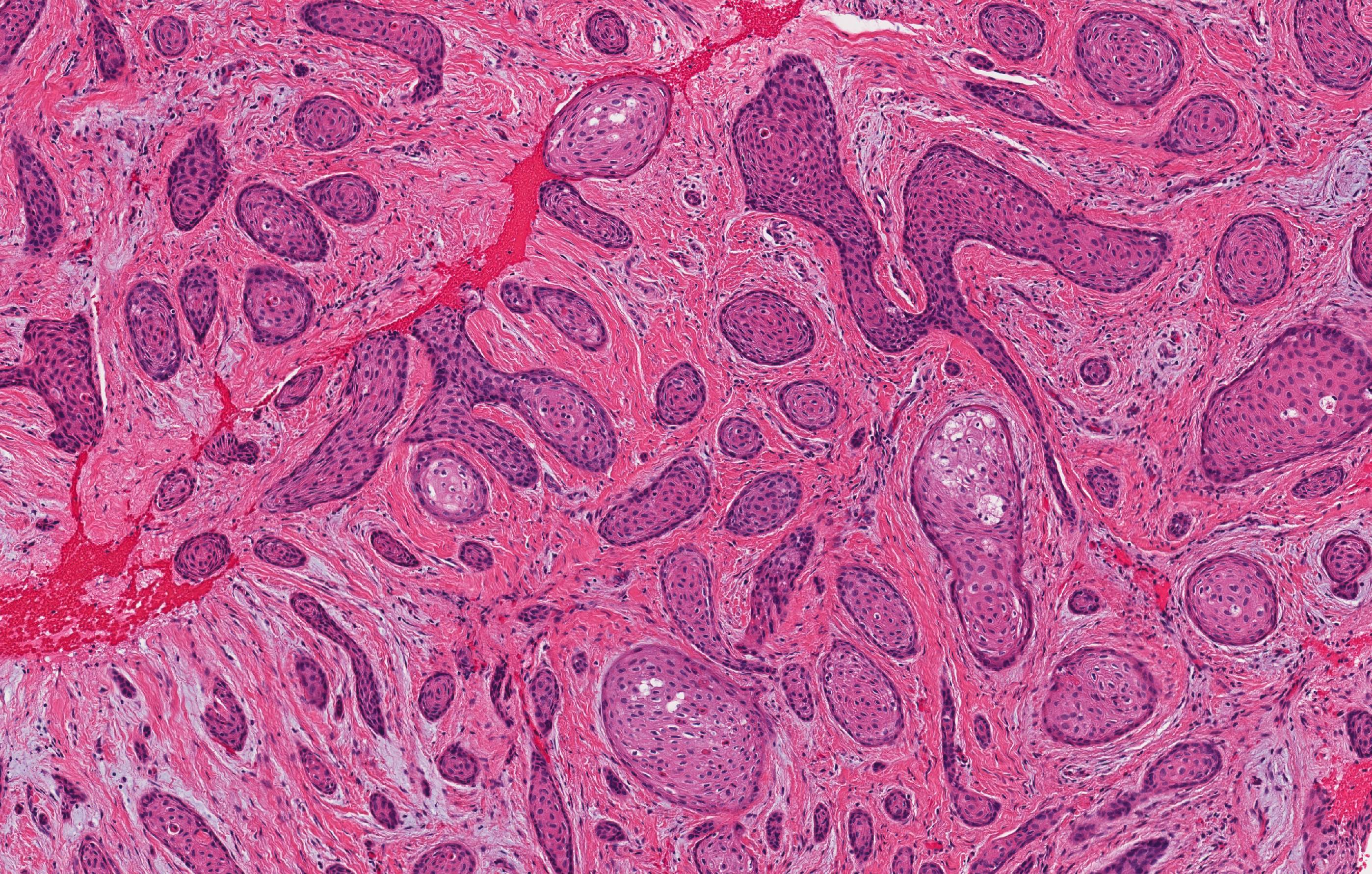
Squamous odontogenic tumor
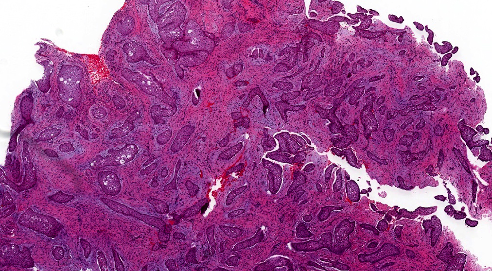
SOT at scanning magnification
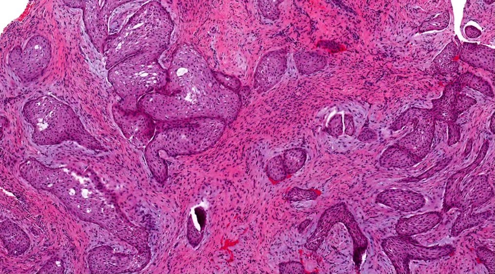
Benign islands of squamous epithelium within fibrous stroma
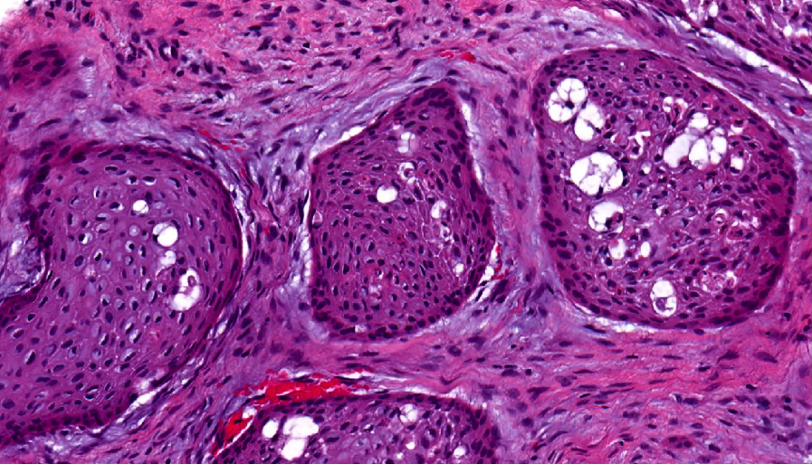
Epithelial vacuolization of squamous islands
Intraepithelial calcifications
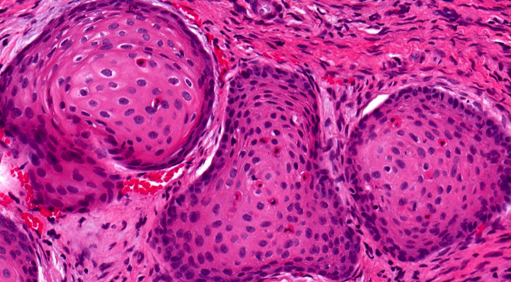
Rounded contours of nests
Differential diagnosis
- Acanthomatous ameloblastoma: peripheral palisading, stellate reticulum
- Mucoepidermoid carcinoma
- Well differentiated squamous cell carcinoma
Additional references
