
Lymph nodes & spleen, nonlymphoma
Pigment / foreign material
Silicone
Last author update: 1 August 2014
Last staff update: 18 July 2022
Copyright: 2003-2025, PathologyOutlines.com, Inc.
PubMed Search: Silicone [title] lymph nodes


Table of Contents
Definition / general | Terminology | Sites | Pathophysiology | Etiology | Clinical features | Diagnosis | Radiology description | Prognostic factors | Case reports | Treatment | Clinical images | Gross description | Microscopic (histologic) description | Microscopic (histologic) images | Cytology description | Positive stains | Negative stains | Electron microscopy description | Differential diagnosis | Additional referencesCite this page: Balakrishna J, Sharabi A. Silicone. PathologyOutlines.com website. https://www.pathologyoutlines.com/topic/lymphnodessiliconeimplant.html. Accessed March 31st, 2025.
Definition / general
- Rare enlargement of regional lymph nodes caused by the presence of silicone carried from tributary organs
- Either an incidental finding or causes painful / enlarged lymph node
- May be associated with granulomatous inflammation (Histol Histopathol 1997;12:1003)
Terminology
- Silicone lymphadenopathy
Sites
- Draining lymph nodes of the implant site, commonly axillary lymph nodes (Respiratory Medicine CME 2011;4:126)
Pathophysiology
- Silicone is widely used in implants, especially augmentation mammoplasty and joint prostheses (Hum Pathol 1980;11:240)
- Once released into tissue, silicone migrates to distant sites through lymphatic channels and bloodstream
- Once it reaches lymph nodes, it elicits a reaction and cause silicone lymphadenopathy
Etiology
- Rupture / leak of the implant, or implant 'bleeds', or releases microparticles into blood / lymphatics
Clinical features
- Enlarged lymph nodes
- Asymptomatic or with pain
Diagnosis
- Biopsy
Radiology description
- Ultrasonogram: hyperechoeic (increased echogenicity of the lymph node mediastinum with dirty acoustic shadowing), beginning in the hilum and progressing outward through the cortex with time and amount of silicone
- In severe cases, snowstorm appearance
Prognostic factors
- Depends upon the amount of silicone and severity of the reaction
Case reports
- 35 year old woman with axillary silicone lymphadenopathy (J Med Case Rep 2009;3:6442)
- 40 year old woman with silicone lymphadenopathy involving intramammary lymph nodes (AJR Am J Roentgenol 1994;162:1089)
- 45 year old woman with axillary silicone lymphadenopathy secondary to augmentation mammaplasty (Indian J Plast Surg 2010;43:206)
- 47 year old woman with silicone lymphadenopathy mimicking lymphoma (J Clin Pathol 2000;53:549)
- 49 year old woman with lymphadenopathy associated with total joint prostheses (J Bone Joint Surg Am 1996;78:588)
- 59 year old woman with silicone migration to the contralateral axillary lymph nodes and breast after highly cohesive silicone gel implant failure (Cases J 2009;2:6420)
- 71 year old woman with siliconoma in internal mammary lymph node (Radiology Case Reports 2011;6(4))
Treatment
- Removal of lymph nodes
Clinical images
Images hosted on other servers:

Rupture of implant


Various images
Gross description
- No specific gross features
- Enlarged and firmer than normal
- Extreme cases show distorted architecture and fibrosis
Microscopic (histologic) description
- Diffuse follicular hyperplasia
- Histiocytes with vacuolated cytoplasm especially inside the sinusoids
- Histiocytes cause foreign body granulomatous reaction, giant cells and empty vacuoles
- Giant cells have refractile and non-birefringent particles
- Asteroid bodies may be seen
- Silicone from orthopedic devices: prominent granulomatous reaction with clumps of granular yellowish refractile material
- Silicone from mammary prostheses: finer vacuolated deposits
Microscopic (histologic) images
Contributed by Dr. Mark R. Wick
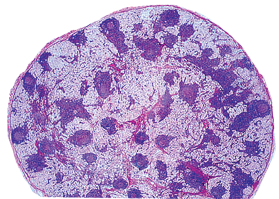
Silicone in axillary lymph node
Images hosted on other servers:

Material consistent with silicone

Liquid silicone droplets

Various images

Subcapsular sinus diffusely expanded

Vacuoles which contain refractile material

Foreign body granuloma

Germinal centers surrounded by sinuses

Silicone leakage

Silicone particles in cystic spaces
Cytology description
- Numerous multi-vacuolated histiocytic cells both scattered individually as well as in aggregates
- Contain clear, refractile but non-polarizable material
- Background may show scattered lymphocytes
Electron microscopy description
- Electron opaque fragmented spicules or flakes
Differential diagnosis
- Fat necrosis
- Lipogranuloma
- Metastatic lobular carcinoma
- Metastatic renal cell carcinoma
- Metastatic signet ring cell carcinoma
- Signet ring cell type lymphoma
- Sinus histiocytosis with massive lymphadenopathy
