
Liver & intrahepatic bile ducts
Other malignancies
Angiosarcoma
Author: Deepali Jain, M.D.

Last author update: 1 February 2012
Last staff update: 8 July 2022
Copyright: 2002-2024, PathologyOutlines.com, Inc.
PubMed Search: Angiosarcoma liver
Table of Contents
Definition / general | Causes | Clinical features | Case reports | Gross description | Gross images | Microscopic (histologic) description | Microscopic (histologic) images | Positive stains | Negative stains | Electron microscopy description | Molecular / cytogenetics description | Differential diagnosisCite this page: Jain D. Angiosarcoma. PathologyOutlines.com website. https://www.pathologyoutlines.com/topic/livertumorangiosarcoma.html. Accessed April 19th, 2024.
Definition / general
- Rare (10 - 30 annual cases in U.S.) but most common hepatic primary sarcoma in adults (2% of all primary liver tumors)
Causes
- 25 - 42% associated with exposure to androgen steroids, arsenic, Thorotrast (radiocontrast agent thorium dioxide, used through 1950s, detect its α particle emissions via autoradiography), vinyl chloride (Wikipedia: Thorotrast [Accessed 30 November 2017])
- Patients with exposure to Thorotrast or vinyl chloride may have synchronous cholangiocarcinoma or hepatocellular carcinoma
- Rarely associated with chemotherapy, copper sulfate, estrogens, hereditary hemochromatosis, phenelzine, radiotherapy
- Cases with above known causes usually have latent period of 20 - 35 years, are accompanied by fibrosis or cirrhosis, have precursor conditions of hypertrophy and atypia of hepatocytes and sinusoidal lining cells but are histologically similar to idiopathic cases
Clinical features
- 75% men, usually age 50+ years; rare in children
- Nonoperative biopsy may cause severe bleeding and death
- Most patients die within 6 months from hepatic failure or intra-abdominal bleeding
- Metastasizes widely, often to lung (vinyl chloride cases usually don't have distant metastases)
Case reports
- 71 year old man with metastatic hepatic epithelioid angiosarcoma (Arch Pathol Lab Med 2001;125:968)
Gross description
- Multicentric, involves right and left lobes
- Diffusely infiltrative, hemorrhagic and gray white solid nodules with blood filled cavities
- Thorotrast associated tumors have subcapsular hepatic and splenic deposits of yellow chalky material
Gross images
Images hosted on other servers:

Vascular lesion with dense fibrosis
Microscopic (histologic) description
- Tumor composed of infiltrative, freely anastomosing vascular channels
- Tumor cells grow along sinusoids adjacent to hepatic cords
- Tumor cells have abundant, pale eosinophilic cytoplasm, poorly defined cell borders, are usually pleomorphic with hyperchromatic nuclei but may be only mildly atypical
- Also variably prominent nucleoli, blood filled cavities present are lined by tumor cells that may be papillary
- 75% have vascular invasion of portal or hepatic vein branches; frequent mitotic activity
- Precursor stage has endothelial hypertrophy and hyperplasia
- Also epithelioid cells with abundant cytoplasm and prominent nucleoli, bizarre tumor giant cells, fibrosarcoma-like spindle cells, cholestatic hepatocellular rosettes with bile plugs, tumor cell phagocytic activity and extramedullary hematopoiesis
- Childhood cases: may have kaposiform areas of spindle cells with PAS+ intracytoplasmic globules; no prominent myxoid areas
- Thorotrast exposed patients: have brown gray refractile but not birefringent granules of Thorotrast free or within macrophages
- Also precursor stage with endothelial hypertrophy and hyperplasia
Microscopic (histologic) images
Images hosted on other servers:
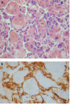
Sinusoidal infiltration
Positive stains
- CD34, CD31, factor VIII related antigen and Ulex europaeus lectin type 1 (may not be present in poorly vasoformative areas)
- Also Prox1 (reflects the lymphatic endothelial phenotype), claudin5
Negative stains
- Keratin (but positive in 12 - 35%)
Electron microscopy description
- Weibel-Palade bodies
Molecular / cytogenetics description
- 50% of vinyl chloride associated cases have A:T to T:A transversion in p53
Differential diagnosis
- Epithelioid hemangioendothelioma:
- Less atypia, less mitotic activity, less necrosis, often prominent fibrous and hyalinized stroma
- Fibrosarcoma:
- Very rare; not particularly vascular; CD31 negative
- Hepatocellular carcinoma:
- Atypical hepatocytes, normal endothelial cells
- Kaposi sarcoma:
- HIV+, have extrahepatic nodules, portal distribution and lack angiosarcomatous foci
- Peliosis hepatis
- Reactive bile ductular proliferation, reactive lymphoid hyperplasia:
- Reactive, lack atypical endothelial lining cells
