
Liver & intrahepatic bile ducts
Infectious nonviral
Echinococcal cyst
Editorial Board Member: Aaron R. Huber, D.O.
Deputy Editor-in-Chief: Catherine E. Hagen, M.D.


Last author update: 9 September 2021
Last staff update: 21 July 2022
Copyright: 2002-2024, PathologyOutlines.com, Inc.
PubMed Search: Echinococcal cyst liver
Table of Contents
Definition / general | Essential features | Terminology | ICD coding | Epidemiology | Sites | Pathophysiology | Etiology | Clinical features | Diagnosis | Laboratory | Radiology description | Radiology images | Prognostic factors | Case reports | Treatment | Clinical images | Gross description | Gross images | Microscopic (histologic) description | Microscopic (histologic) images | Virtual slides | Positive stains | Videos | Sample pathology report | Differential diagnosis | Additional references | Board review style question #1 | Board review style answer #1 | Board review style question #2 | Board review style answer #2Cite this page: Aghighi M, Fazlollahi L. Echinococcal cyst. PathologyOutlines.com website. https://www.pathologyoutlines.com/topic/liverechinococcal.html. Accessed April 19th, 2024.
Definition / general
- Cestode (tapeworm) infection widespread across the world
- Common cause of hepatic cysts; caused by the larval form of Echinococcus tapeworms: Echinococcus granulosus (most common, causes cystic disease), Echinococcus multilocularis (less common, causes alveolar disease), Echinococcus vogeli (polycystic disease) and Echinococcus oligarthus (extremely rare, unicystic form)
Essential features
- Cestode (tapeworm) infection widespread across the world
- Echinococcus granulosus is the most common (classic hydatid cyst)
- E. granulosus cysts consist of inner, middle and outer layers
- Inner layer is a germinal layer with brood capsules and protoscolices (adult tapeworm heads)
- Middle layer consists of hyalinized, laminated and acellular material
- Outer layer consists of granulation tissue and fibrosis
Terminology
- Hydatid disease
ICD coding
- ICD-10: B67. 90 - echinococcosis, unspecified
Epidemiology
- Common cause of hepatic cysts worldwide, particularly in sheep and cattle in farming areas in Middle East, Greece, Australia, North Africa and parts of South America
- In U.S., usually in immigrants from above areas
- Reference: World J Gastroenterol 2012;18:1425
Sites
- 60 - 70% in liver; also brain, lung, other sites
- Frequently communicates with biliary tract (Iran J Med Sci 2013;38:2)
- Rare in breast (0.27% of cases) but should be considered in the differential diagnosis of a breast mass in endemic regions (J Gynecol Obstet Biol Reprod (Paris) 1986;15:187)
Pathophysiology
- Mucosal attachment of tapeworm to small intestine in definitive hosts, such as dogs (CDC: Echinococcosis [Accessed 4 March 2022])
- Ingestion of eggs in contaminated feces can infect humans
- Larval oncospheres are released from eggs and are transported to liver by portal vein
- Oncospheres grow into cysts enlarging at slow pace of about 10 - 15 mm per year
- Cysts are composed of protoscolices and daughter cysts (J Clin Transl Hepatol 2016;4:39)
Etiology
- Liver as primary site of infection:
- E. granulosus
- E. multilocularis
Clinical features
- Often asymptomatic for years due to slow growth but hepatic and pulmonary symptoms are most common
- Symptoms can be secondary to cyst rupture or compression of other structures
- Bacterial infection, bile duct compression, cholangitis, rupture into peritoneal or pleural cavities
- Portal hypertension secondary to portal vein compression
- Breast masses (Folia Med (Plovdiv) 2020;62:23)
- Rupture of cysts in the lung may cause intense cough or vomiting of cystic materials (Curr Opin Pulm Med 2010;16:257)
- Clinically may resemble carcinoma
- Parasitic flatworm (tapeworm) of class Cestoda (i.e., a cestode) (Pritt: Creepy Dreadful Wonderful Parasites Blog - Answer to Case 514 [Accessed 4 March 2022])
- Member of family Taeniidae due to presence of an armed rostellum (i.e., 2 rows of hooklets) (Pritt: Creepy Dreadful Wonderful Parasites Blog - Answer to Case 514 [Accessed 4 March 2022])
- Echinococcus granulosus is a complex of closely related organisms (Pritt: Creepy Dreadful Wonderful Parasites Blog - Answer to Case 514 [Accessed 4 March 2022])
- E. granulosus sensu stricto is most common species causing human disease worldwide
- E. canadensis and E. ortleppi also cause human disease
- Species are differentiated via molecular means
- Alveolar echinococcosis is caused by infection with larval stage of E. multilocularis, a 1 - 4 millimeter long tapeworm found in foxes, coyotes, dogs (definitive hosts)
- E. multilocularis human infection is less common but has more aggressive and invasive growth that resembles a tumor and is not contained within a large parent cyst (Pritt: Creepy Dreadful Wonderful Parasites Blog - Answer to Case 514 [Accessed 4 March 2022])
- E. oligarthra and E. vogeli are rare causes of human echinococcosis in South and Central America (Pritt: Creepy Dreadful Wonderful Parasites Blog - Answer to Case 514 [Accessed 4 March 2022])
- Breast: slow growing lesion, may mimic fibroadenoma, phylloides or rarely intracystic carcinoma; may become infected and be indistinguishable radiologically from an abscess
Diagnosis
- Usually made by ultrasound or CT scan supported by positive hydatid serology
Laboratory
- Monoclonal antibodies to hydatid antigens detection by immunoelectrophoresis, enzyme linked immunosorbent assay (ELISA) and immunoblots
- Immunoblots have the highest sensitivity, followed by ELISA and immunoelectrophoresis
- Antigen assays have more specificity but lower sensitivity than antibody assays
- Unruptured cysts may not produce antibody response (J Clin Transl Hepatol 2016;4:39)
Radiology description
- Ultrasound / CT scan reveal cysts with septations
- Breast: on mammography, lesions may be well circumscribed, oval or spherical densities with smooth lobulated margins; peripheral calcification may be present
- Breast: on ultrasound, may see double wall sign (cyst wall as 2 echogenic layers), snowstorm sign (due to movement of scolices within the cyst), waterlily sign (floating membranes due to detached endocyst / daughter cyst), scroll sign (due to folding of the detached endocyst) (J Clin Ultrasound 2014;42:502, Singapore Med J 2010;51:e72)
Radiology images
Images hosted on other servers:

Breast: well defined opacity


Breast: axial and longitudinal sections
Prognostic factors
- Although mortality is uncommon, fatalities have been reported in rare cases of anaphylactic shock or cardiac tamponade (Acta Cardiol 2005;60:39)
- Ruptured cystic disease may require lifelong antiparasitic therapy to prevent recurrence
- Most common complications are mechanical and superinfection (Am J Trop Med Hyg 2019;101:628)
Case reports
- 8 year old girl with cystic echinococcosis involving left ventricle (Medicine (Baltimore) 2019;98:e15267)
- 25 year old man with abdominal pain, syncope and hypotension (N Engl J Med 2015;372:265)
- 31 year old year old woman with painless breast lump for 1 year (Int J Surg Case Rep 2015;7C:115)
- 32 year old man with an Echinococcus cyst in liver and pancreas (BMC Infect Dis 2019;19:661)
- 62 year old woman with a huge Echinococcus cyst in liver resembling malignancy (J Med Case Rep 2017;11:113)
- Echinococcus found in a single liver cyst (Pritt: Creepy Dreadful Wonderful Parasites Blog - Case of the Week 514 [Accessed 4 March 2022])
- Objects seen in liver cyst aspirate (Pritt: Creepy Dreadful Wonderful Parasites Blog - Case of the Week 537 [Accessed 4 March 2022])
Treatment
- Surgical resection
- Cyst rupture during surgery may cause urticaria, anaphylaxis or recurrence of infection
- Puncture, aspiration, infusion of protoscolicidal agent, reaspiration
- Preferred technique due to lower risk of anaphylaxis or recurrence (BMC Surg 2019;19:95)
Clinical images
Images hosted on other servers:

Breast cyst
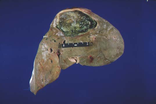
Gross description
- Echinococcus granulosus: typical cyst is spherical, up to 30 cm or more in diameter, has a fibrous rim and frequently contains several daughter cysts
- Echinococcus multilocularis: multilocular, necrotic, cystic cavities containing thick pasty material; fibrous rim is absent
- Reference: Zhonghua Bing Li Xue Za Zhi 2021;50:650
Gross images
Contributed by Ladan Fazlollahi, M.D., M.P.H.
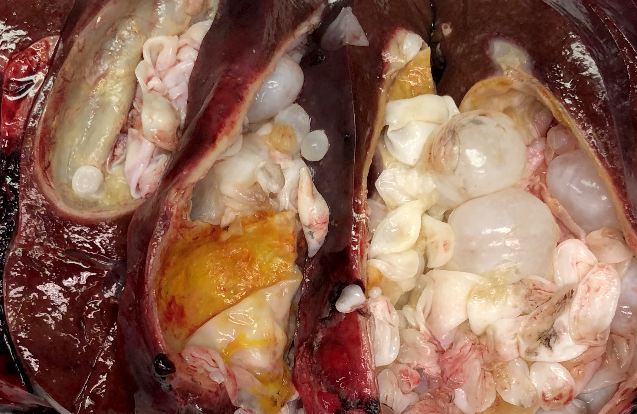
Gross section of hydatid cyst
Images hosted on other servers:
Hydatid cyst with fluid and daughter cysts

Breast: excised cyst

Breast: excised cyst with endocysts
Microscopic (histologic) description
- E. granulosus
- Cyst wall has 3 structural components:
- Outer acellular laminated membrane (1 mm thick)
- Germinal membrane (a transparent nucleated lining)
- Protoscolices, attached to the membrane and budding from it
- Protoscolices are ovoid and contain hooklets (birefringent under polarized light) and a sucker
- Outer fibrotic layer with granulation tissue with increased eosinophils also exists
- Cyst wall has 3 structural components:
- E. multilocularis
- Irregular cysts with laminated membrane without germinal membrane or protoscolices
- Invasion of liver parenchyma can create inflammatory / granulomatous reaction or extensive peripheral necrosis and fibrosis
- Reference: Zhonghua Bing Li Xue Za Zhi 2021;50:650
Microscopic (histologic) images
Contributed by Ladan Fazlollahi, M.D., M.P.H. and Bobbi Pritt, M.D.
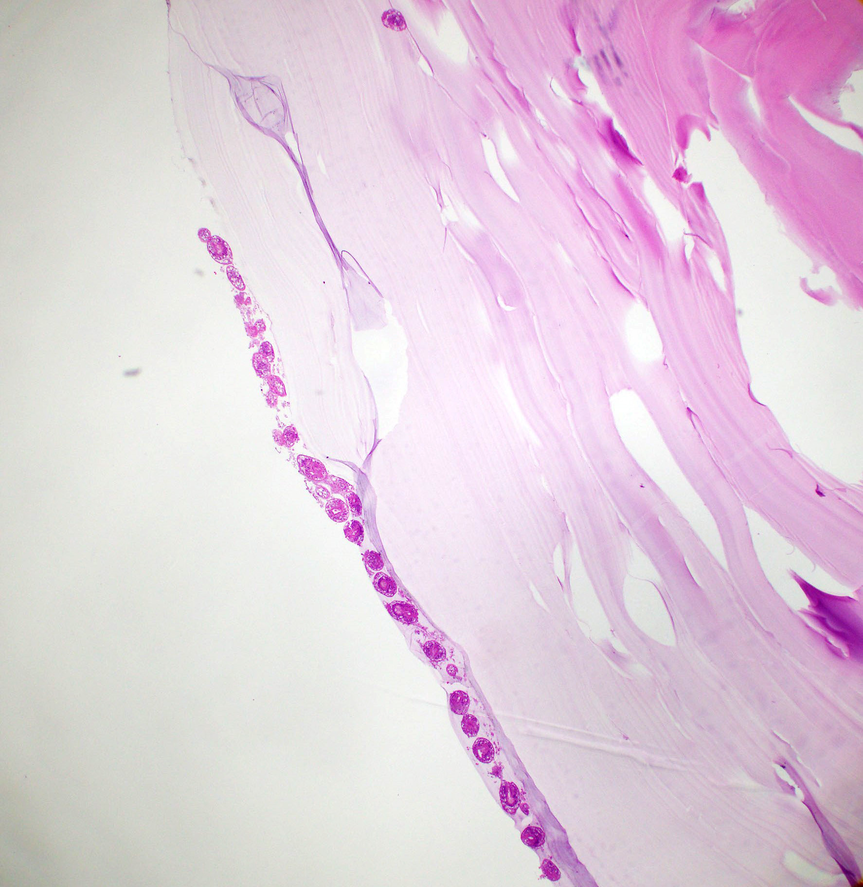
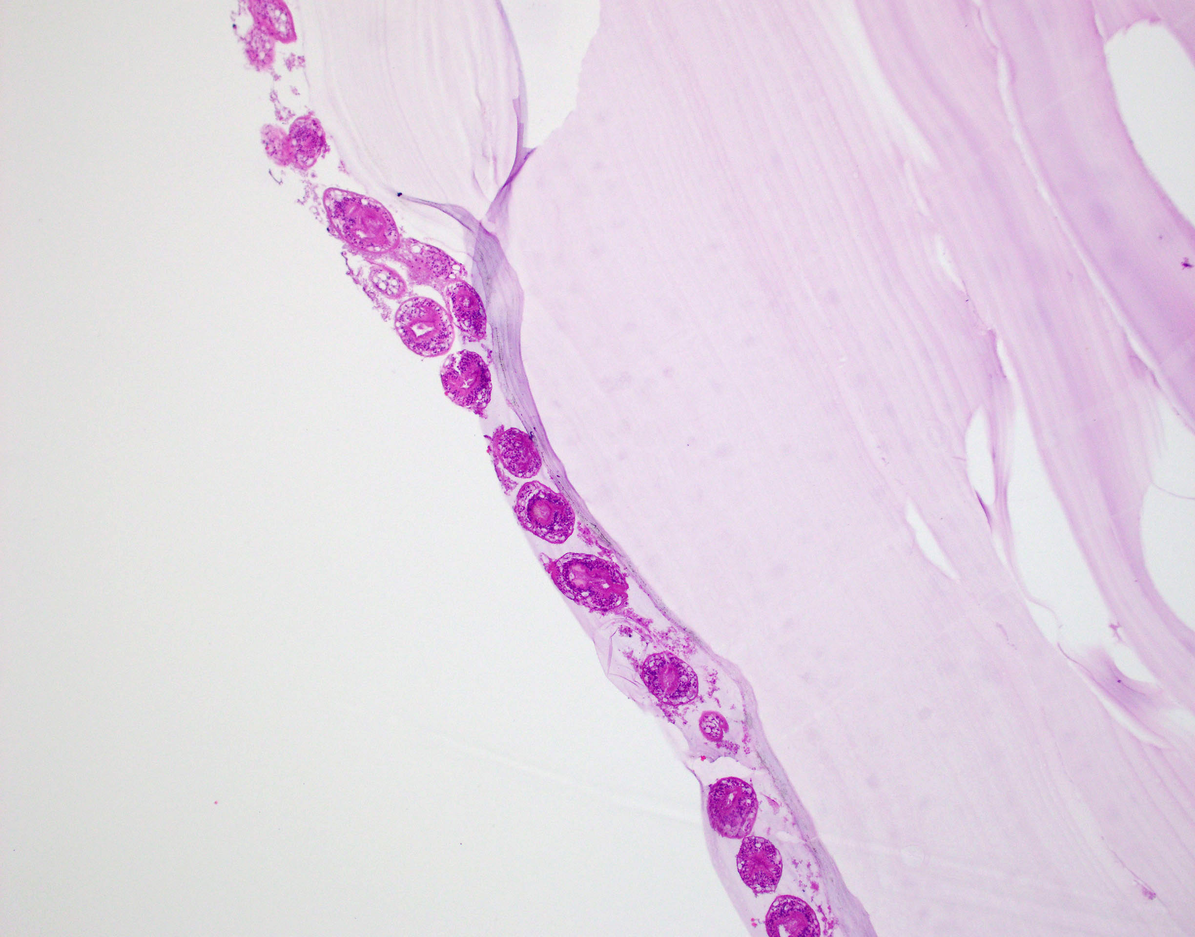
Hydatid cyst wall
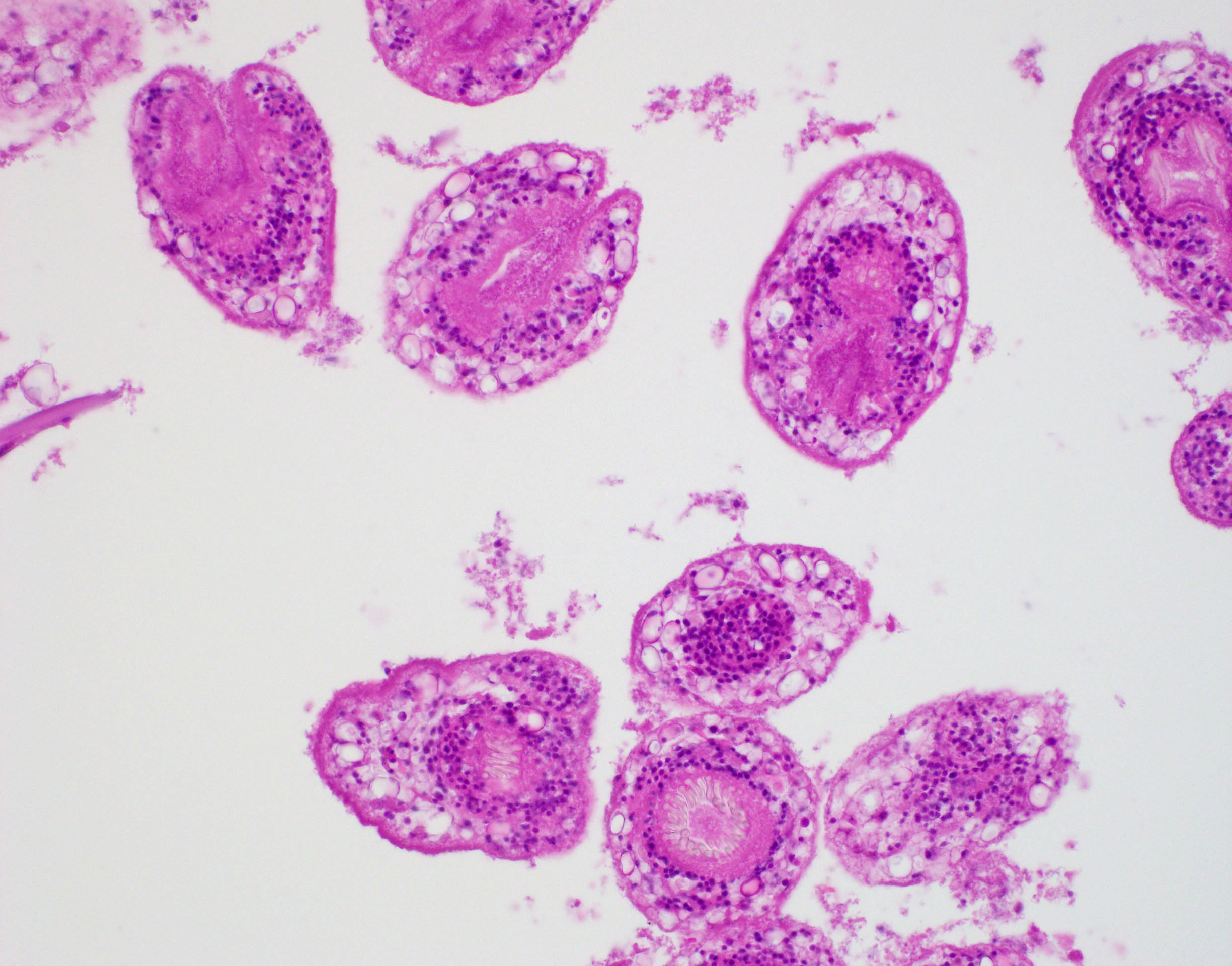
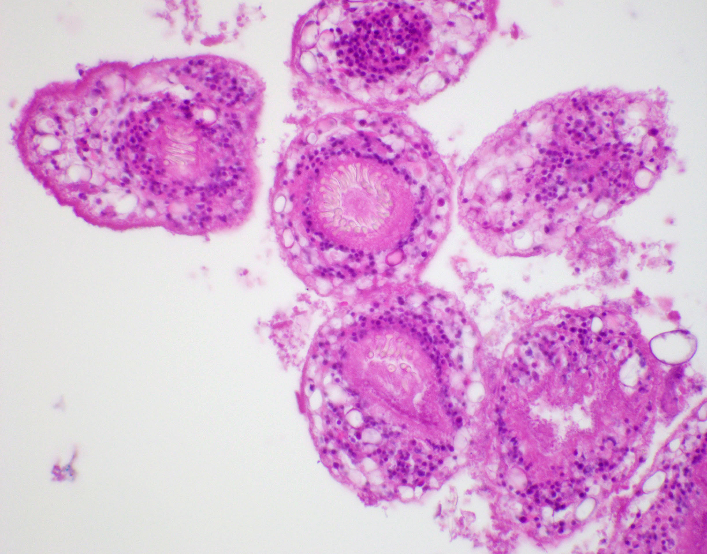
Hydatid cyst

Hydatid cyst

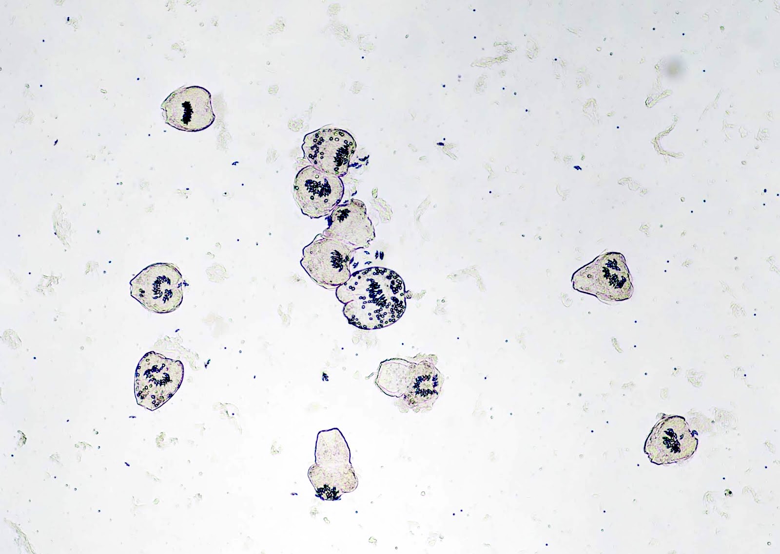
Numerous free hooklets


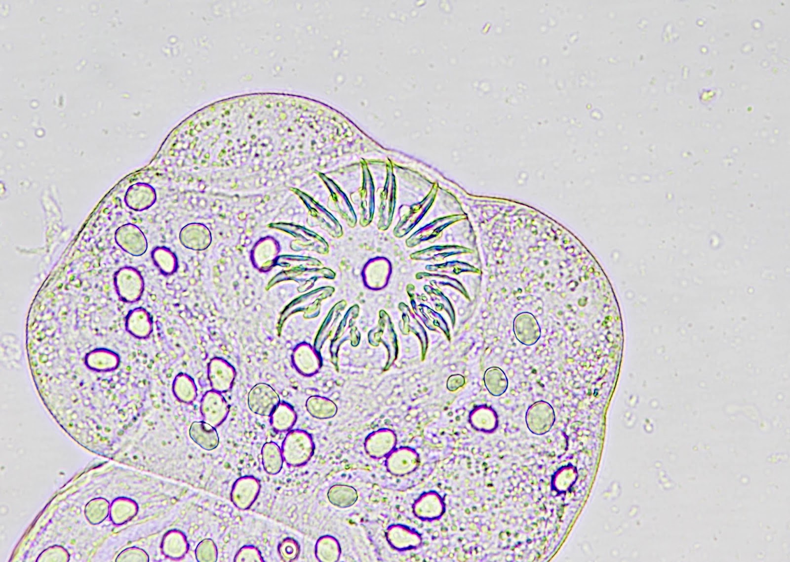
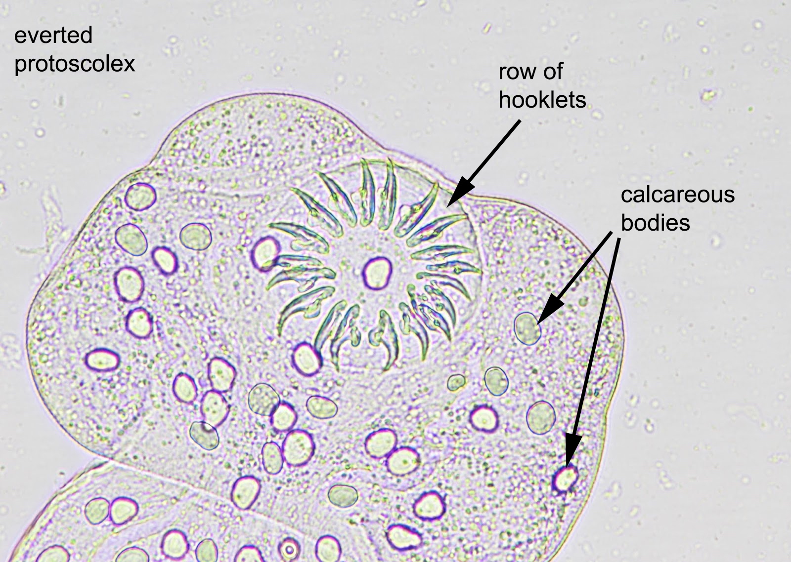
Classic protoscoleces with an armed rostellum
Virtual slides
Images hosted on other servers:


Hydatid cyst
Positive stains
- Hooklets are acid fast positive on Ziehl-Neelsen stain; also stain with GMS (J Cytol Histol 2016;7:1000422)
Videos
Bird's eye view of Echinococcus
Sample pathology report
- Liver, excision:
- Echinococcal cyst
- Comment: There are degenerated echinococcal cysts that contain abundant debris with protoscolices fragments.
Differential diagnosis
- Other tapeworm infections: cysticercosis (Taenia solium)
- Fibropolycystic liver disease:
- Lacks 3 layered cyst, protoscolices
- Numerous cystic lesions similar to solitary cysts, covered by cuboidal to flat biliary epithelium
- Amoebic or pyogenic abscess:
- Lacks 3 layered cyst, protoscolices
- Nuclear fragments with inflammatory cells
- Organisms with foamy cytoplasm and eccentric round nucleus
- Ingested red blood cells pathognomonic of Entamoeba histolytica
- Trophozoites look like macrophages
Additional references
Board review style question #1
A 45 year old man presented with weakness, weight loss and abdominal pain for a month. An ultrasound showed a cystic lesion in liver. The cystic lesion was resected and revealed cysts consisting of inner, middle and outer layers. Which of the following is the best diagnosis?
- Cystic hepatocellular carcinoma
- Echinococcus granulosus cysts
- Hemorrhagic cysts
- Polycystic liver disease
Board review style answer #1
Board review style question #2
A 65 year old woman presented with fever, jaundice, weight loss and abdominal discomfort. She lives in the countryside with her 2 dogs. CT scan of liver revealed large cystic mass, suspected to represent an echinococcal cyst. Which of the following is the most likely microscopic feature?
- Cystic lesion with inner, middle and outer layers and protoscolices
- Ingested red blood cells
- Numerous cystic lesions similar to solitary cysts, covered by cuboidal epithelium
- Organisms with foamy cytoplasm and eccentric round nucleus
Board review style answer #2
A. Cystic lesion with inner, middle and outer layers and protoscolices
Comment Here
Reference: Echinococcal cyst
Comment Here
Reference: Echinococcal cyst
