
Liver & intrahepatic bile ducts
Viral hepatitis
Adenovirus hepatitis
Editorial Board Members: Monika Vyas, M.D., Wei Chen, M.D., Ph.D.


Last author update: 10 January 2023
Last staff update: 10 January 2023
Copyright: 2002-2024, PathologyOutlines.com, Inc.
PubMed Search: Adenovirus hepatitis

Table of Contents
Definition / general | Essential features | ICD coding | Epidemiology | Sites | Etiology | Clinical features | Diagnosis | Laboratory | Radiology description | Radiology images | Prognostic factors | Case reports | Treatment | Gross description | Gross images | Microscopic (histologic) description | Microscopic (histologic) images | Positive stains | Negative stains | Sample pathology report | Differential diagnosis | Board review style question #1 | Board review style answer #1 | Board review style question #2 | Board review style answer #2Cite this page: Ilchenko V, Mannan R. Adenovirus hepatitis. PathologyOutlines.com website. https://www.pathologyoutlines.com/topic/liveradenovirus.html. Accessed April 26th, 2024.
Definition / general
- Adenoviruses are widely distributed viruses that usually cause self limited infections; but in an immunocompromised host they can cause severe infections with injuries to multiple organs, including the liver
- While severe acute liver failure due to adenovirus infection is rare, it is characterized by rapid progression and is frequently fatal
- Such cases were reported in patients after allogeneic hematopoietic stem cell transplantation (HSCT), liver transplantation, chemotherapy regimens and from other causes (Transpl Infect Dis 2021;23:e13496, Infection 2014;42:105)
- However, adenovirus hepatitis can also occur in immunocompetent adults (J Investig Med High Impact Case Rep 2022;10:23247096221079192)
Essential features
- Usually immunocompromised transplant recipients
- Grossly characterized by the presence of coagulative necrosis in liver parenchyma
- Diagnosis made on liver biopsy with confirmation by adenovirus immunostaining
ICD coding
- ICD-10: B97.0 - adenovirus as the cause of diseases classified elsewhere
Epidemiology
- Usually occurs in immunocompromised adults and children
- More frequent in the patients from these groups (Infection 2014;42:105):
- Liver transplant recipients
- Bone marrow transplant recipients
- Patients on (or after) oncology related chemotherapy regimens
- Patients with severe combined immunodeficiency
- HIV infected patients
- Heart, kidney and other transplant recipients
Sites
- Liver
Etiology
- Human adenovirus serotypes C1, C2, C5 and other (Transpl Infect Dis 2021;23:e13496)
- It can be either a primary infection or reactivation of latent infection
Clinical features
- In immunocompetent patients, it causes mild illness with respiratory symptoms, keratoconjunctivitis, gastroenteritis, etc.
- Fever is the most common presenting symptom (Am J Surg Pathol 2017;41:810)
- Other nonspecific signs (e.g., abdominal pain, memory and cognitive disorder, convulsions) were reported (Transpl Infect Dis 2021;23:e13496)
- In immunocompromised patients, severe disease may occur, including fulminant hepatitis
Diagnosis
- Diagnosis of adenovirus hepatitis can be made on liver biopsy with confirmation by adenovirus immunostaining (Am J Surg Pathol 2017;41:810)
- Detection of viral DNA by PCR on blood or broncoalveolar lavage (Transpl Infect Dis 2021;23:e13496, BMC Infect Dis 2021;21:152)
Laboratory
- Viral DNA detection by PCR on blood
- Positive viral culture
- Positive serology for adenovirus
- Elevated serum total bilirubin, lactate dehydrogenase (Transpl Infect Dis 2021;23:e13496)
- Elevated serum alanine aminotransferase (ALT), aspartate aminotransferase (AST) (Am J Surg Pathol 2017;41:810)
Radiology description
- Abdominal CT may reveal small or large hypodense areas in liver, areas of necrosis (Case Rep Infect Dis 2012;2012:463569)
Radiology images
Images hosted on other servers:

Liver hypodense areas
Prognostic factors
- Rapid progression with massive liver necrosis is usually fatal; patients die secondary to organ failure (Am J Surg Pathol 2017;41:810)
- Surviving patients had limited necrosis at the time of initial diagnosis; these patients also had only relatively mild peak elevations in their serum AST and ALT (mean: 140 and 133, respectively) (Am J Surg Pathol 2017;41:810)
Case reports
- 2 year old boy after a liver transplantation for hepatoblastoma (J Pediatric Infect Dis Soc 2015;4:e1)
- 35 year old immunocompetent woman with fever (J Investig Med High Impact Case Rep 2022;10:23247096221079192)
- 48 year old woman after remission of large B cell lymphoma and autologous HSCT; 51 year old man with acute myeloid leukemia after 4 allogeneic HSCTs (Transpl Infect Dis 2021;23:e13496)
- 59 year old man with T cell prolymphocytic leukemia who had focal liver lesions on abdominal CT with suspicion of metastatic disease (Ann Hepatol 2014;13:827)
- 66 year old man on a chemotherapy regimen for mantle cell lymphoma developed fulminant adenovirus hepatitis (Pathol Int 2018;68:259)
Treatment
- Supportive care (Am J Surg Pathol 2017;41:810)
- Antiviral therapy can be helpful (J Investig Med High Impact Case Rep 2022;10:23247096221079192, Liver Transpl 2022;28:505)
Gross description
- Liver parenchyma demonstrates reddish brown color on the cut surface (Pathol Int 2018;68:259)
- Heterogeneous areas of coagulative necrosis present in liver parenchyma (Am J Surg Pathol 2017;41:810)
Gross images
Images hosted on other servers:
Areas of coagulative necrosis
Microscopic (histologic) description
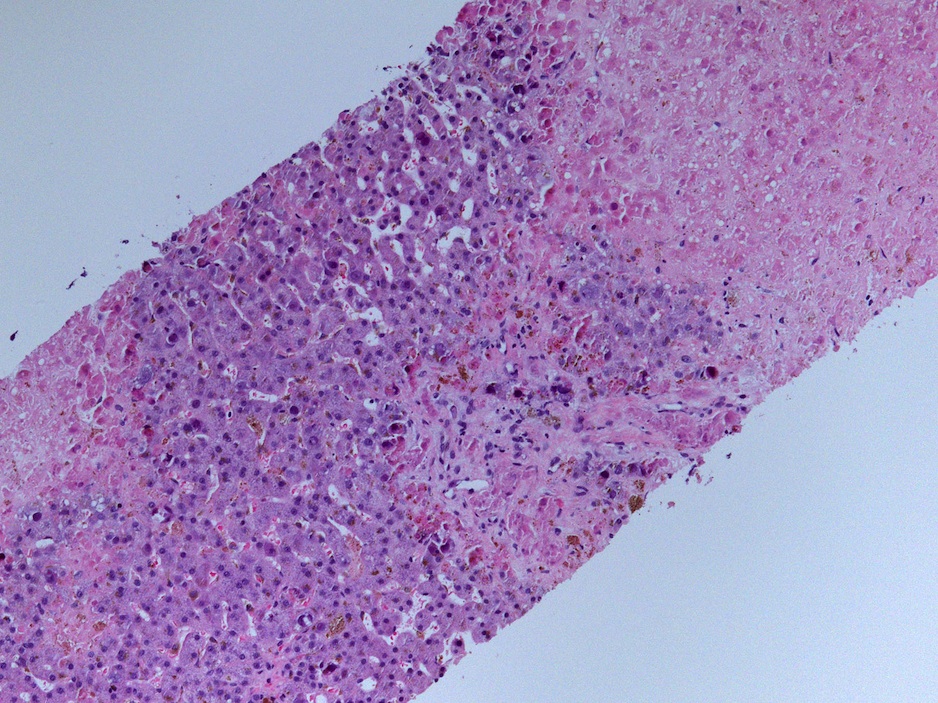
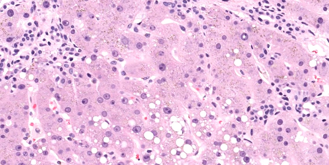
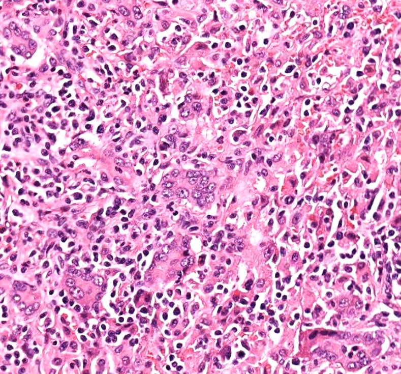
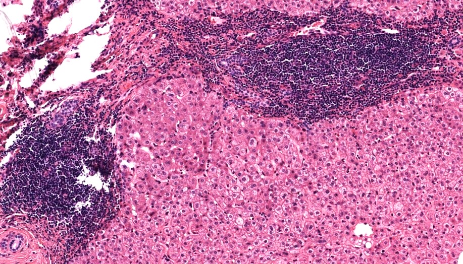
- Focal to massive coagulative necrosis in liver without particular zonal distribution (Am J Surg Pathol 2017;41:810)
- Intranuclear viral inclusions within hepatocytes with smudged or glassy appearance (Am J Surg Pathol 2017;41:810)
- Viral inclusions within the biliary epithelium (Am J Surg Pathol 2017;41:810)
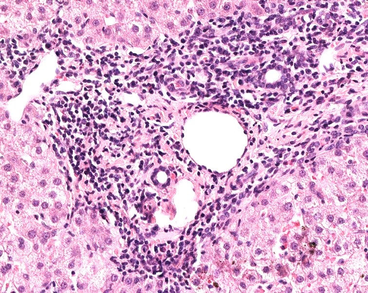
- Mild or absent inflammation, portal tract granulomas may be present (Am J Surg Pathol 2017;41:810)
- Immunostaining for adenovirus demonstrates strong nuclear staining and is used for confirmation of the diagnosis because immunocompromised patients can have herpes simplex, cytomegalovirus and other viral infections
Microscopic (histologic) images
Contributed by Vladyslav Ilchenko, M.D. and Case #354
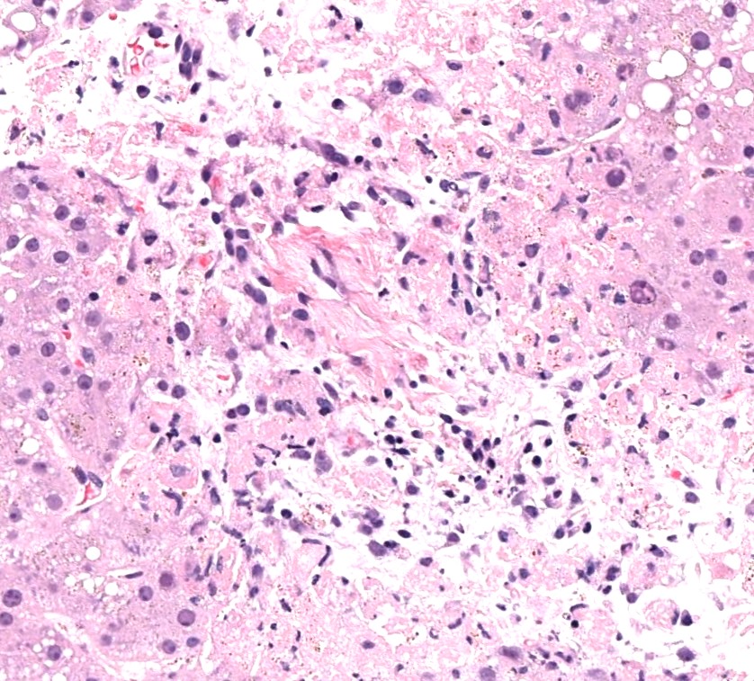
Coagulative necrosis
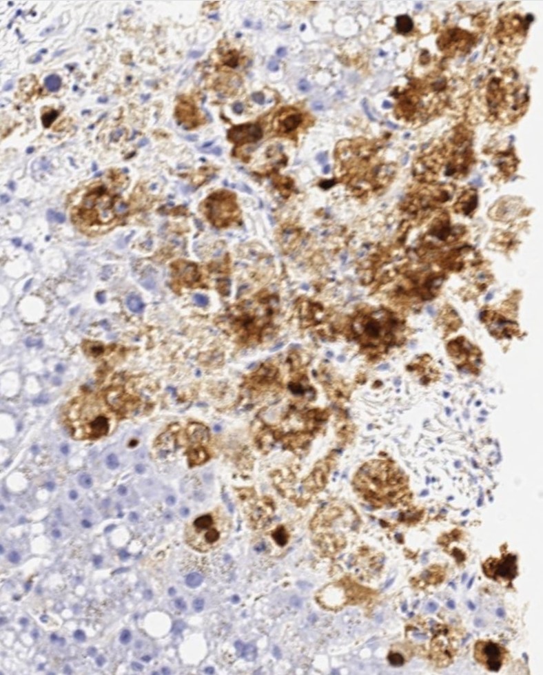
Adenovirus IHC
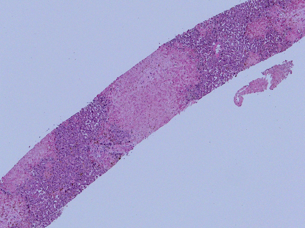
Large necrotic focus
Nuclear inclusion bodies
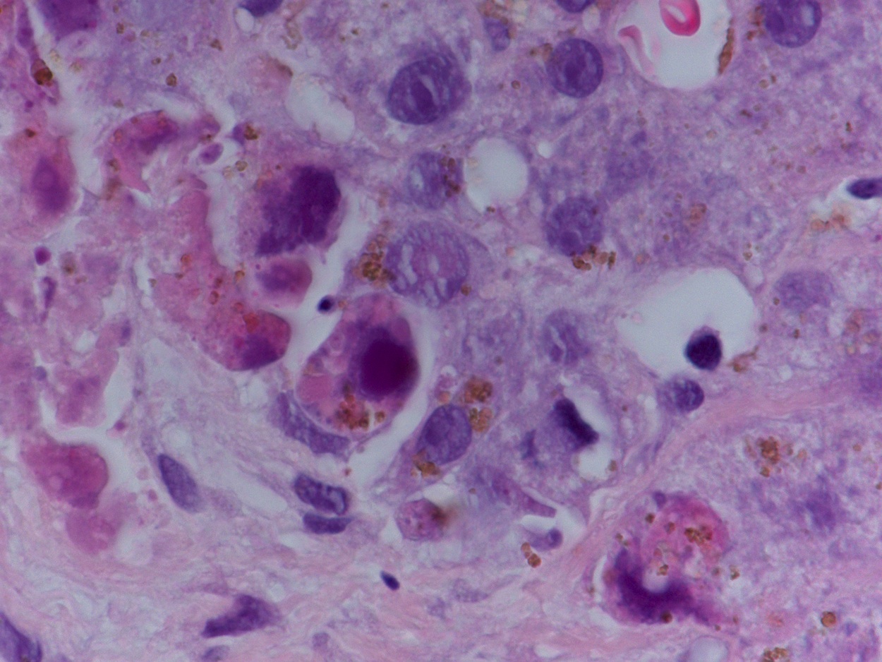
Nuclear and cytoplasmic viral inclusion bodies
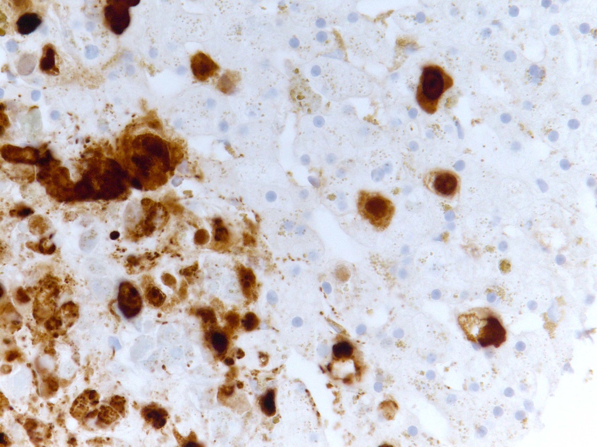
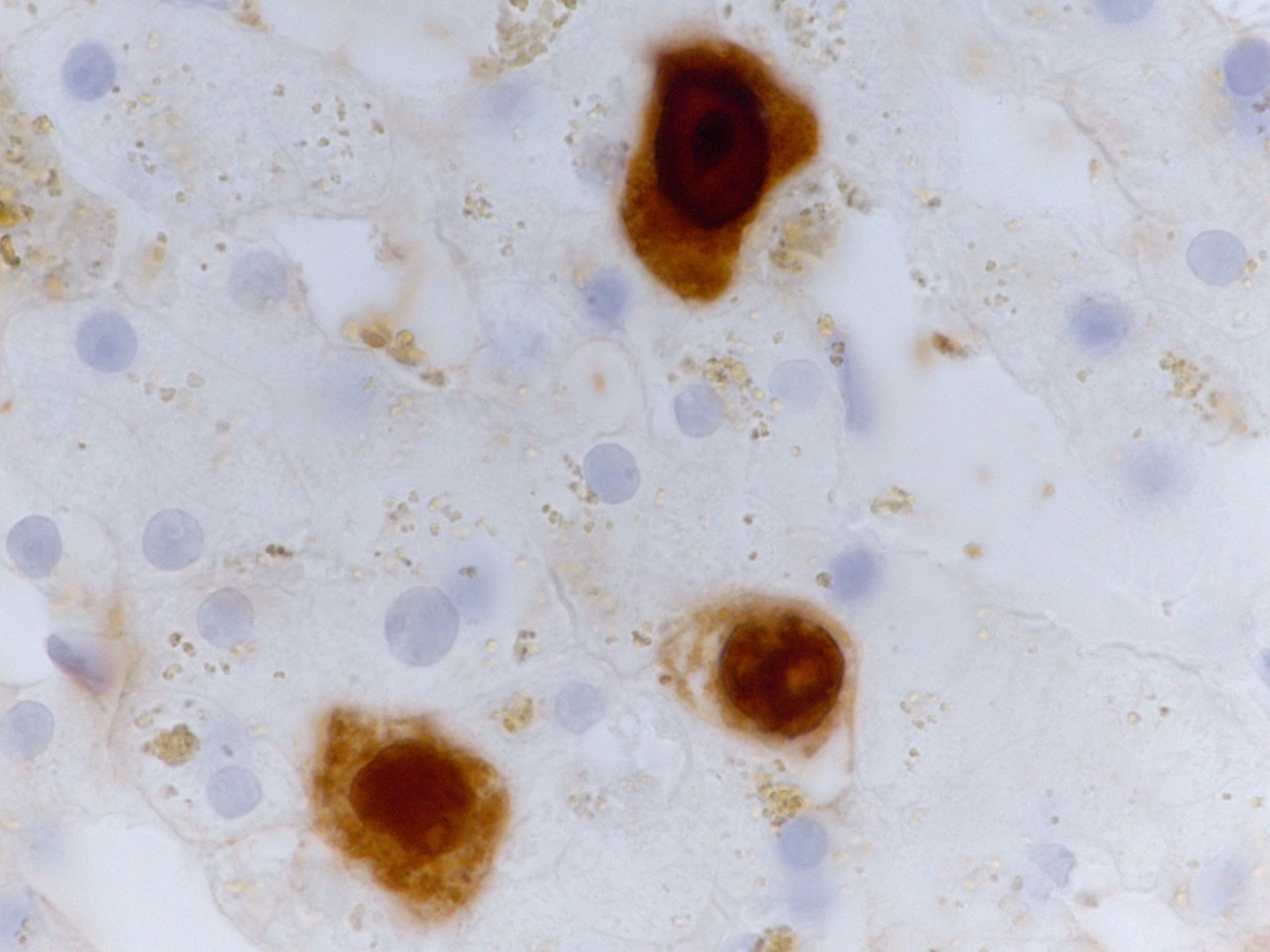
Adenovirus immunostain
Positive stains
- Adenovirus immunohistochemistry (nuclear staining)
Negative stains
Sample pathology report
- Liver, biopsy:
- Hepatitic injury pattern with focal coagulative necrosis, consistent with adenovirus hepatitis (positive adenovirus immunohistochemistry); negative for portal fibrosis (see comment)
- Comment: The patient's clinical history of stem cell transplantation for acute myeloid leukemia and positive serology for adenovirus is noted.
Differential diagnosis
- Cytomegalovirus hepatitis:
- Neutrophilic microabscesses may be present
- Characteristic intranuclear / cytoplasmic inclusions for CMV
- Positive CMV immunostain, both intranuclear and cytoplasmic
- Herpes simplex virus hepatitis:
- Hemorrhagic necrosis may be present
- Multinucleation of hepatocyte nuclei
- Positive immunostain is helpful
- Varicella zoster virus hepatitis:
- Histologic features are nonspecific
- Immunostain and PCR can be helpful
- Drug induced liver injury:
- May show cholestatic injury pattern
- Lacks viral inclusions
Board review style question #1
Which of the following denotes classical morphological findings in the liver biopsy from a patient with adenovirus hepatitis?
- Hepatocytes with enlarged, smudged nuclei and intranuclear inclusion bodies
- Lymphoid aggregates in portal tracts, epithelial damage of small bile ducts
- Neutrophilic microabscess
- Presence of multinucleated giant cells
- Prominent mononuclear infiltrate within portal tracts and sinusoids
Board review style answer #1
A. Hepatocytes with enlarged, smudged nuclei and intranuclear inclusion bodies
Comment Here
Reference: Adenovirus hepatitis
Comment Here
Reference: Adenovirus hepatitis
Board review style question #2
What immunohistostaining pattern is typical for liver cells in adenovirus hepatitis?
- Cytoplasmic inclusions staining
- Diffuse staining of cytoplasm
- Partial or complete membrane staining
- Strong nuclear staining
Board review style answer #2
