

Kidney tumor
Childhood tumors
Rhabdoid tumor
Editorial Board Member: Bonnie Choy, M.D.
Deputy Editor-in-Chief: Maria Tretiakova, M.D., Ph.D.


Last author update: 11 November 2022
Last staff update: 11 November 2022
Copyright: 2003-2024, PathologyOutlines.com, Inc.
PubMed Search: Rhabdoid tumor kidney
Table of Contents
Definition / general | Essential features | Terminology | ICD coding | Epidemiology | Sites | Pathophysiology | Etiology | Clinical features | Diagnosis | Prognostic factors | Case reports | Treatment | Gross description | Gross images | Microscopic (histologic) description | Microscopic (histologic) images | Cytology description | Positive stains | Negative stains | Electron microscopy description | Molecular / cytogenetics description | Sample pathology report | Differential diagnosis | Additional references | Board review style question #1 | Board review style answer #1 | Board review style question #2 | Board review style answer #2Cite this page: D'Hooghe E, Vujanic GM. Rhabdoid tumor. PathologyOutlines.com website. https://www.pathologyoutlines.com/topic/kidneytumorrhabdoid.html. Accessed April 25th, 2024.
Definition / general
- Highly malignant undifferentiated tumor composed of noncohesive cells with large, eccentric nuclei, prominent nucleoli and intracytoplasmic inclusions; characterized by loss of INI1 immunostaining and SMARCB1 genetic abnormalities (in ~95% of cases)
- The most aggressive renal tumor of young age
Essential features
- Noncohesive cells with large, eccentric nuclei and prominent nucleoli
- Loss of INI1 (~95% of cases) or BRG1 (~5% of cases) immunostaining
- Young age (< 2 years, rarely > 5 years)
Terminology
- Other terms: malignant rhabdoid tumor, atypical teratoid rhabdoid tumor, rhabdoid tumor predisposition syndrome
ICD coding
- ICD-O: 8963/3 - malignant rhabdoid tumor
- ICD-11: 2C90.Y & XH3RF3 - other specific malignant neoplasms of kidney, except renal pelvis & rhabdoid tumor, NOS
Epidemiology
- 1 - 2% of childhood renal tumors
- Median age at presentation is ~1 year (Pediatr Blood Cancer 2018;65:e26746, Pediatr Blood Cancer 2011;56:733)
- 80% diagnosed before the age of 2 years
- Can be congenital
- Exceptionally rare > 5 years of age
- ~15% are associated with atypical teratoid rhabdoid tumor in the brain
- M:F = 1.5:1
Sites
- Kidney
- Extrarenal rhabdoid tumors share the same histologic, immunohistochemical and molecular features
Pathophysiology
- The majority (~95%) of rhabdoid tumors of kidney have genetic changes resulting in the loss of function of SMARCB1, a tumor suppressor gene encoding BAF47 (INI1), which is a core member of the BAF chromatin remodeling complex
- The few remaining cases show a loss of function of the SMARCA4 gene, which encodes BRG1, the helicase / ATPase protein of the BAF complex (Pediatr Dev Pathol 2018;21:6)
Etiology
- Predisposition factor: rhabdoid tumor predisposition syndromes (RTPS)
- Germline inactivation of one allele of a gene occurs
- If it occurs in the SMARCB1 gene: RTPS1
- If it occurs in the SMARCA4 gene: RTPS2 (Fam Cancer 2021;20:305)
- Children with RTPS develop tumors at a younger age
- Not clear whether germline mutation influences prognosis
Clinical features
- Usually presents as an abdominal mass or hematuria
- In ~20% of patients it is associated with hypercalcemia
- Rarely stage 1 (< 10%); bilateral presentation rare
- Metastases occur in ~80% of patients, mainly in the lungs, liver and brain
- Associated with rhabdoid tumor predisposition syndromes (RTPS1 and RTPS2) in ~30% of patients (Fam Cancer 2021;20:305)
Diagnosis
- Imaging features not diagnostic (J Pediatr Surg 2008;43:1301)
- Diagnosed as a renal mass but if distant metastases detected, should be suspected
- Biopsy (with INI1 immunostain) diagnostic
Prognostic factors
- Very aggressive tumor with poor prognosis
- Widespread, hematogeneous and lymphatic metastases to bone, brain and other sites
- > 75% of patients die within 1 year, ~90% within 2 years of diagnosis
- Age at diagnosis is a highly significant factor: infants < 6 months of age have a very poor prognosis (an 8.8% 4 year survival)
- Survival for children ≥ 2 years of age is 41.1% (J Clin Oncol 2005;23:7641, Pediatr Blood Cancer 2011;56:733)
Case reports
- 28 week gestation fetus with INI1 negative sarcoma diagnosed as malignant rhabdoid tumor, presented as hydrops fetalis metastatic to the placenta (J Matern Fetal Neonatal Med 2021;34:3790)
- 8 month old girl presented with malignant rhabdoid tumor of the kidney and spine (J Korean Neurosurg Soc 2014;55:57)
- 3 year old boy presented with WT1 positive renal malignant tumor and loss of INI1 expression (Transl Androl Urol 2020;9:2275)
- 5 year old girl presented with malignant rhabdoid tumor of the kidney and multicystic dysplasia (J Korean Med Sci 2010;25:785)
- 11 year old boy presented with rhabdoid tumor predisposition syndrome with renal tumor 10 years after brain tumor (Pathol Int 2021;71:155)
Treatment
- No established standard treatment
- Most patients are treated with intensive multimodal regimens; surgical resection, intensive multidrug chemotherapy regimens and local radiotherapy or high dose chemotherapy followed by autologous stem cell rescue (Cancer Manag Res 2022;14:479)
Gross description
- Unilateral and unicentric tumor
- Rare bilateral cases
- Sharply demarcated from the renal parenchyma
- Necrotic areas may be seen (Histopathology 1996;28:333)
Gross images
Contributed by Ellen D’Hooghe, M.D. and Gordan M. Vujanic, M.D., Ph.D.
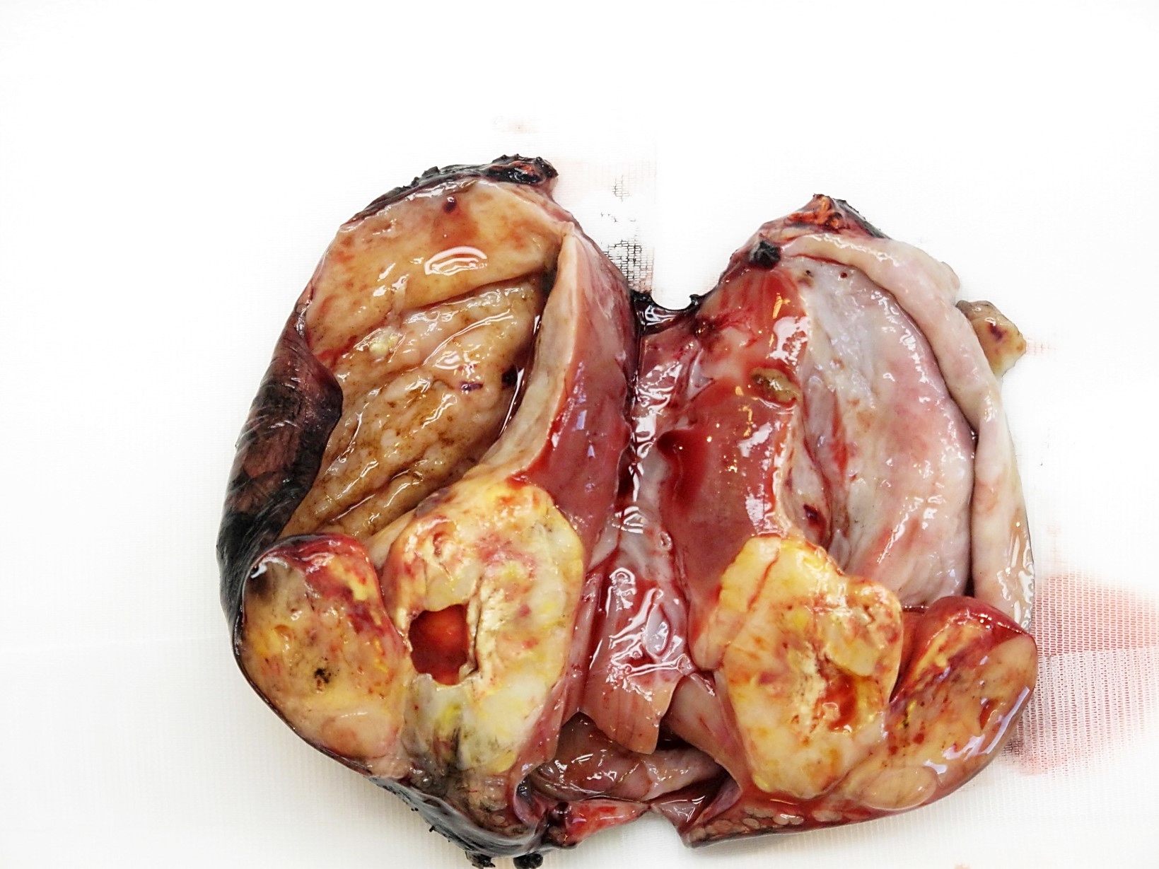
Solid, fleshy tumor
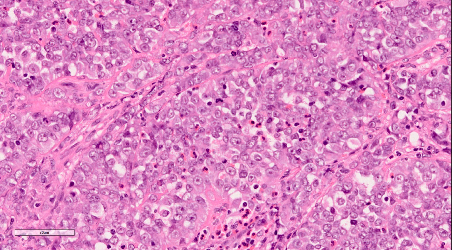
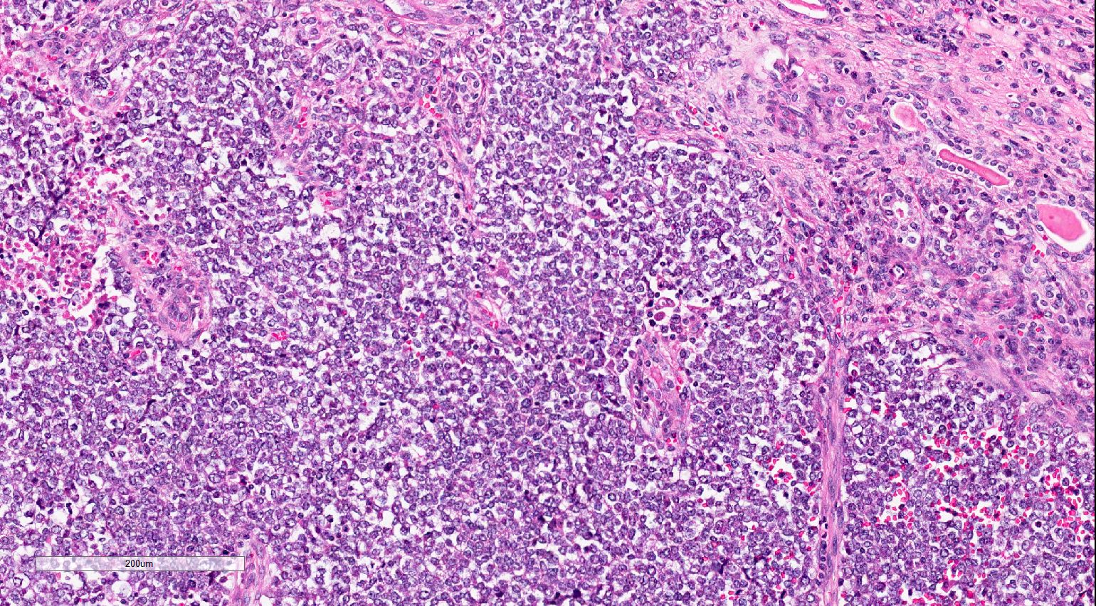
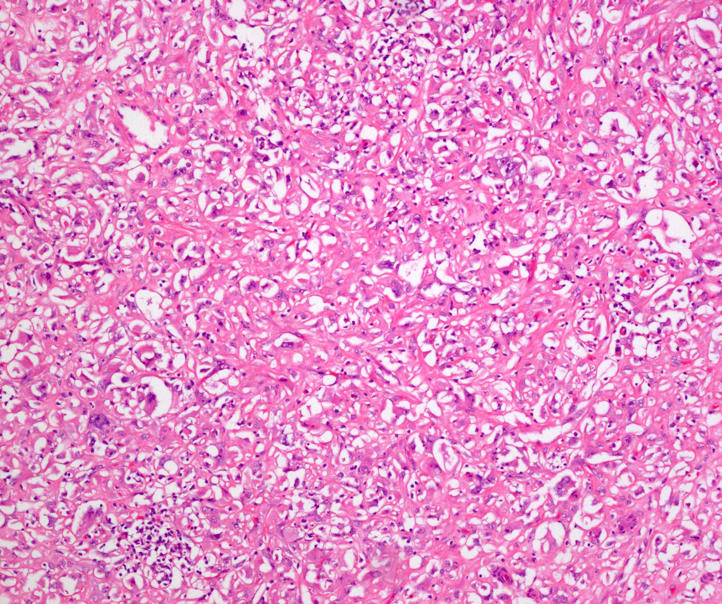
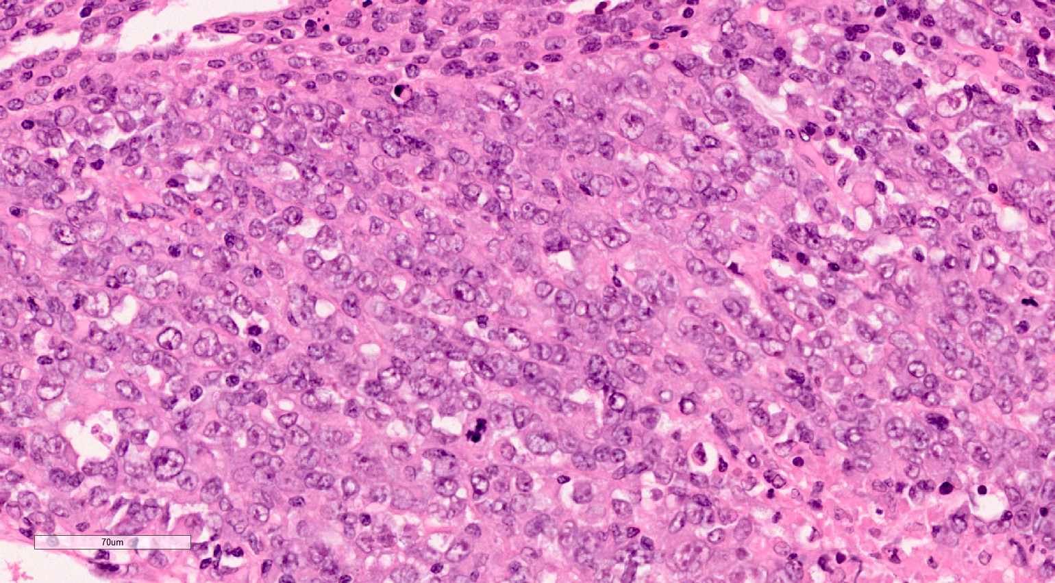
Microscopic (histologic) description
- Classical, diffuse pattern: sheets of noncohesive, monotonous looking tumor cells
- Tumor cells have abundant eosinophilic cytoplasm which may contain hyaline inclusions (in ~20% of cases)
- Nuclei are large, vesicular and contain 1 - 2 prominent nucleoli
- High mitotic activity; mild to moderate cellular pleomorphism
- Rhabdoid tumors of kidney may exhibit other patterns too: sclerosing, myxoid, epithelioid, spindled, clear cell sarcoma-like, microcystic, solid lymphomatoid (Am J Surg Pathol 1989;13:439, Histopathology 1996;28:333)
- Different patterns may be present in the same tumor
- Extremely invasive; the lymphatics and blood vessels of the adjacent parenchyma are usually stuffed with tumor cells
- Staging criteria for rhabdoid tumor of the kidney are the same criteria as nephroblastoma (Histopathology 2022;80:1026)
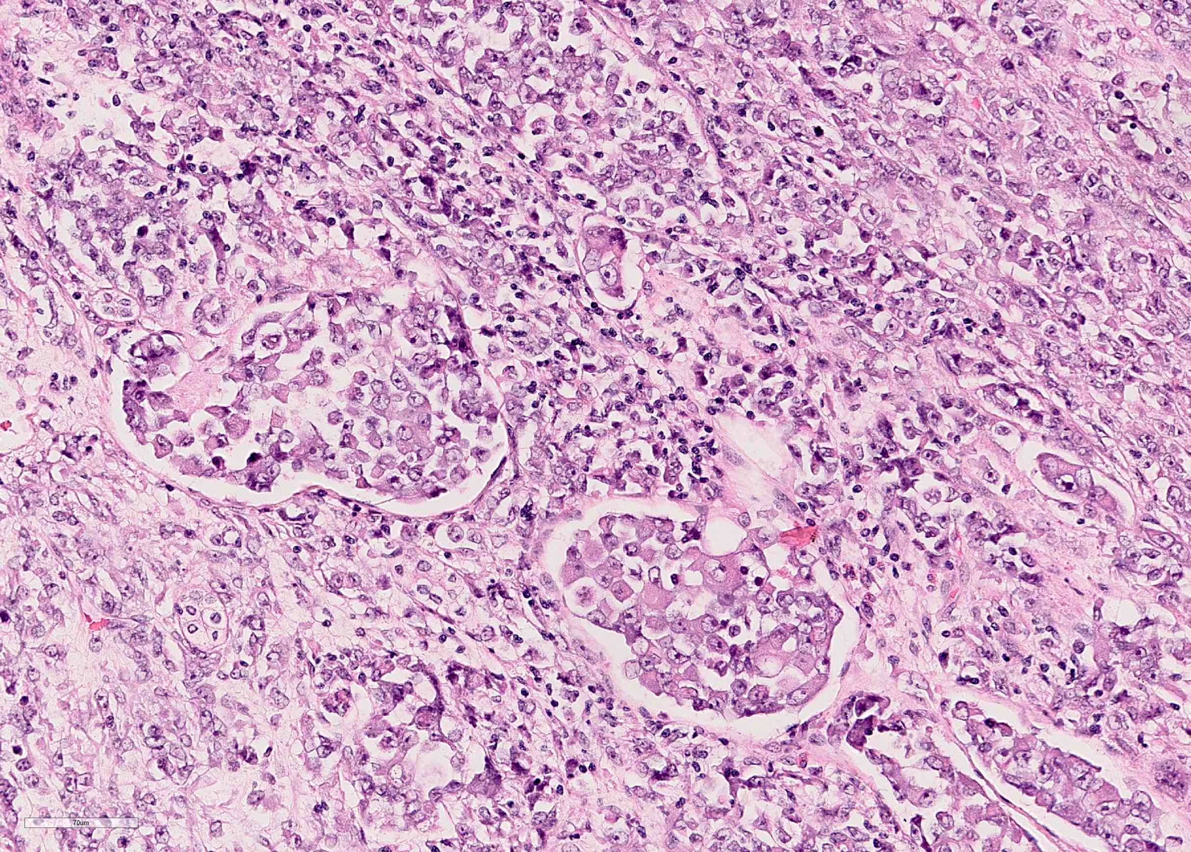
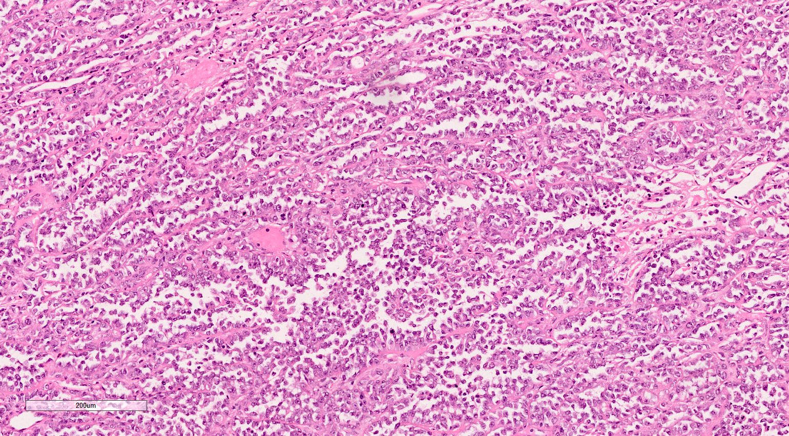
Microscopic (histologic) images
Contributed by Ellen D’Hooghe, M.D. and Gordan M. Vujanic, M.D., Ph.D.

Classic pattern
Classic pattern associated with eosinophils

Hypercellular tumor
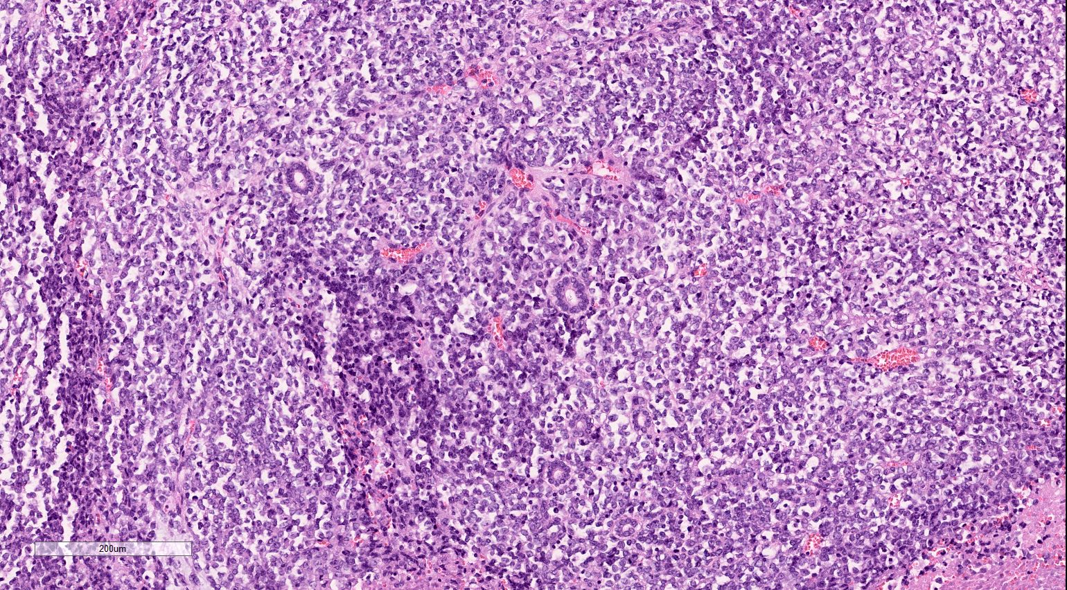
Entrapped normal renal tubules
Epithelioid pattern
Intracytoplasmic
inclusions
Lymphomatoid pattern
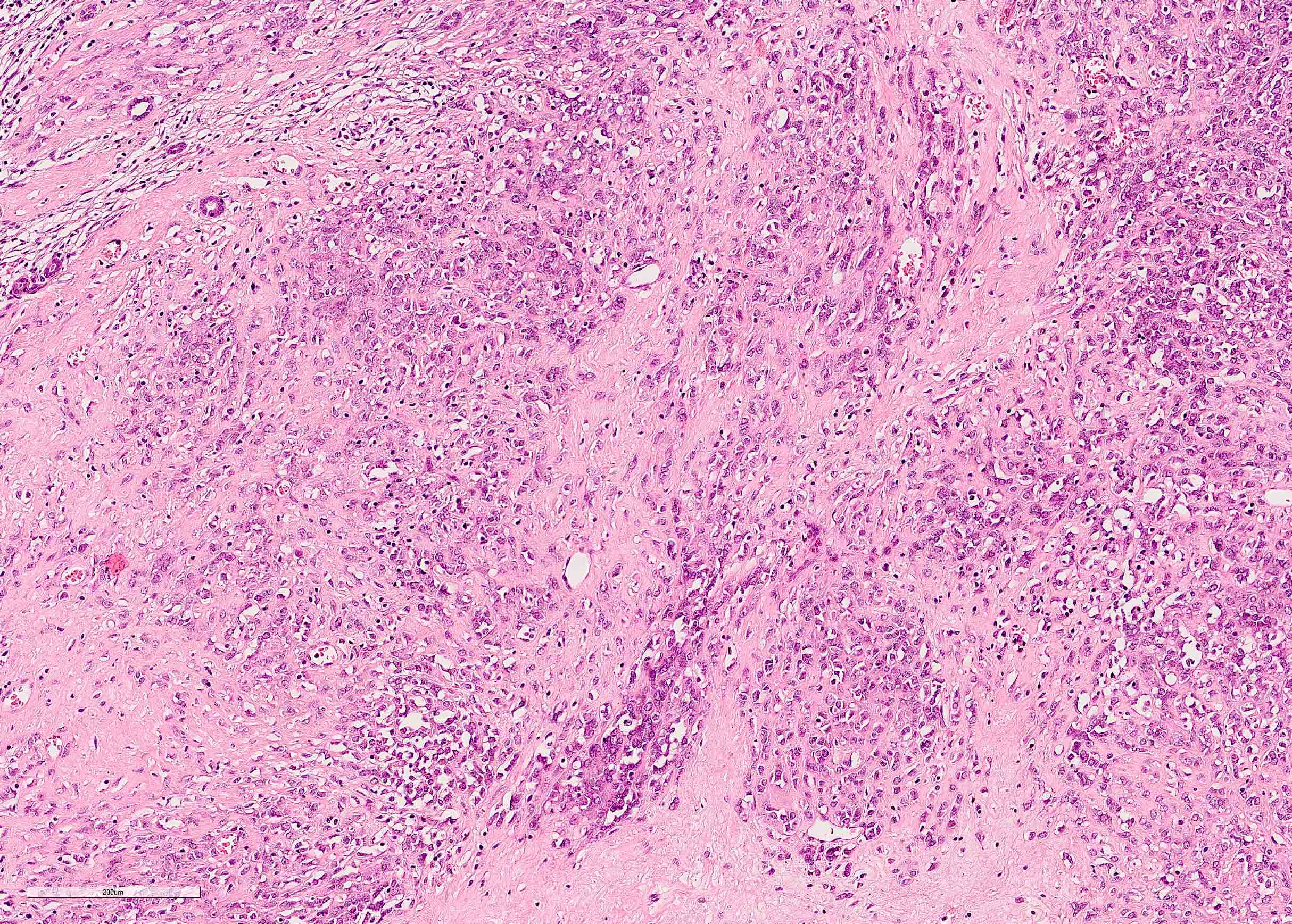
Sclerosing pattern
Prominent sclerosing pattern

Spindling growth pattern
Pseudoalveolar pattern
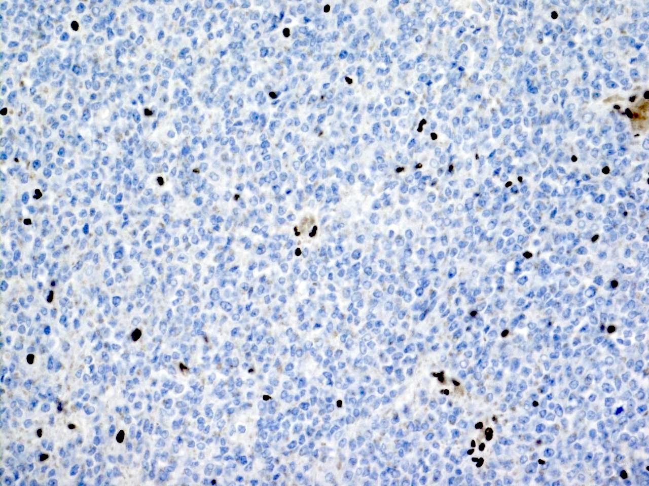
Loss of nuclear INI1
Cytology description
- Highly cellular with clusters and noncohesive tumor cells (Diagn Cytopathol 2021;49:711)
- Sometimes arranged in an alveolar fashion attached to fibrovascular stroma (Acta Cytol 2003;47:1055)
- Large cells with round to oval nuclei and often a single nucleolus
- Scant to moderate pale eccentric cytoplasm, vacuolation or intracytoplasmic inclusions / densities can be seen
- Mostly mononuclear cells but also some bi and multinuclear cells (Cancer Cytopathol 2011;119:49)
- Epithelioid cells but also spindle cells are described
- Atypical mitotic figures and necrosis might be present
- International Society of Paediatric Oncology (SIOP) and Children’s Oncology Group (COG) do not recommend the use of fine needle aspiration biopsy in diagnosis
Positive stains
- Vimentin (almost always)
- CD99
- Cytokeratin
- EMA
- Desmin
- Neurofilaments (frequent and patchy) (Cancers (Basel) 2020;12:729)
- WT1 can be positive (Acta Histochem 2013;115:70)
- PAX8 can be focally positive (Appl Immunohistochem Mol Morphol 2018;26:721)
Negative stains
- Loss of nuclear INI1 / SMARCB1 staining (Pathology 2008;40:664)
- Rarely, loss of nuclear BRG1 / SMARCA4 staining (Am J Hum Genet 2010;86:279)
- MyoD1
- Myogenin
Electron microscopy description
- Hyaline globules composed of whorls of intermediate filaments
Molecular / cytogenetics description
- Alterations in SMARCB1 (located on 22q11.2) include:
- Whole gene deletion (~22%)
- Intragenic duplication and deletions (~24%)
- Nonsense mutations (~24%)
- Frameshift mutations (~20%)
- Rare SMARCB1 wild type tumors have SMARCA4 loss instead (Pediatr Dev Pathol 2018;21:6)
- ~33% of patients with a newly diagnosed rhabdoid tumor of kidney have a germline mutation in SMARCB1
- Missense mutations are virtually absent
- Splice mutations are uncommon
- Otherwise, a small number of somatic mutations
Sample pathology report
- Kidney, total nephrectomy, stage 2:
- Rhabdoid tumor (see comment)
- Comment: Tumor shows diffuse growth of poorly cohesive cells, which have large nuclei with prominent nucleoli. Some cells show eosinophilic, intracytoplasmic inclusions. Tumor is infiltrating the renal sinus and the perirenal fat. Immunohistochemistry for INI1 shows a loss of nuclear staining in the tumor cells.
Differential diagnosis
- Renal medullary carcinoma:
- INI1 not retained (as in rhabdoid tumors of kidney)
- Older age, associated with sickle cell disorder, reticular or cribriform pattern
- Ewing sarcoma:
- Wilms tumor:
- INI1 is retained
- Usually other component present (epithelial and stromal)
- Collecting duct carcinoma:
Additional references
Board review style question #1
What is the diagnostic immunostaining for this entity?
- BCOR
- INI1
- PAX8
- WT1
Board review style answer #1
Board review style question #2
Which of the following morphological features is characteristic for rhabdoid tumor?
- Anaplasia
- Cells with large nuclei and prominent nucleoli
- Epithelial structures
- Heterologous muscle differentiation
Board review style answer #2
