Page views in 2023: 17,634
Page views in 2024 to date: 6,433
Cite this page: Zynger D. Staging. PathologyOutlines.com website. https://www.pathologyoutlines.com/topic/kidneytumormalignantstaging.html. Accessed April 24th, 2024.
Definition / general
- All carcinomas of the renal cortex are covered by this staging system
- These topics are not covered: urothelial carcinoma, lymphoma, sarcoma and Wilms tumor
Essential features
- AJCC 7th edition staging was sunset on December 31, 2017; as of January 1, 2018, use of the 8th edition is mandatory
Primary Tumor (pT)
- pTX: primary tumor cannot be assessed
- pT0: no evidence of primary tumor
- pT1a: ≤ 4 cm, limited to the kidney
- pT1b: > 4 cm and ≤ 7 cm, limited to the kidney
- pT2a: > 7 cm and ≤ 10 cm, limited to the kidney
- pT2b: > 10 cm, limited to the kidney
- pT3a: invades renal vein / branches, perirenal fat, renal sinus fat or pelvicaliceal system
- pT3b: extends into vena cava below the diaphragm
- pT3c: extends into vena cava above the diaphragm or invades vena cava wall
- pT4: invades beyond Gerota fascia, including direct extension to adrenal gland
Regional lymph nodes (pN)
- pNX: cannot be assessed
- pN0: no regional lymph node metastasis
- pN1: regional lymph node metastasis
Notes:
- Regional lymph nodes=hilar, caval, aortic
AJCC prognostic stage groups
- Stage group I: T1a-1b N0 M0
- Stage group II: T2a-2b N0 M0
- Stage group III: T1a-3c N1 M0, T3a-3c NX-0 M0
- Stage group IV: T4 any N M0, any T any N M1
Registry data collection variables
- Histologic subtype
- WHO / ISUP grade
- Tumor size
- Perinephric fat invasion, renal sinus tissue invasion
- Venous involvement (specify renal vein, branches of renal vein, inferior vena cava)
- Lymphovascular invasion
- Adrenal gland involvement (specify direct extension pT4 or metastasis pM1)
- Presence and percentage of sarcomatoid features
- Presence of rhabdoid features
- Histologic tumoral necrosis
Histologic grade (G)
- GX: cannot be assessed
- G1: nucleoli inconspicuous or absent and basophilic at 40x objective
- G2: nucleoli conspicuous and eosinophilic at 40x but not prominent at 10x objective
- G3: nucleoli conspicuous and eosinophilic at 10x objective
- G4: marked nuclear pleomorphism, multinucleated tumor giant cells, rhabdoid differentiation or sarcomatoid differentiation
Histopathologic type
- Clear cell renal cell carcinoma
- Multilocular cystic renal neoplasm of low malignant potential
- Papillary renal cell carcinoma
- Hereditary leiomyomatosis renal cell carcinoma associated renal cell carcinoma/fumarate hydratase deficient renal cell carcinoma
- Clear cell papillary renal cell carcinoma
- Chromophobe renal cell carcinoma
- Collecting duct renal cell carcinoma
- Renal medullary carcinoma
- MiT family translocation renal cell carcinoma
- Succinate dehydrogenase deficient renal cell carcinoma
- Mucinous and tubular spindle cell carcinoma
- Tubulocystic renal cell carcinoma
- Acquired cystic disease associated renal cell carcinoma
- Renal cell carcinoma, unclassified
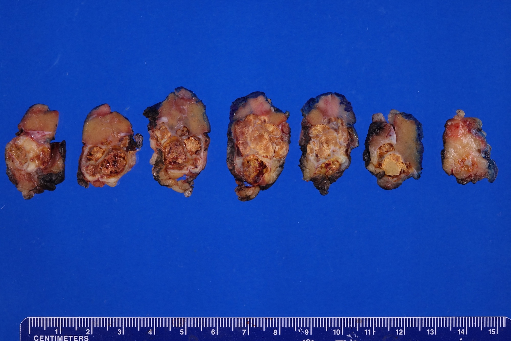
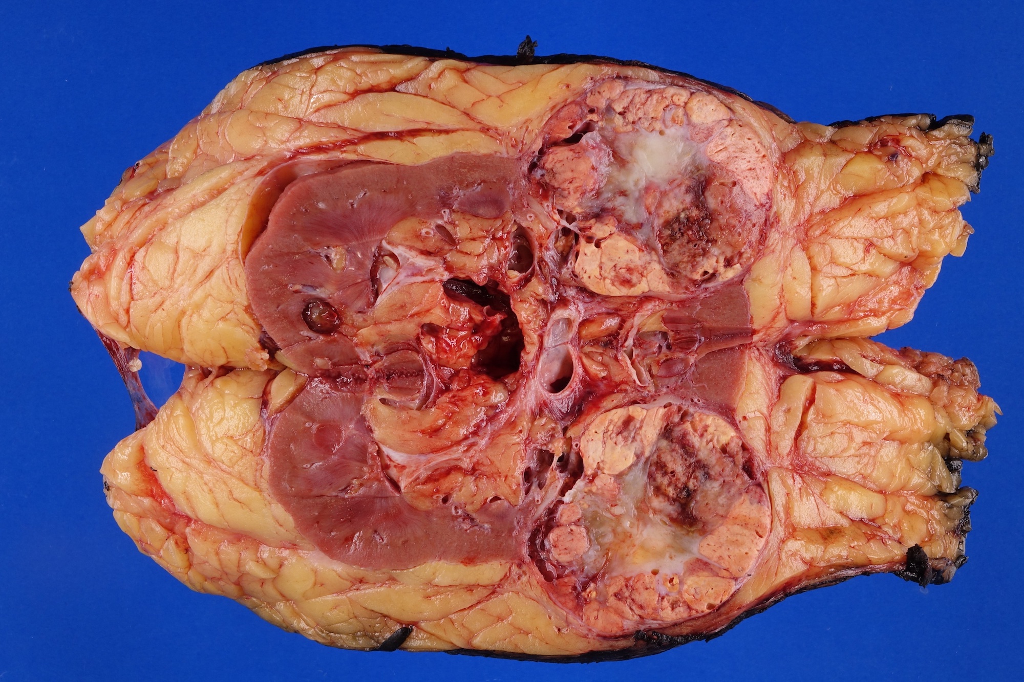
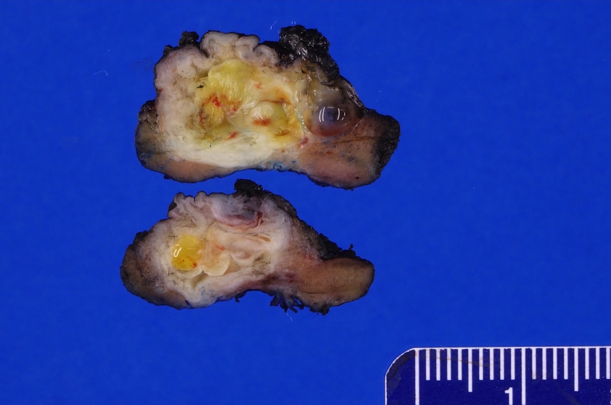
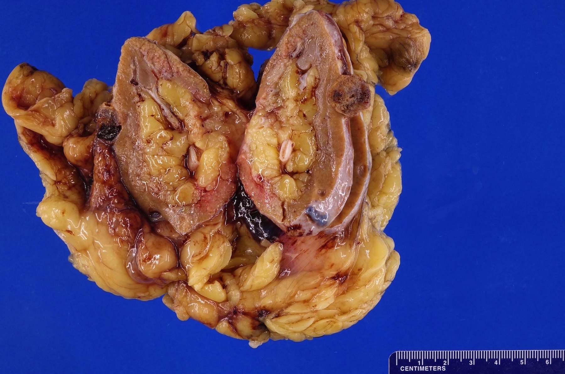
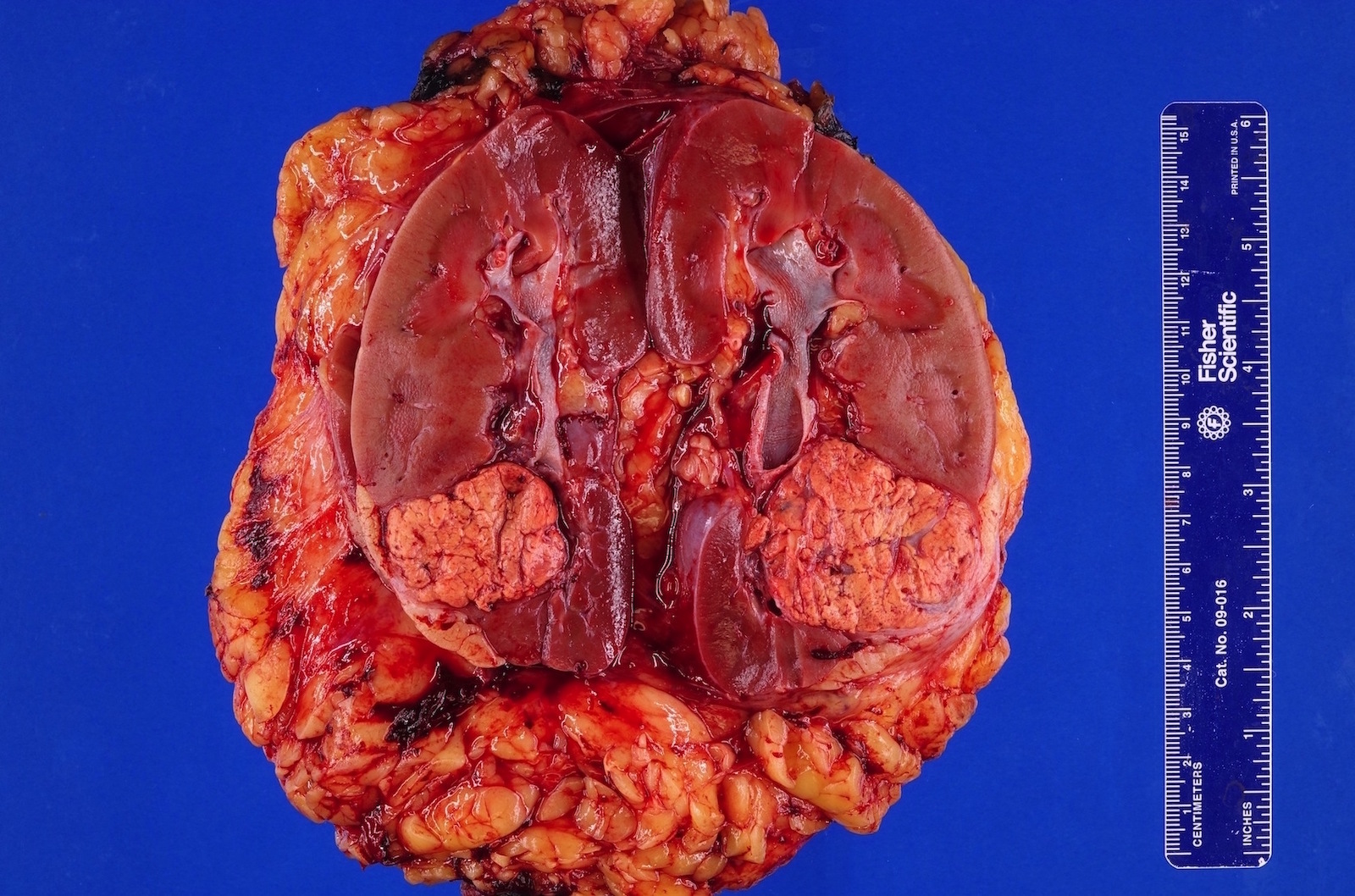
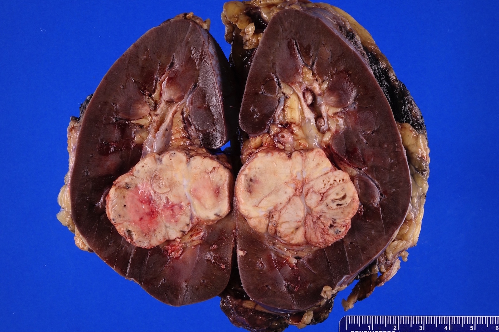
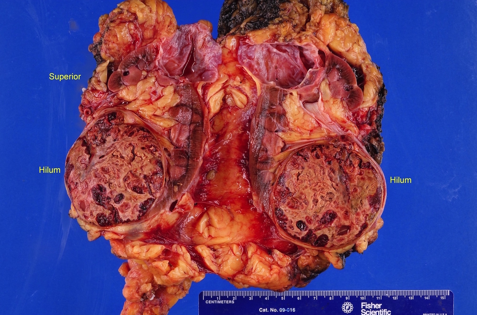
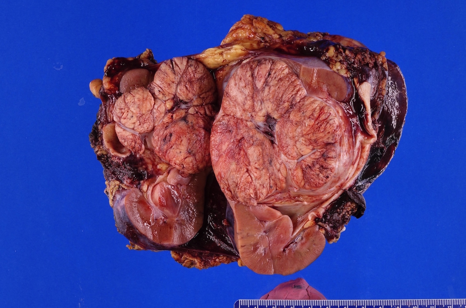
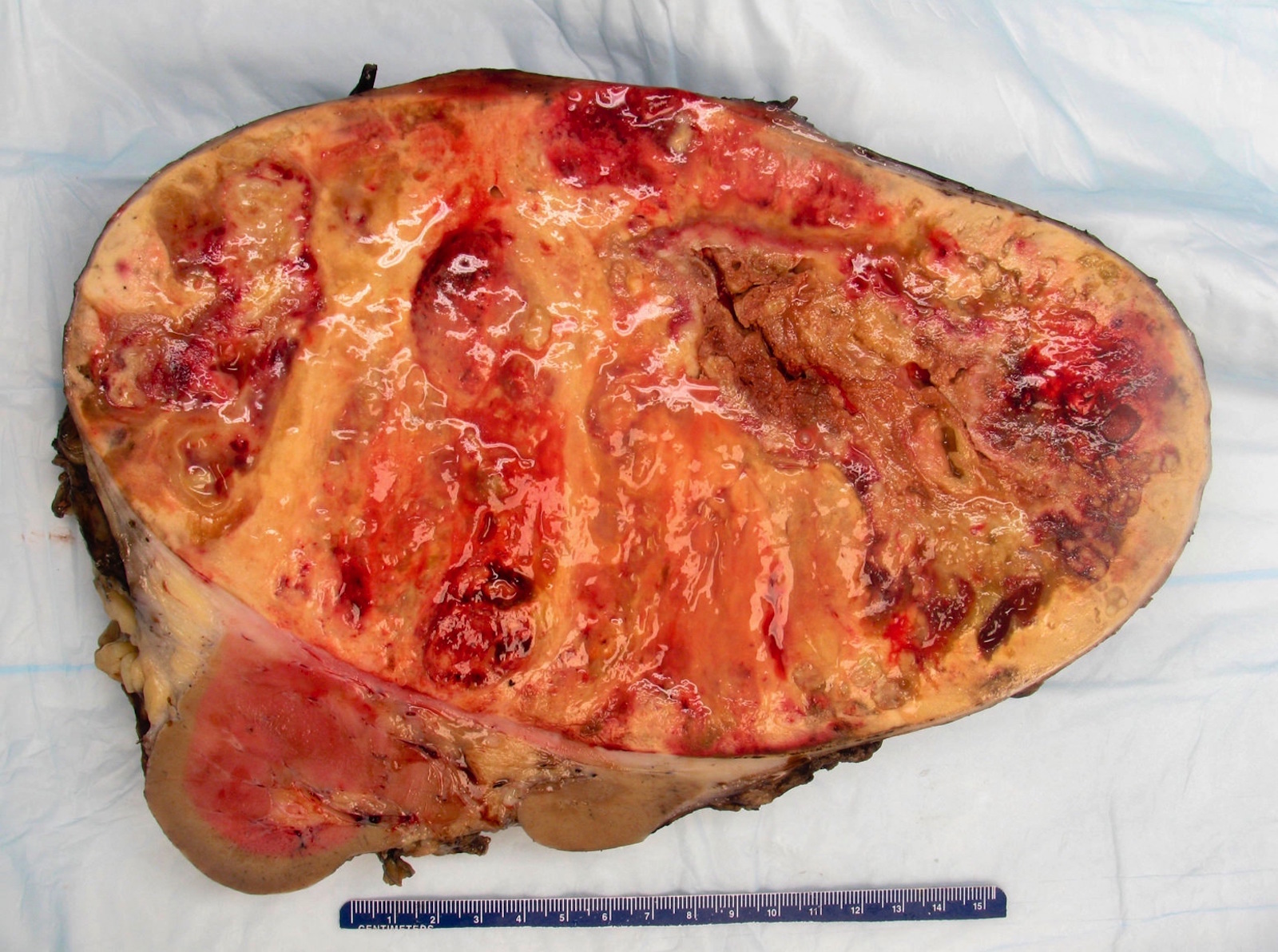
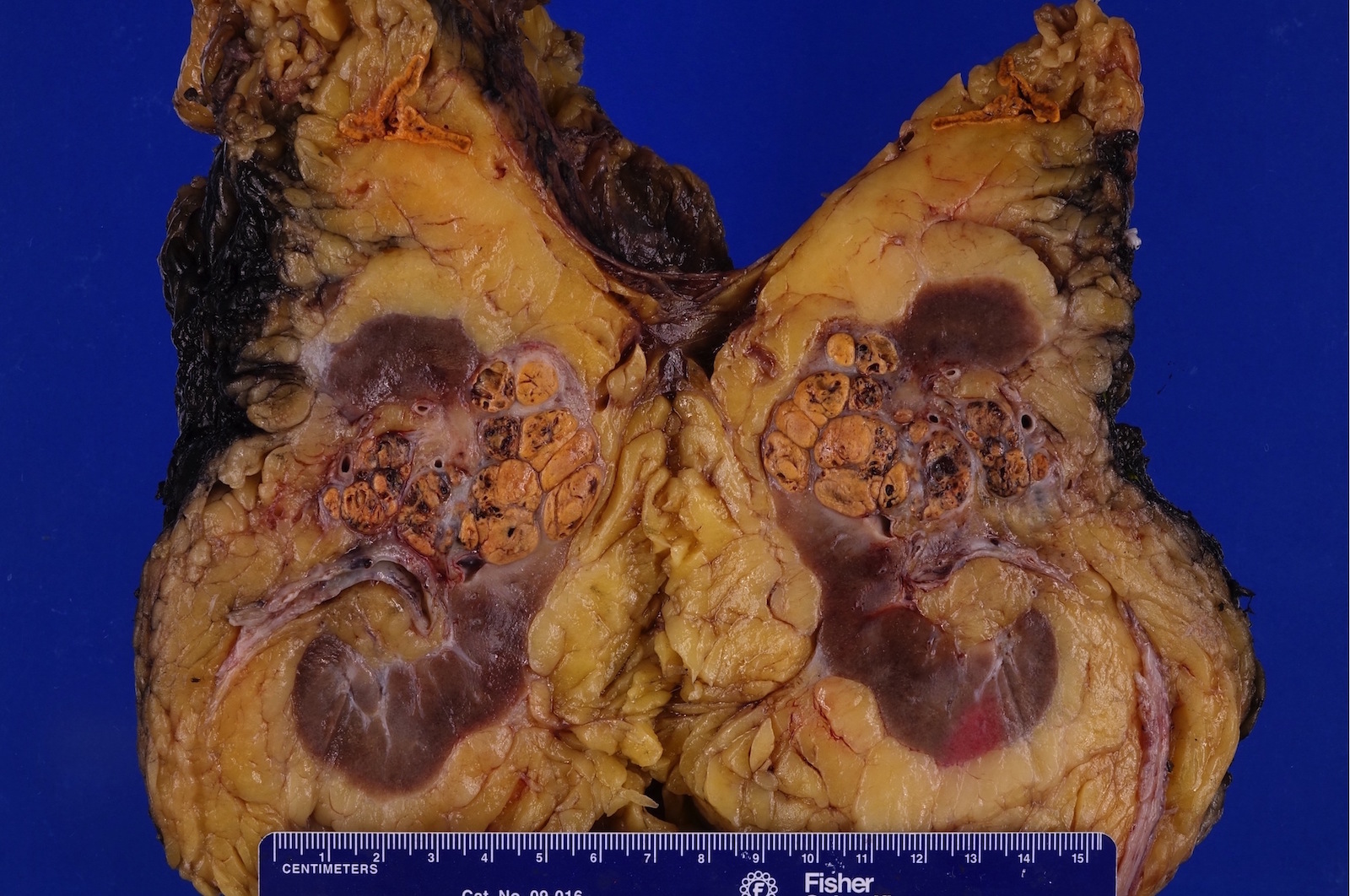
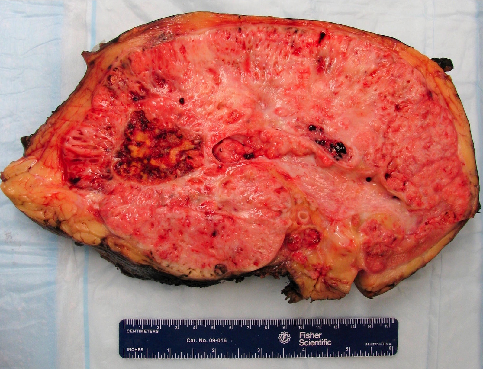
Gross images
Contributed by Debra Zynger, M.D.

Clear cell RCC (pT1a)
Clear cell papillary RCC (pT1a)
ACDA RCC (pT1a)
Clear cell RCC (pT1b)
Chromophobe RCC (pT1b)
Papillary type 2 RCC (pT2a)
Chromophobe RCC (pT2a)
Papillary RCC (pT2b)
Clear cell RCC (pT3a)
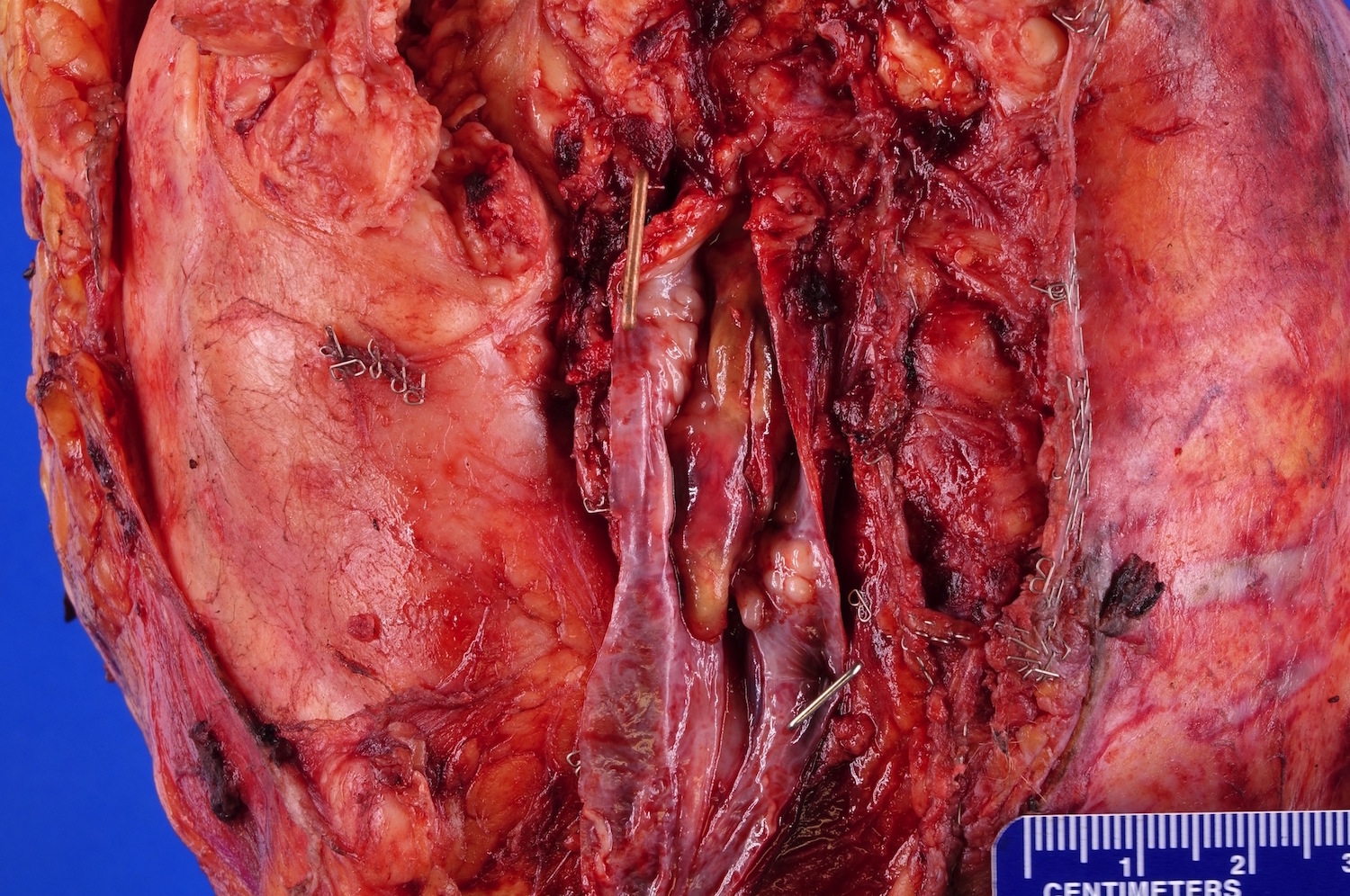
Clear cell RCC (pT3b)
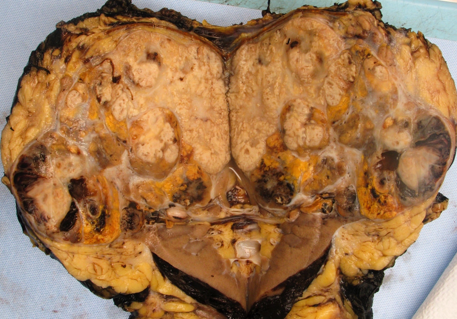
Clear cell RCC (pT4)
Frozen section images
Contributed by Debra L. Zynger, M.D.
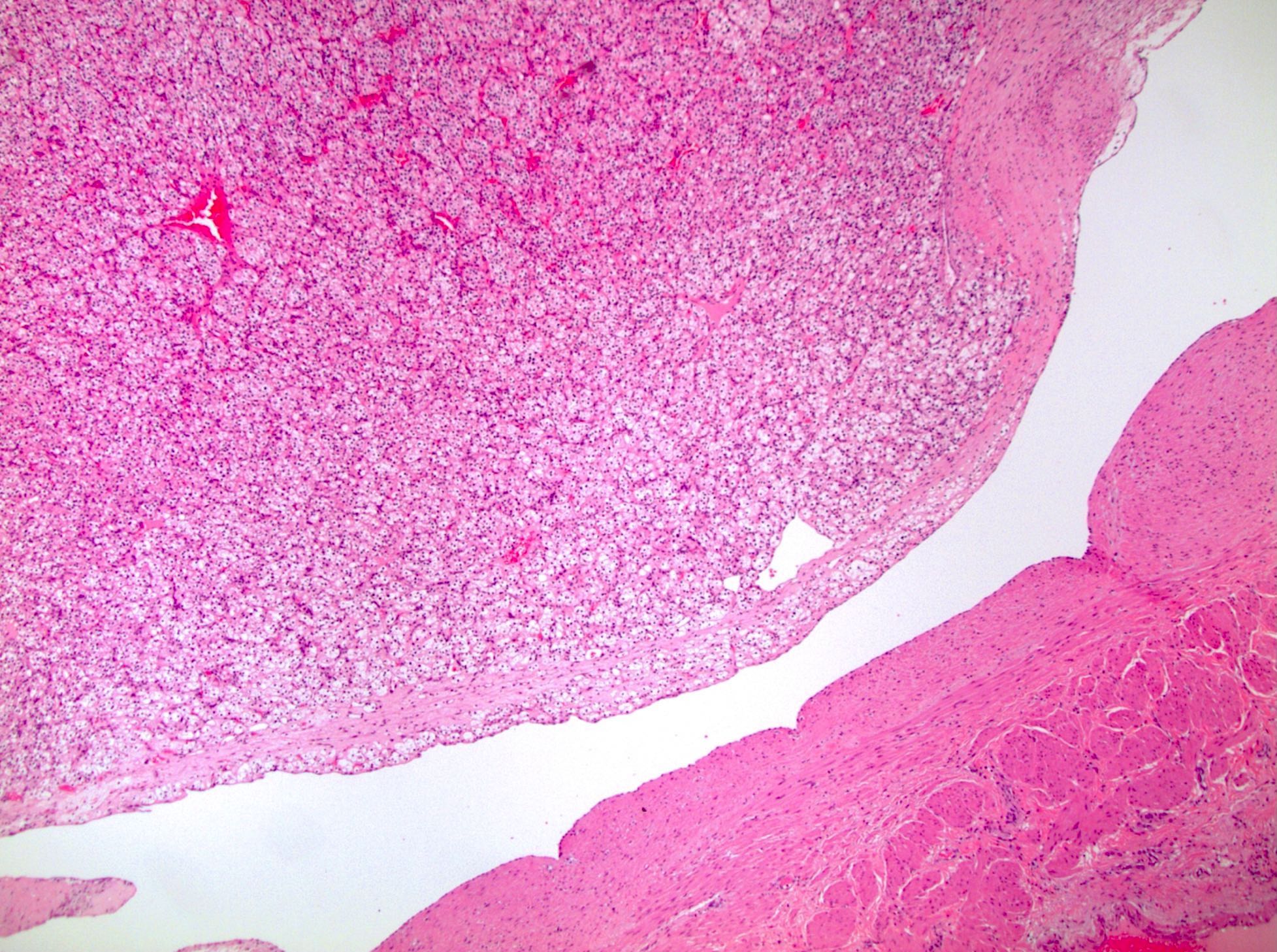
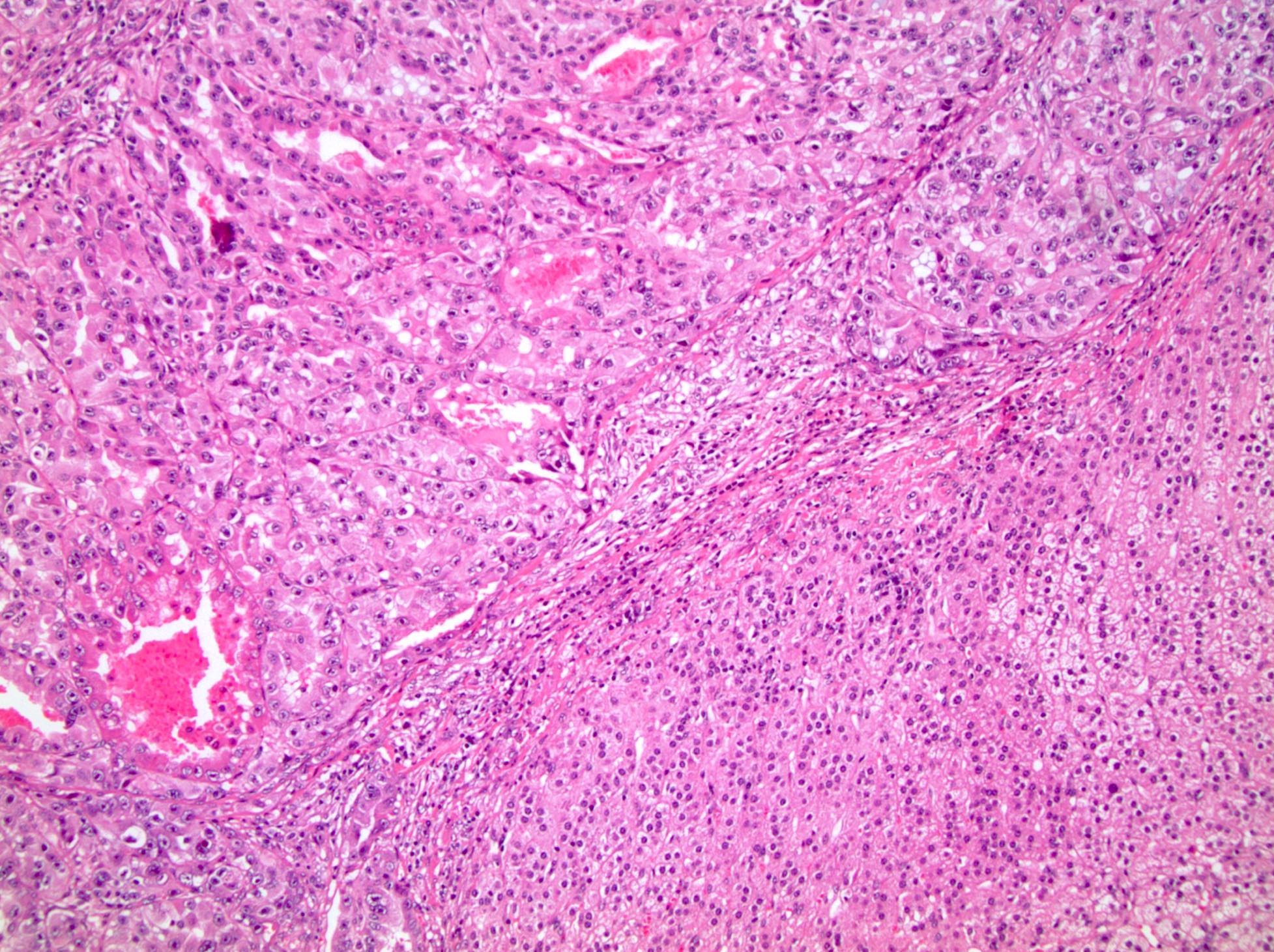
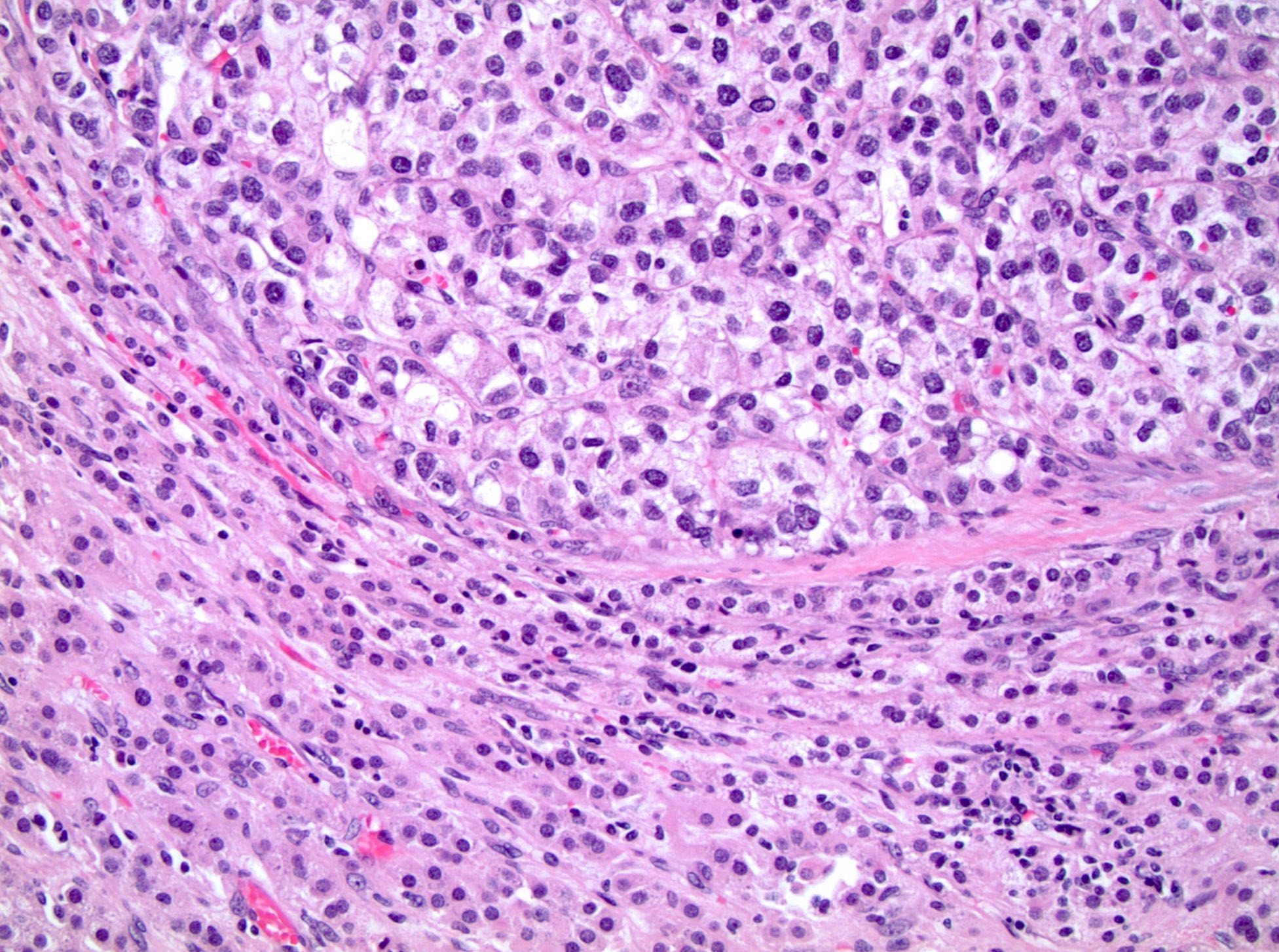
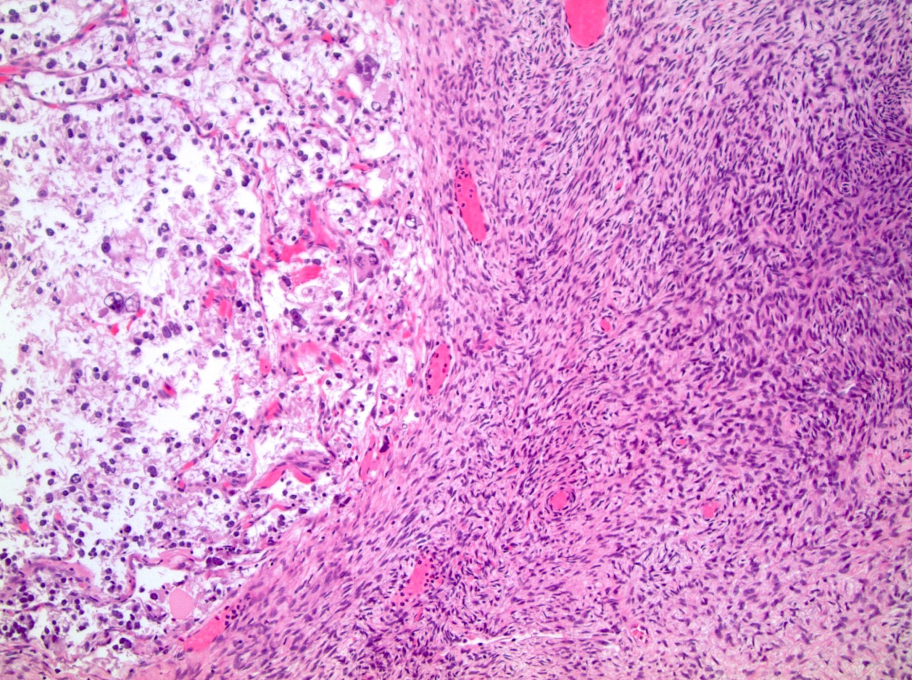
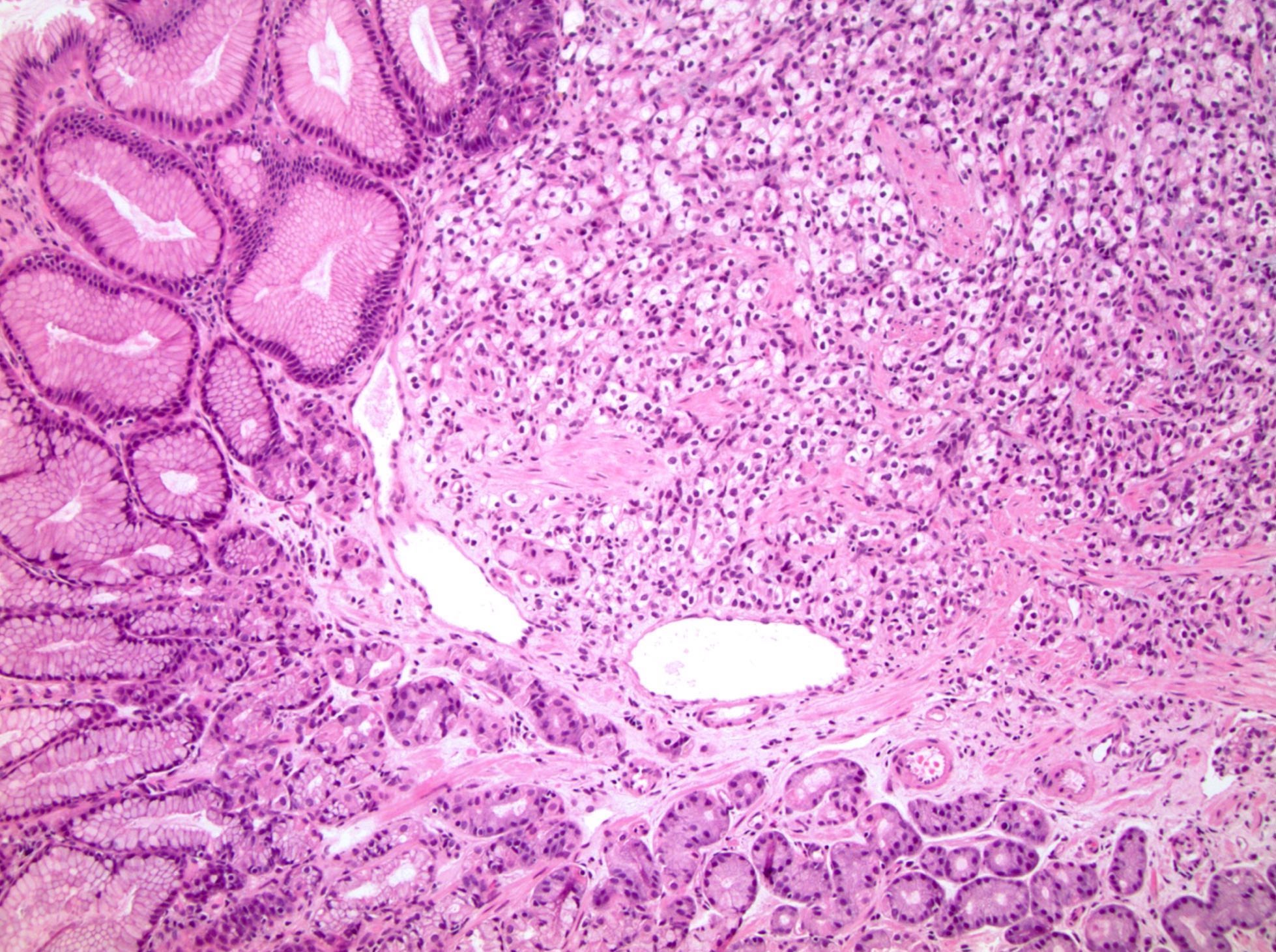
Microscopic (histologic) images
Contributed by Debra Zynger, M.D.

Renal sinus fat invasion (pT3a)
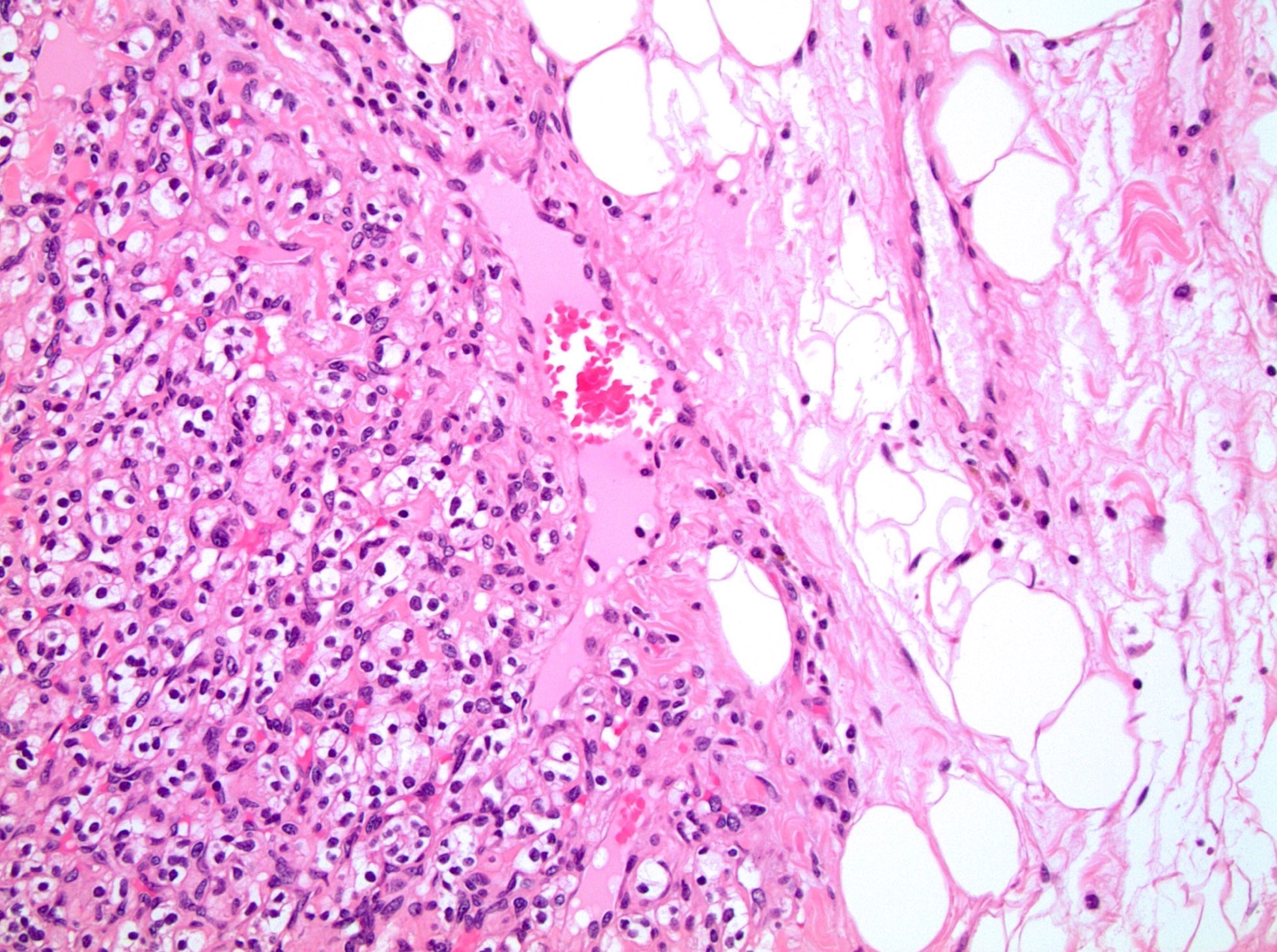
Perinephric fat invasion (pT3a)
Renal vein invasion (pT3a)
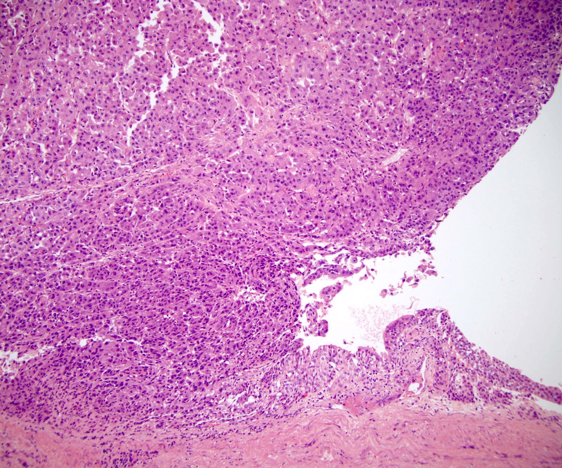
Pelvicaliceal invasion (pT3a)
Direct extension to adrenal (pT4)
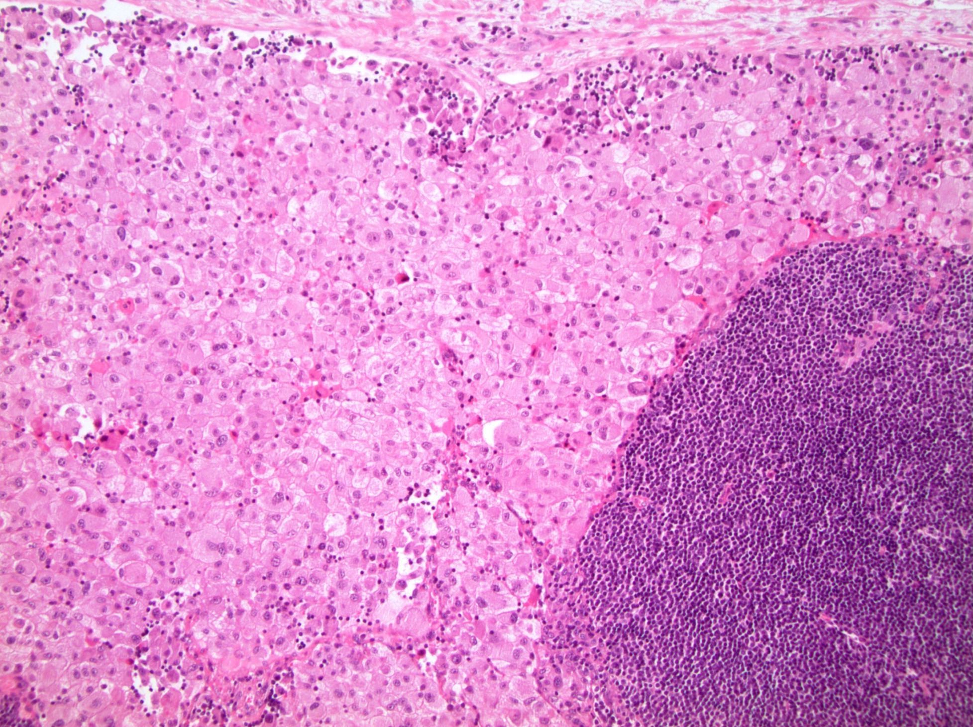
Periaortic lymph node metastasis (pN1)
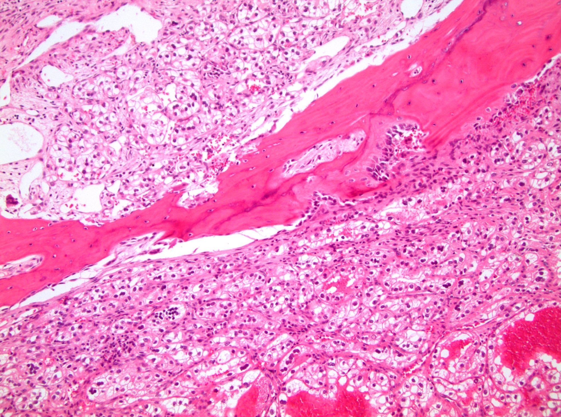
Bone metastasis (pM1)
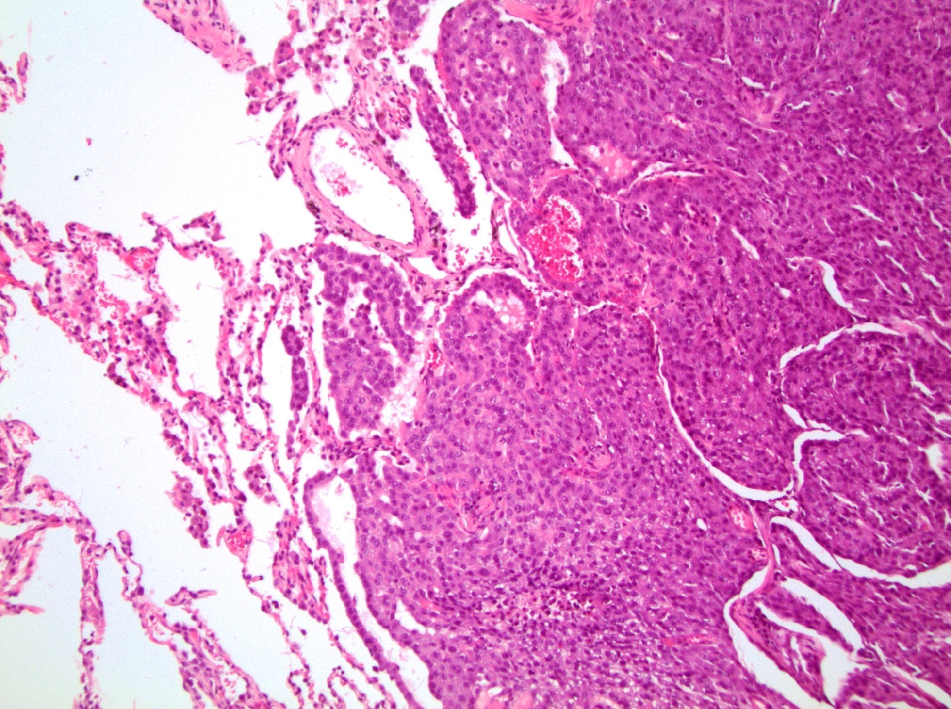
Lung metastasis (pM1)
Ovary metastasis (pM1)
Stomach metastasis (pM1)
Board review style question #1
This partial nephrectomy of renal cell carcinoma, papillary type 2 contains a 2.5 cm tumor limited to the cortex. Which is the correct pT category?
- pT1a
- pT1b
- pT2a
- pT2b
- pT3a
Board review style question #2
This radical nephrectomy of renal cell carcinoma, clear cell type contains a tumor which involves the renal sinus fat and perirenal fat. Which is the correct pT category?
- pT1a
- pT1b
- pT2a
- pT2b
- pT3a
Back to top