

Kidney tumor
Renal cell carcinoma - common
Papillary oncocytic variant
Last author update: 1 June 2015
Last staff update: 12 December 2024 (update in progress)
Copyright: 2003-2024, PathologyOutlines.com, Inc.
PubMed Search: Adult papillary renal tumor oncocytic cells


Table of Contents
Definition / general | Terminology | Sites | Clinical features | Case reports | Treatment | Gross description | Gross images | Microscopic (histologic) description | Microscopic (histologic) images | Positive stains | Negative stains | Electron microscopy description | Molecular / cytogenetics description | Differential diagnosisCite this page: Andeen NK, Tretiakova M. Papillary oncocytic variant. PathologyOutlines.com website. https://www.pathologyoutlines.com/topic/kidneytumormalignantadultpapillaryrenal.html. Accessed December 23rd, 2024.
Definition / general
- Most experts believe it is a variant of type 1 papillary renal cell carcinoma
- NOT considered a distinct entity by ISUP (Am J Surg Pathol 2013;37:1469) or WHO classification
Terminology
- Synonymous term: oncocytic papillary renal cell carcinoma (OPRCC) (Ann Diagn Pathol 2006;10:133, Int J Clin Exp Pathol 2013;6:1392)
- May be the same entity as oncocytic papillary renal cell carcinoma with inverted nuclear pattern (Pathol Int 2009;59:137)
Sites
- Kidney
Clinical features
- Usually male, median age 71 years (Am J Surg Pathol 2005;29:1576)
- Low stage, indolent clinical behavior
Case reports
- 81 year old woman with oncocytic papillary renal cell carcinoma (Int J Urol 2009;16:765)
Treatment
- Resection
Gross description
- Median 3 cm, well circumscribed, no extrarenal extension
Gross images
Contributed by Nicole K. Andeen, M.D.
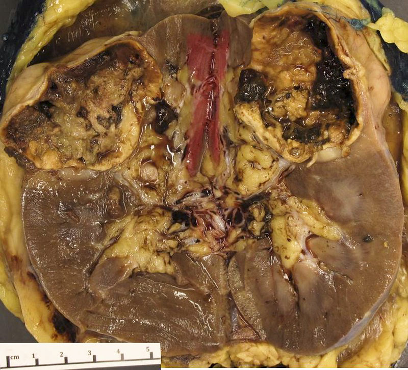
Papillary renal cell carcinoma with oncocytic cells
Microscopic (histologic) description
- Thin, nonfibrotic papillae lined by single layer of oncocytic cells
- Low grade, round, regular, nonoverlapping nuclei with central nucleolus (Am J Surg Pathol 2005;29:1576)
- Abundant granular eosinophilic cytoplasm
- Variable foamy macrophages within papillae
- Hemosiderin accumulation, focal necrosis
- May have luminal orientation of tumor nuclei (Pathol Int 2009;59:137) or solid oncocytoma-like areas (Ann Diagn Pathol 2006;10:133)
Microscopic (histologic) images
Contributed by Nicole K. Andeen, M.D. and Maria Tretiakova, M.D., Ph.D.
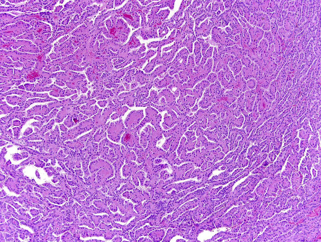


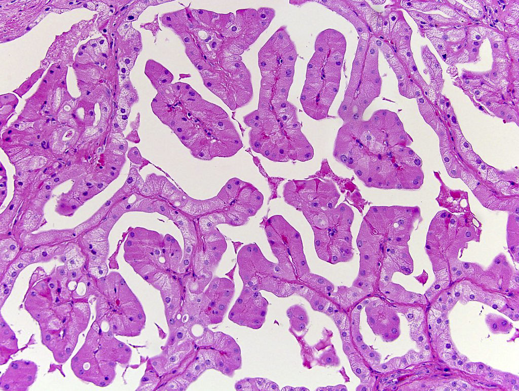
Papillary type 1 oncocytic renal cell carcinoma
Contributed by Semir Vranić, M.D., Ph.D., University of Sarajevo (Bosnia)
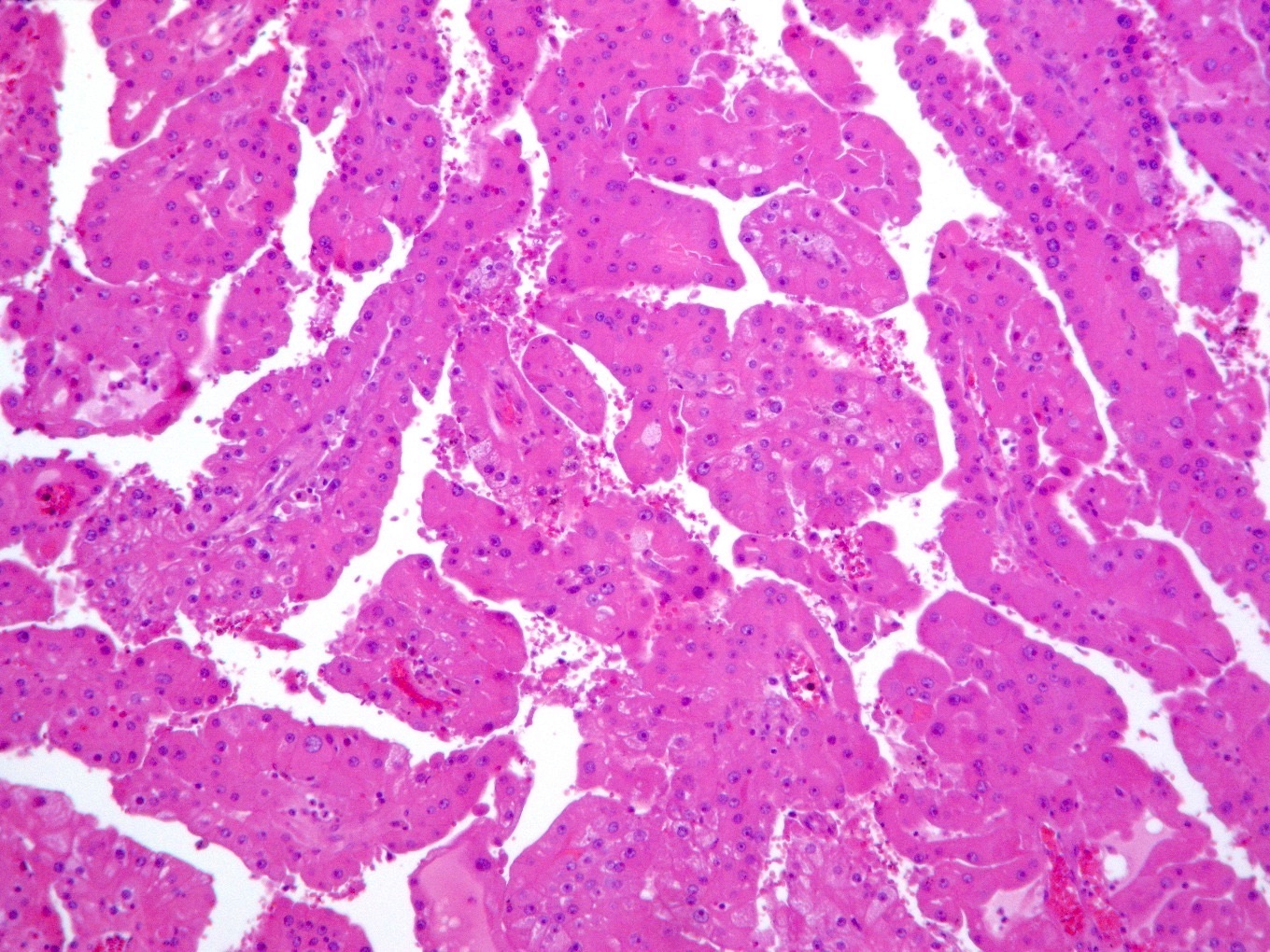
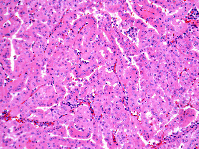

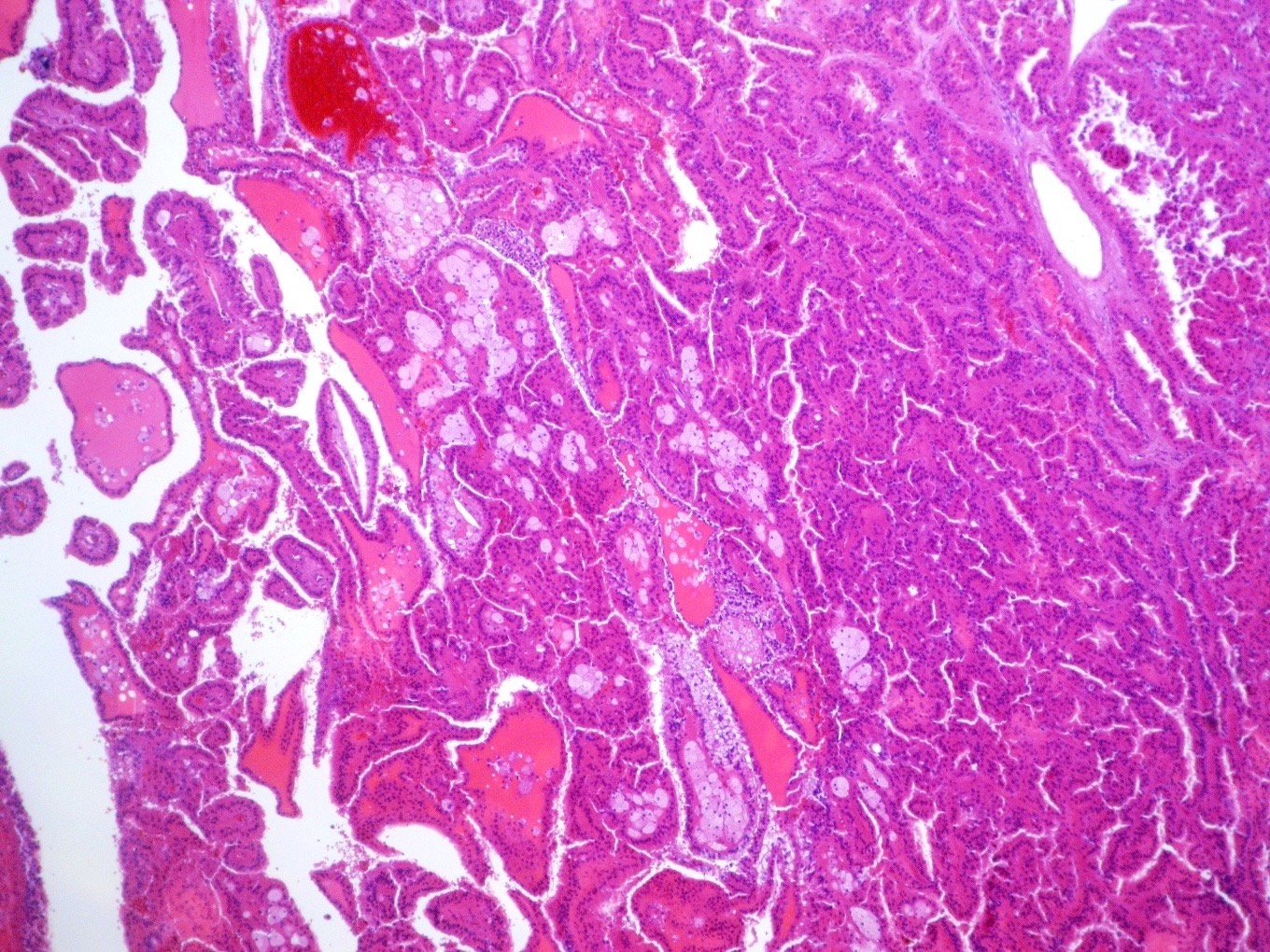
Oncocytic "variant" of papillary renal cell carcinoma
Images hosted on other servers:

Racemase, CD10, vimentin, MET
Positive stains
- AMACR, vimentin, mitochondrial antigen, CD10 (79%),
- Variable CK7 (17% - 100%), variable RCC (Int J Clin Exp Pathol 2013;6:1392, Am J Surg Pathol 2014;38:887)
Negative stains
- c-kit / CD117, low Ki67 proliferative rate
Electron microscopy description
- Abundant mitochondria with lamellar cristae (Ann Diagn Pathol 2006;10:133)
Molecular / cytogenetics description
- Majority have trisomy 7 and 17, loss of Y (Hum Pathol 2008;39:96, Int J Clin Exp Pathol 2013;6:1392, Ann Diagn Pathol 2006;10:133,, Am J Surg Pathol 2014;38:887)
- Gene expression studies show closer relationship to type 1 than type 2 papillary RCC (Cancer Res 2005;65:5628)
Differential diagnosis
- Oncocytoma: no papillary architecture, no necrosis; negative for vimentin, CD10, AMACR, CK7; positive for c-kit / CD117
- Papillary adenoma with oncocytic cells: 15 mm (1.5 cm) or less (size criteria)
- Papillary type 1 tumors: single layer of cells lining papillae but with pale basophilic cytoplasm, strong diffuse CK7 positivity
- Papillary type 2 tumors: higher nuclear grade, crowded and pseudostratified nuclei

