

Kidney tumor
Childhood tumors
MiT family translocation renal cell carcinomas
Last author update: 1 September 2016
Last staff update: 8 April 2024 (update in progress)
Copyright: 2003-2024, PathologyOutlines.com, Inc.
PubMed Search: MiT family translocation renal cell carcinoma


Table of Contents
Definition / general | Essential features | Terminology | Epidemiology | Sites | Pathophysiology | Clinical features | Prognostic factors | Case reports | Treatment | Gross description | Gross images | Microscopic (histologic) description | Microscopic (histologic) images | Positive stains | Negative stains | IHC panels | Electron microscopy description | Molecular / cytogenetics description | Differential diagnosisCite this page: Andeen NK, Tretiakova M. MiT family translocation renal cell carcinomas. PathologyOutlines.com website. https://www.pathologyoutlines.com/topic/kidneytumormalignantASPLTFE3.html. Accessed April 25th, 2024.
Definition / general
- Harbor gene fusions involving members of the MiT family of transcription factors, including TFE3 and TFEB
- Histologically diverse, often have papillary, alveolar and nested growth pattern with clear and eosinophilic cells and psammoma bodies
- More common in children / young adults
Essential features
- Papillary with clear and eosinophilic cells but wide morphologic spectrum
- Diagnosis often relies on IHC or FISH for translocation (Am J Clin Pathol 2012;137:761)
Terminology
- Renal cell carcinomas / RCCs associated with Xp11 translocations have gene fusions involving TFE3, which has multiple gene partners; RCCs with t(6:11) translocations have MALAT1-TFEB gene fusions
- MiT refers to microphthalmia associated transcription factors
Epidemiology
- 3% of adult renal cell carcinomas (Clin Cancer Res 2009;15:1170)
- 19 of 46 (40%) of renal cell carcinomas in children/young adults were TFE3+ vs. 1.6% - 4% of adult RCCs (Int J Surg Pathol 2011;19:170)
- Exposure to cytotoxic chemotherapy is a risk factor (Semin Diagn Pathol 2015;32:103)
Sites
- Kidney
Pathophysiology
- Overexpression of TFE3 or TFEB activates multiple downstream targets, including those normally activated by MiT family transcriptions factors; thus often expresses the cysteine protease cathepsin K, may express melanocytic markers and is less likely to express epithelial markers like cytokeratins
Clinical features
- In adults, strong female predominance (22 of 28 cases in one series), usually age 35 or less, usually high stage at diagnosis (Am J Surg Pathol 2007;31:1149)
Prognostic factors
- Aggressive clinical course (Am J Clin Pathol 2007;128:70)
- t(6;11)(p21;q12) appears to have low malignant potential in contrast to other translocation carcinomas (Am J Surg Pathol 2005;29:230, Int Urol Nephrol 2009;41:553)
Case reports
- 12 year old boy with nodal involvement at diagnosis (Afr J Paediatr Surg 2011;8:317)
- 20 year old woman with metastasis to placenta (Int J Surg Pathol 2011;19:80)
- Mini review with gross and radiographic images (SpringerPlus 2014;3:245)
Treatment
- Resection
- mTOR and tyrosine kinase inhibitors may be beneficial (Semin Diagn Pathol 2015;32:103)
- Selective MET inhibitors appear to have modest antitumural activity (Cancer 2012;118:5894)
Gross description
- Tan yellow, frequently hemorrhagic and necrotic; not distinct from other RCCs
Gross images
Contributed by Debra L. Zynger, M.D.
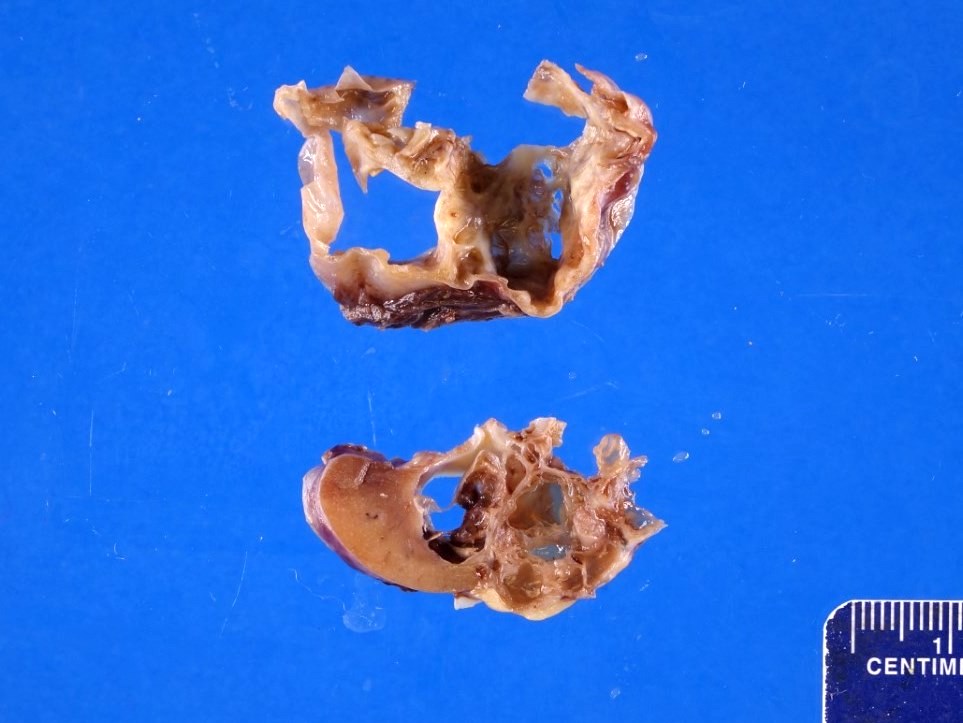
Cystic tumor
Microscopic (histologic) description
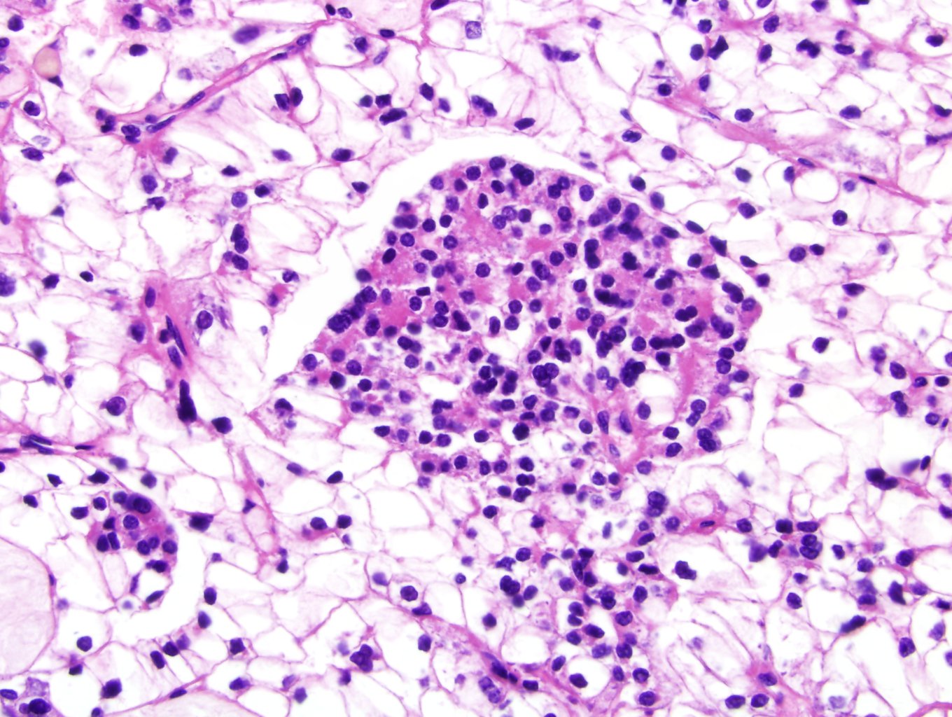
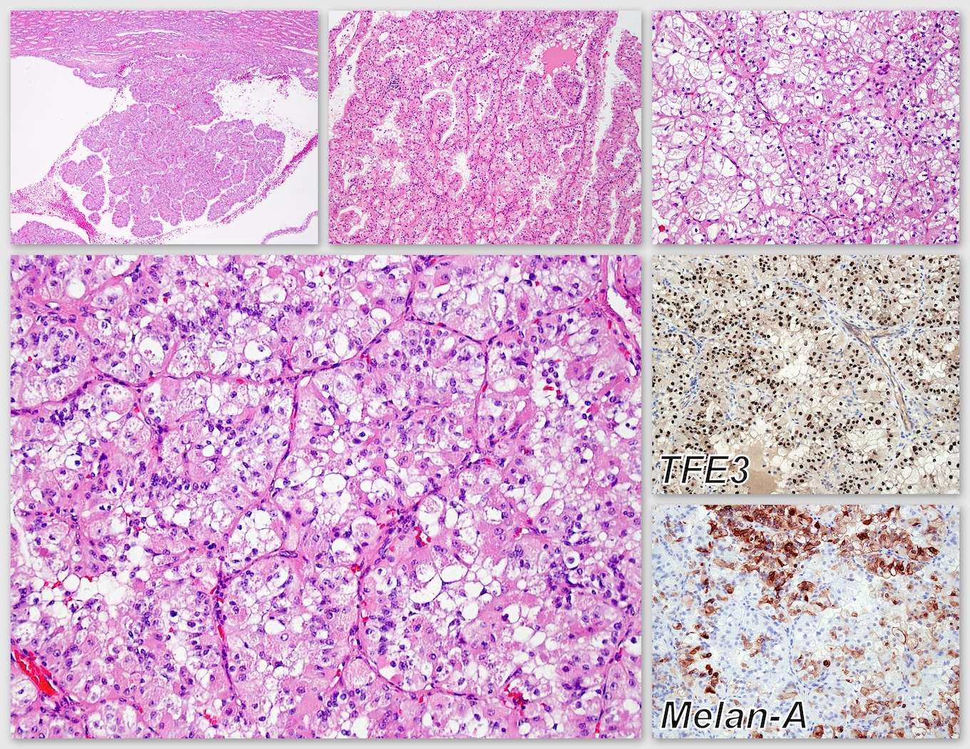
- TFE3 rearranged carcinomas usually have papillary and solid alveolar growth pattern, composed of clear to eosinophilic discohesive pseudostratified cells with voluminous cytoplasm and high grade nuclei
- TFE3 rearranged carcinomas also frequently have psammoma bodies and may contain melanin pigment, similar to a pigmented perivascular epithelioid tumor (PEC)oma (Semin Diagn Pathol 2015;32:103)
- t(6:11) rearranged carcinomas are characteristically biphasic, with small cells clustered around basement membrane material (reminiscent of Call-Exner bodies in adult granulosa cell tumor) and larger epithelioid cells (Semin Diagn Pathol 2015;32:103)
Microscopic (histologic) images
Contributed by Nicole K. Andeen, M.D.
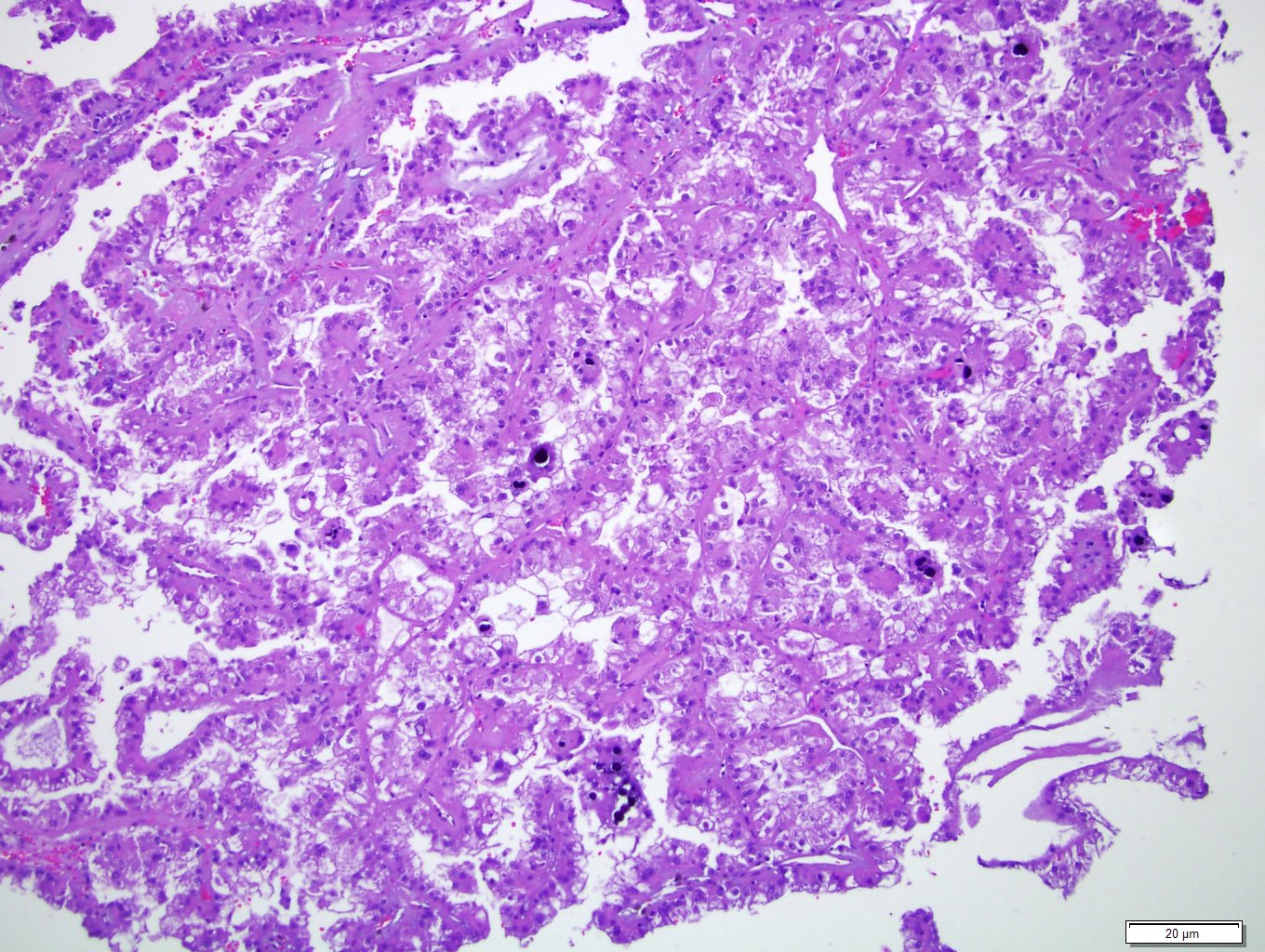

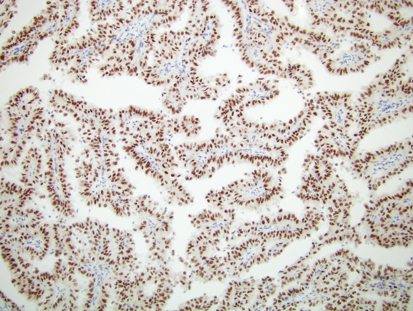
TFE3 rearranged carcinoma with papillary and alveolar growth pattern,
composed of clear to eosinophilic discohesive pseudostratified cells
with voluminous cytoplasm, high grade nuclei and psammoma bodies; there
is strong diffuse nuclear positivity for TFE3 by immunohistochemistry
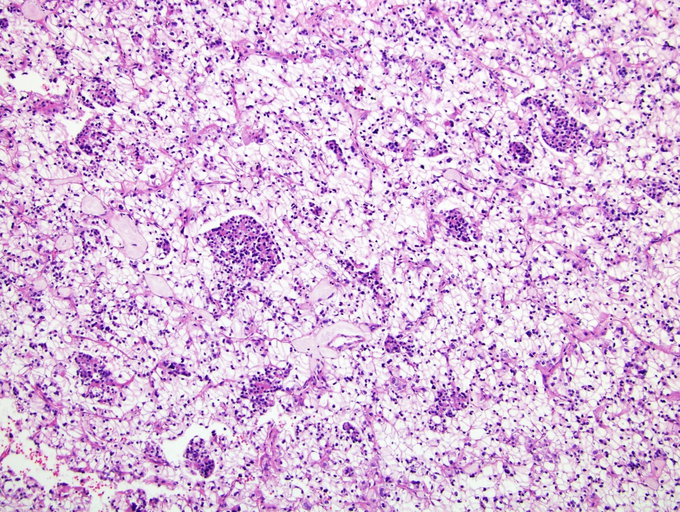
TFEB rearranged carcinoma with biphasic
appearance: larger epithelioid cells
and small cells clustered around
basement membrane material
Contributed by Sean Williamson, M.D. and David J. Grignon, M.D.
Translocation carcinoma (adults)
Positive stains
- TFE3 or TFEB (strong nuclear staining), PAX8, cathepsin K, CD10, AMACR, vimentin, E-cadherin
- May express melanocytic markers HMB45 and MelanA, more commonly in t(6:11) carcinomas and infrequently in Xp11 (WHO 2016; Semin Diagn Pathol 2015;32:103)
- Weak TFE3 staining in adults may not be specific (Am J Surg Pathol 2012;36:654)
Negative stains
- Variable cytokeratin (only 30% - 50% positive, less than other RCC types) and EMA (50%, frequently only focal) (Am J Surg Pathol 2002;26:1553, Am J Surg Pathol 2008;32:656, Am J Surg Pathol 2010;34:1295)
- Carbonic anhydrase / CAIX usually negative except areas of necrosis (Am J Clin Pathol 2010;134:873)
- CD45, calretinin, smooth muscle actin
IHC panels
| Hale | KIT | CK7 | S100A1 | VIM | CAIX | AMACR | SDH | TFE3 | |
| Chromophobe RCC | +++ | +++ | +++ | - | - | - | - | +++ | - |
| Clear cell RCC | - | - | - | - | +++ | +++ | - | +++ | - |
| Oncocytoma | - | +++ | rare | +++ | - | - | - | +++ | - |
| Papillary RCC | - | - | +++ | - | +++ | - | +++ | +++ | - |
| Translocation RCC | - | - | - | - | - | - | ++ | +++ | +++ |
| SDH deficient RCC | - | - | - | - | - | - | - | - | - |
References: Pathol Res Pract 2015;211:303, Ann Diagn Pathol 2020;44:151448, Am J Surg Pathol 2014;38:e6, Arch Pathol Lab Med 2019;143:1455, Transl Androl Urol 2019;8:S123, Hum Pathol 2020 Jul 13 [Epub ahead of print]
Electron microscopy description
- Features of clear cell carcinoma, including cell junctions, numerous mitochondria, microvilli, basement membrane, abundant glycogen
Molecular / cytogenetics description
- Fluorescence in situ hybridization (FISH) with a TFE3 or TFEB breakapart probe is highly sensitive and specific; it is diagnostic when indeterminate by morphology and immunohistochemistry
- TFE3 (on Xp11) has been reported with multiple gene partners, including the two most common, ASPL (17q25) and PRCC (1q21), and less commonly NONO (Xq12), PSF/SFPQ (1p34), CLTC (17q23) (WHO 2016, Am J Surg Pathol 2007;31:1149, Expert Rev Anticancer Ther 2010;10:843)
- t(6;11)(p21;q12): translocation between TFEB and MALAT1 genes result in overexpression of TFEB
- t(X;17)(p11.2;q25), with balanced translocation of TFE3 gene at Xp11.2 and ASPL gene at 17q25, is present in renal neoplasms, whereas in alveolar soft part sarcoma, this translocation is unbalanced, der(17)t(X;17)(p11.2;q25) (Am J Pathol 2001;159:179)
- Melanotic Xp11 renal cell carcinoma and PSF/SFPQ-TFE3 perivascular epithelioid cell tumors may share the same genetic abnormality and may be in the same clinicopathologic spectrum (Am J Surg Pathol 2015;39:1181)
Differential diagnosis
- Clear cell papillary renal cell carcinoma: typically a homogeneous low grade population of clear cells without eosinophilic cells, no calcifications, often branched ductular structures, secretory cells with nuclei aligned above basement membrane (resembling secretory endometrium); CK7+, CAIX+, CD10-, AMACR-
- Clear cell renal cell carcinoma: older patients, no true papillae (although pseudopapillary areas due to poor cell cohesion can be seen in high grade clear cell RCC); frequently keratin / EMA+, vimentin+, TFE3-, diffuse CAIX+, 3p deletion present
- Epithelioid angiomyolipoma: always negative for PAX8
- Chromophobe renal cell carcinoma: diffuse CD117 staining favors chromophobe
- Papillary renal cell carcinoma: predominantly papillary, no nested alveolar patterns, no extensive areas of clear cells; CK7+, TFE3-, trisomy 7 and 17
