

Kidney nontumor / medical renal
Monoclonal gammopathy of renal significance (MGRS) / paraprotein-related kidney disease
Light chain deposition disease
Author: Nikhil Sangle, M.D.

Last author update: 1 December 2014
Last staff update: 9 April 2024 (update in progress)
Copyright: 2003-2024, PathologyOutlines.com, Inc.
PubMed Search: Light chain deposition disease kidney pathology
Table of Contents
Definition / general | Case reports | Treatment | Microscopic (histologic) description | Microscopic (histologic) images | Immunofluorescence description | Immunofluorescence images | Negative stains | Electron microscopy description | Electron microscopy images | Differential diagnosisCite this page: Sangle N. Light chain deposition disease. PathologyOutlines.com website. https://www.pathologyoutlines.com/topic/kidneylightchaindepositiondisease.html. Accessed April 25th, 2024.
Definition / general
- Monoclonal gammopathy characterized by overproduction and deposition of nonamyloid immunoglobulin light chains in various organs (eMedicine: Light-Chain Deposition Disease [Accessed 17 January 2018])
- Uncommon; occasionally also heavy chains
- See also myeloma
- 80% male, usually older adults
- 60% have associated myeloma or other lymphoplasmacytic disorder, although it may not become apparent until years later
- Renal failure with heavy proteinuria; also Fanconi anemia with aminoaciduria, glucosuria and phosphaturia
- Also cardiac, hepatic and neural damage and deposition in soft tissues and other organs of histiocytes and fibroblasts containing crystals (Am J Surg Pathol 1993;17:461)
- May recur in renal transplants
- Variable 5 year survival; ~ 70%, less if coexisting myeloma
Case reports
- 41 year old man with no clinical plasma cell dyscrasia (Ultrastruct Pathol 2012;36:134)
- 49 year old woman with biopsy proven resolution after autologous stem cell transplantation (Nephrol Dial Transplant 2010;25:2020)
- 53 year old woman with recurrent light chain deposition disease post renal transplant (Clin Transplant 2012;26:64)
- 66 year old man presenting with severe jaundice (Oman Med J 2012;27:56)
- Middle aged woman with myeloma and type II diabetes (Arch Pathol Lab Med 1983;107:319)
Treatment
- Chemotherapy with bortezomib, a proteasome inhibitor, thalidomide (Med Oncol 2012;29:1197, Int J Hematol 2011;93:673)
- Also autologous stem cell transplantation (Int J Lab Hematol 2012;34:347, J Nephrol 2011;24:246)
Microscopic (histologic) description
- Enlarged glomeruli with PAS+ material in thickened capillary walls and mesangial nodules
- Occasional fibroepithelial crescents
- Thickened tubular basement membranes with glassy (crystalline) appearance
- Also crystals within histiocytes; weakly positive on silver stain
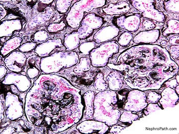
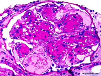
Microscopic (histologic) images
Contributed by NephroPath
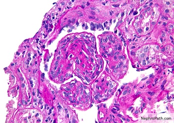
Light chain deposits along GBM
sclerosis and
tubular injury
on silver stain
Mesangial nodular sclerosis on PAS
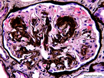
Mesangial nodular sclerosis on silver stain
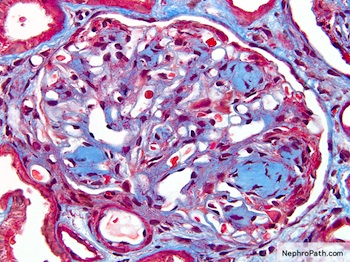
Mesangial nodular sclerosis on trichrome stain
Images hosted on other servers:

Various images
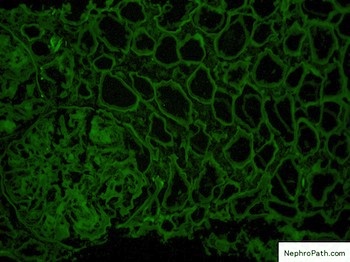
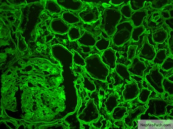
Immunofluorescence description
- Granular deposits of kappa (80%) or lambda (20%) light chains (not both) along glomerular and tubular basement membranes, in mesangium, vessel walls and interstitium
Immunofluorescence images
Contributed by NephroPath
Negative staining for lambda light chain on IF
Positive GBM and
TBM staining
for kappa light
chain on IF
Negative stains
- Congo red, thioflavin T and amyloid P protein
Electron microscopy description
- Diffuse electron dense, finely granular material in glomerular basement membrane, mesangium, tubular and vascular basement membranes
- Immunoelectron microscopy may be useful for diagnosis (Hum Pathol 2003;34:270)
Electron microscopy images
Contributed by NephroPath
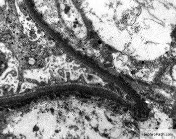
Light chain deposits along GBM

Light chain deposits along TBM
Differential diagnosis
- AL amyloidosis: fibrillar deposits, usually lambda light chains, Congo Red+, thioflavin T+, amyloid P protein+
- Diabetes: severe arteriolar hyalinosis, fibrin caps, capsular drops, strongly positive with silver stain
- Drug related crystals
