

Bone & joints
Other nonneoplastic
Ganglion cyst
Editorial Board Members: Jose G. Mantilla, M.D., Borislav A. Alexiev, M.D.


Last author update: 15 March 2021
Last staff update: 1 May 2023
Copyright: 2002-2024, PathologyOutlines.com, Inc.
PubMed Search: Ganglion [title] joints (y_5[Filter]) AND (ffrft[Filter])
Table of Contents
Definition / general | Terminology | ICD coding | Epidemiology | Sites | Pathophysiology | Etiology | Clinical features | Clinical images | Diagnosis | Radiology description | Radiology images | Prognostic factors | Case reports | Treatment | Gross description | Gross images | Microscopic (histologic) description | Microscopic (histologic) images | Virtual slides | Cytology description | Cytology images | Electron microscopy description | Videos | Sample pathology report | Differential diagnosis | Additional references | Board review style question #1 | Board review style answer #1 | Board review style question #2 | Board review style answer #2Cite this page: Serinelli S, de la Roza G. Ganglion cyst. PathologyOutlines.com website. https://www.pathologyoutlines.com/topic/jointsganglion.html. Accessed April 25th, 2024.
Definition / general
- Most common soft tissue mass found in the hand and wrist (StatPearls: Ganglion Cyst [Accessed 2 March 2021])
- Cystic structure containing mucoid material
Terminology
- Ganglion cyst (synonym)
- Mucous cyst (when occurring at the distal interphalangeal joint) (StatPearls: Ganglion Cyst [Accessed 2 March 2021])
ICD coding
- ICD-10: M67.40 - ganglion, unspecified site
Epidemiology
- 60 - 70% of the soft tissue masses in the hand and wrist (StatPearls: Ganglion Cyst [Accessed 2 March 2021])
- F:M = 3:1
- Can occur at any age
- Most common: 20 - 50 years
- Risk factors:
- Repetitive microinjuries due to overuse of the joint
- Previous traumas
Sites
- Dorsal aspect of the wrist, from the scapholunate ligament or scapholunate articulation → 70%
- Volar aspect of the wrist, from the radiocarpal joint or scaphotrapezial joint → 20%
- Distal interphalangeal joint, hip, knee, ankle, foot, others → 10%
- Some studies found the volar location to be more common than the dorsal (J Ultrasound Med 2019;38:2155)
Pathophysiology
- Cystic fluid analysis: gelatinous material containing mainly hyaluronic acid and lesser amounts of glucosamine, globulins and albumen (Curr Rev Musculoskelet Med 2008;1:205)
- Since no epithelial lining exists in these structures, they should not be classified as true cysts
Etiology
- Unclear
- Numerous theories (Curr Rev Musculoskelet Med 2008;1:205):
- Displacement of synovial tissue during embryogenesis
- Herniation of synovial capsule / fluid from joints into the surrounding tissues → reaction between fluid and local tissue results in the creation of the cyst
- Proliferation of pluripotential mesenchymal cells
- Myxoid degeneration of connective tissue after trauma: repetitive injury to the capsular and ligamentous structures → production of hyaluronic acid from fibroblasts → accumulation of mucin jelly-like material to form the cyst (most likely)
- Inflammatory etiology
Clinical features
- Firm, rubbery, superficial mass (Curr Rev Musculoskelet Med 2008;1:205)
- May form suddenly or gradually (J Hand Surg Am 2015;40:546)
- May be a history of trauma
- Symptoms (Curr Rev Musculoskelet Med 2008;1:205):
- Majority are asymptomatic (StatPearls: Ganglion Cyst [Accessed 2 March 2021])
- May be painful or tender but usually the pain is not debilitating (Lindberg: Diagnostic Pathology - Soft Tissue Tumors, 2nd Edition, 2015)
- Pain may be due to nerve compression / involvement or less frequently, due to inflammatory changes related to complications by rupture, hemorrhage or infection (Insights Imaging 2016;7:179)
- May cause decreased range of motion, decreased strength and paresthesias
Clinical images
Images hosted on other servers:

Dorsal wrist ganglion

Ganglion: intraoperative view

Volar wrist ganglion
Diagnosis
- Clinical presentation is usually adequate for diagnosis
- Cyst typically transilluminates on the exam
- Ultrasound is used for a definitive diagnosis
- MRI is useful to rule out a possible solid tumor or in case of occult dorsal ganglion cyst (which is not clinically observed or palpated but is found on imaging studies or intraoperatively) (J Wrist Surg 2019;8:276)
Radiology description
- Ultrasound: well defined, unilocular or multilocular, noncompressible and anechoic or hypoechoic fluid collection (J Ultrasound Med 2019;38:2155)
- MRI: well circumscribed, thin walled cyst, typically hypo to isointense on T1 weighted images and homogenously hyperintense on T2 (StatPearls: Ganglion Cyst [Accessed 2 March 2021], Insights Imaging 2016;7:179)
Radiology images
Images hosted on other servers:

Ultrasound: dorsal wrist ganglion

MRI: volar wrist ganglion
Prognostic factors
- Benign condition
- Prognosis for most patients is excellent
- Ganglion cysts spontaneously recede in more than half of patients (ISRN Orthop 2013;2013:940615)
- Recurrence rate is approximately 10 - 15% after surgery (Adv Clin Exp Med 2019;28:95)
- Recurrence after surgery is unpredictable and independent of patient demographic factors or surgical techniques
- Aspiration has higher rates of recurrence (ISRN Orthop 2013;2013:940615)
Case reports
- 51 year old woman with a recurrent right foot ganglion cyst (J Am Podiatr Med Assoc 2020;110:Article9)
- 54 year old man with posttraumatic extensive ganglion cyst to the knee (Clin Pract 2011;1:e61)
- 88 year old woman with a large multilocular ganglion cyst of the right temporomandibular joint (J Oral Maxillofac Surg 2016;74:1783)
Treatment
- Indications for treatment include pain, stiffness, weakness and cosmetic appearance (J Hand Surg Am 2015;40:546)
- Three general treatment approaches:
- Observation
- Aspiration: often combined with some form of injection, electrocautery or multiple puncture
- Excision: open or arthroscopic
Gross description
- Cystic structure
- Typically, 1 - 2 cm in size
- Either uni or multilocular
- Usually attached to the underlying tendon sheath or joint capsule through a stalk (Lindberg: Diagnostic Pathology - Soft Tissue Tumors, 2nd Edition, 2015)
- Does not communicate with the joint cavity (Goldblum: Rosai and Ackerman's Surgical Pathology, 11th Edition, 2018)
- Contains viscous / gelatinous fluid
- Often excised in fragments
Gross images
Images hosted on other servers:

Ganglion
Microscopic (histologic) description
- Uni or multilocular cystic structure
- Dense collagenous walls with foci of myxoid changes (Lindberg: Diagnostic Pathology - Soft Tissue Tumors, 2nd Edition, 2015)
- No true epithelial lining
- Lumen may contain myxoid fluid
- There is no nuclear atypia or mitotic activity
- Inflammation / hemorrhage may be observed if the cyst has previously been ruptured
Microscopic (histologic) images
Contributed by Serenella Serinelli, M.D., Ph.D.
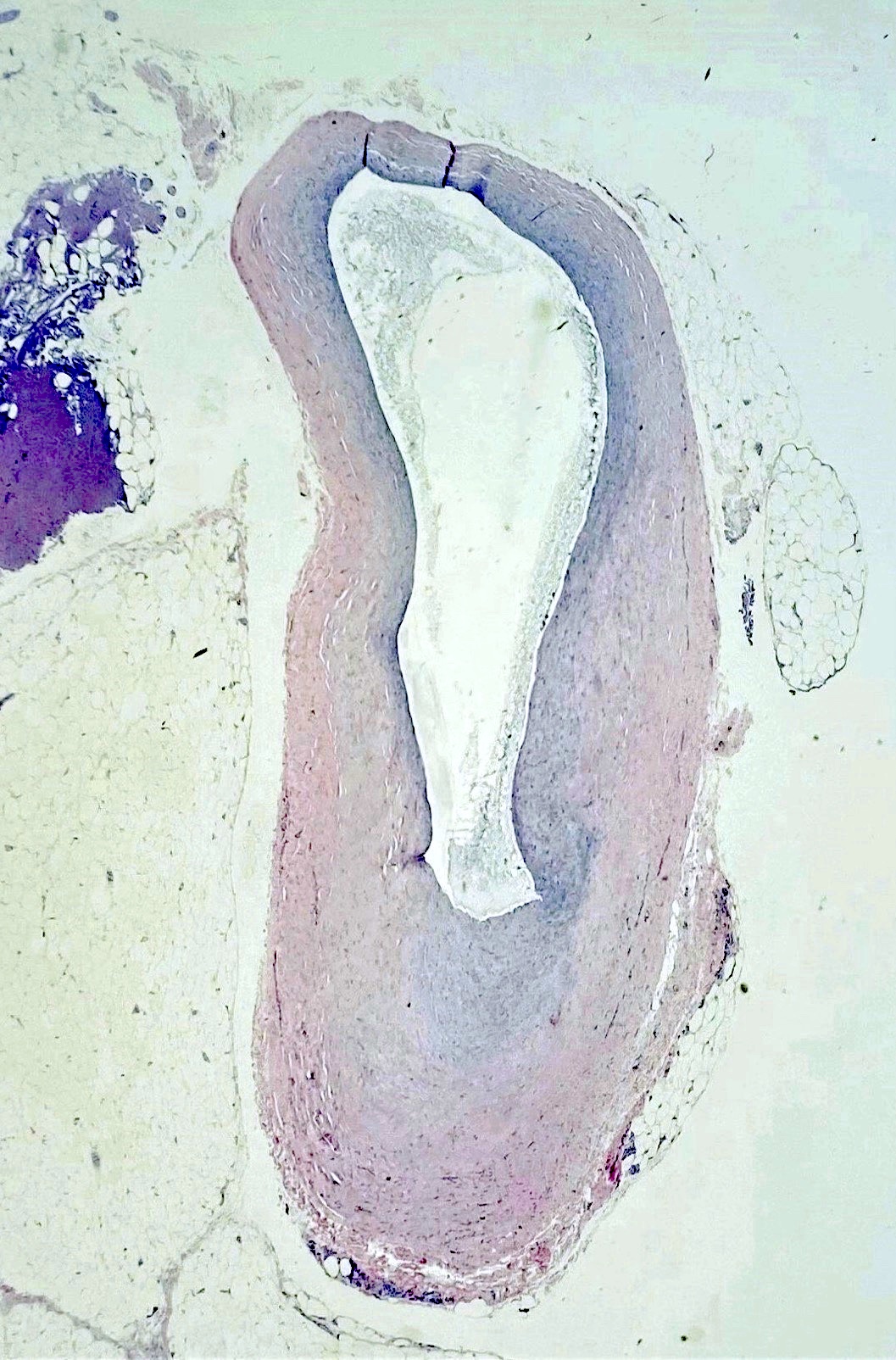
Unilocular ganglion cyst
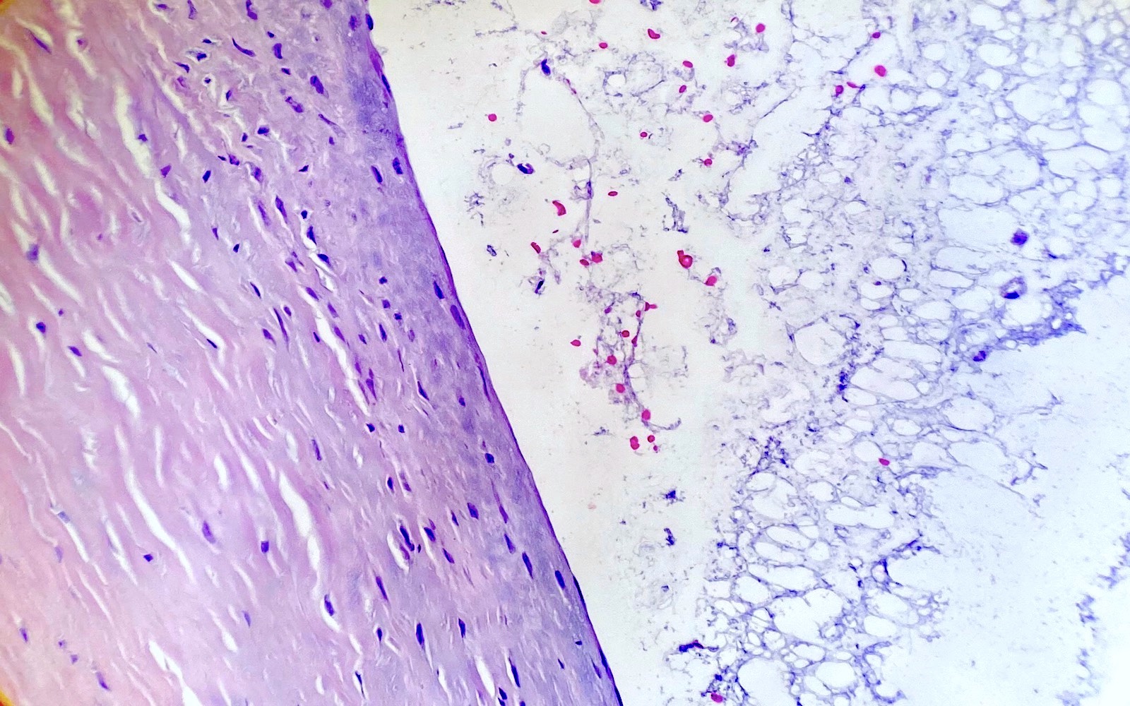
Cyst wall and content
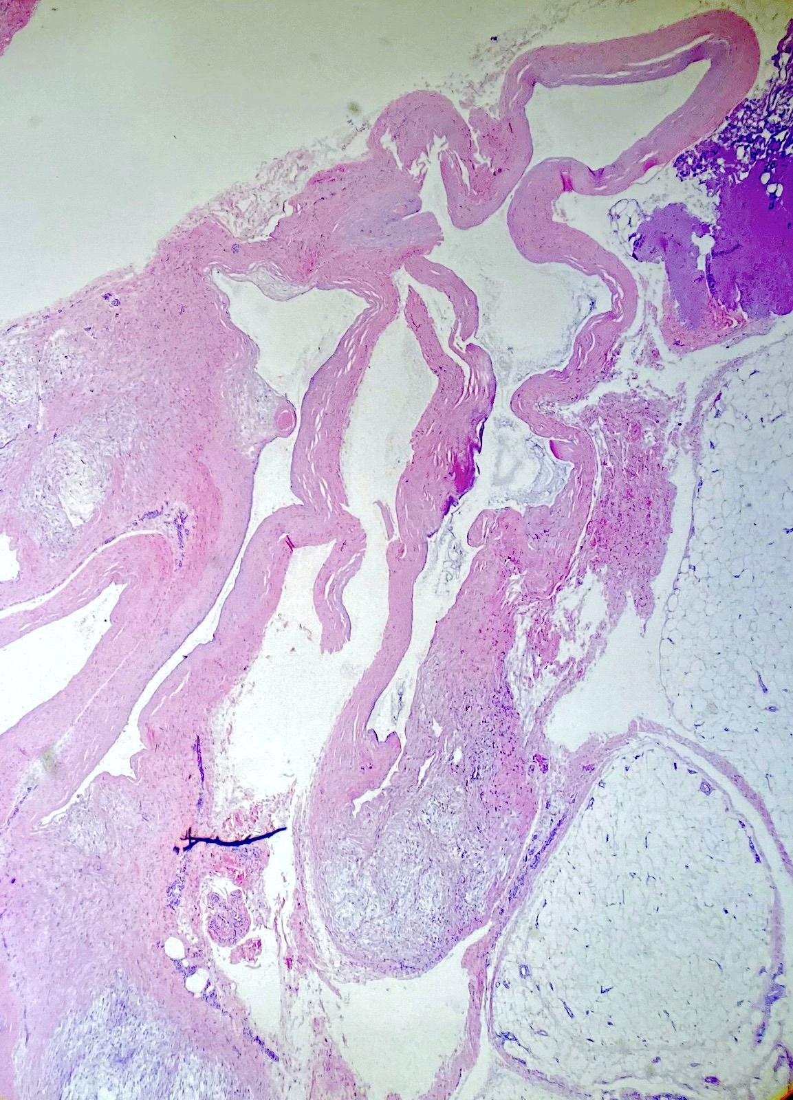
Multilocular ganglion cyst
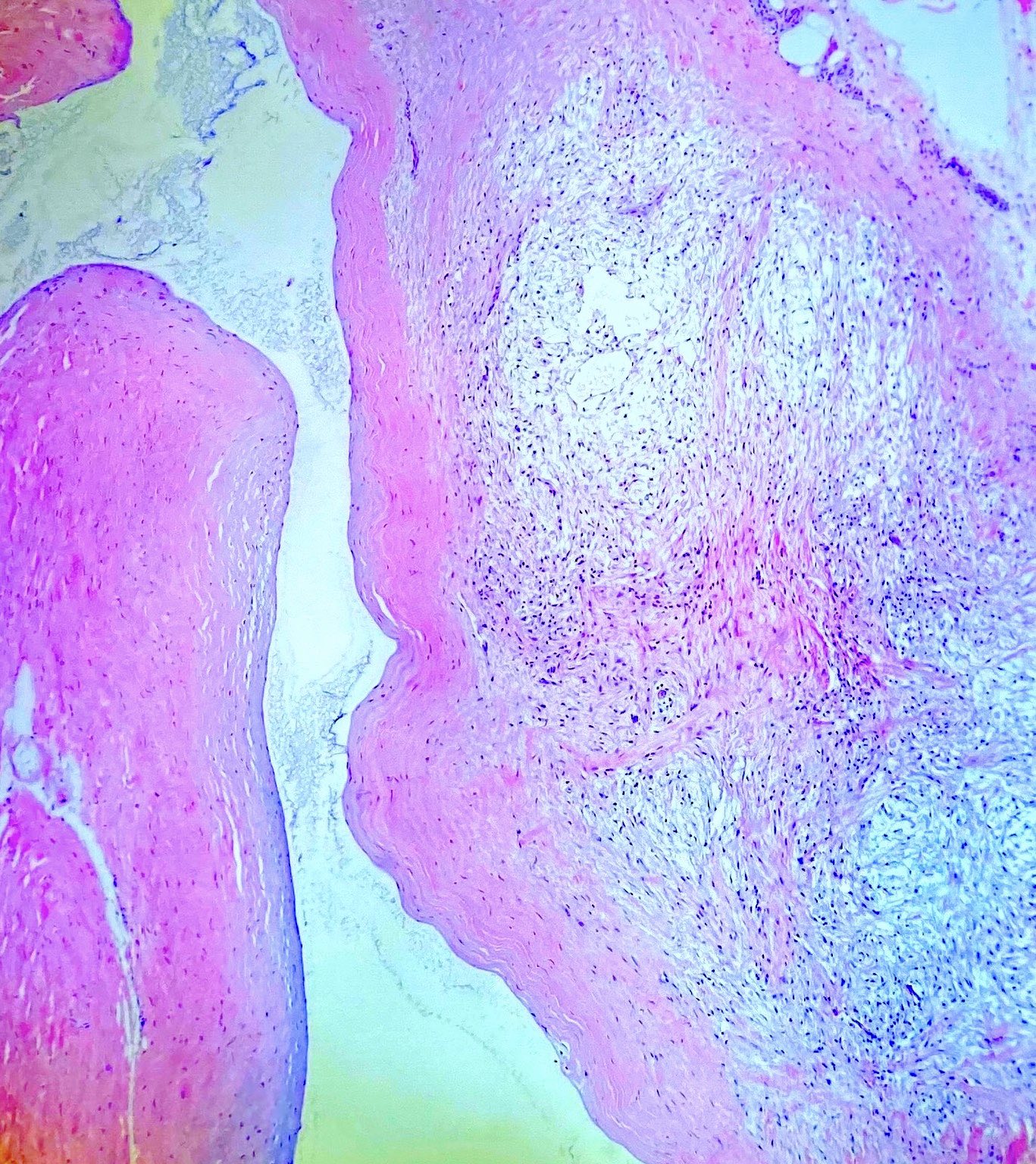
Myxoid changes
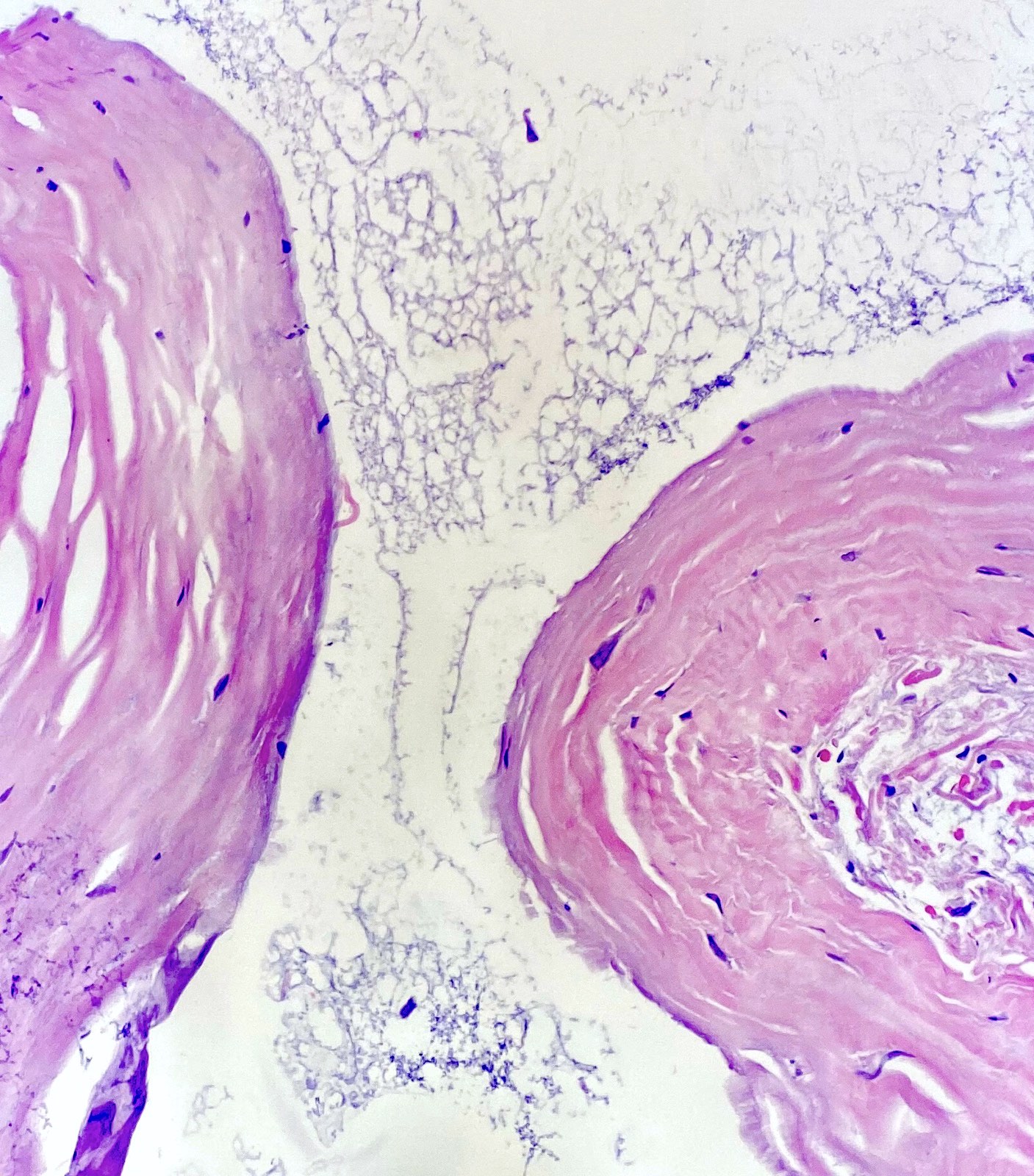
Absence of synovial lining
Virtual slides
Images hosted on other servers:

Ganglion cyst
Cytology description
- Aspiration of a ganglion can be used as a preoperative diagnostic tool or a therapeutic procedure
- Paucicellular myxoid material that contains scattered / clustered histiocytes (Am J Clin Pathol 2005;123:858)
- Myxoid material of the cyst usually forms thick folds on the slide
Cytology images
Images hosted on other servers:

Ganglion cyst mucoid material

Histiocytes in ganglion cyst
Electron microscopy description
- Wall is composed of randomly oriented sheets of collagen arranged in loose layers
- Rare cells are present in the collagen sheets and appear to be fibroblasts or mesenchymal cells (Curr Rev Musculoskelet Med 2008;1:205)
Videos
Ganglion cyst features
Sample pathology report
- Soft tissue, left dorsal wrist, excision:
- Ganglion cyst
Differential diagnosis
- Myxoma:
- Lobulated, gelatinous cut surfaces
- Most commonly occurring within large muscles (thigh, shoulder, upper arm)
- Low grade myxofibrosarcoma:
- Multinodular cut surfaces
- At least focal nuclear atypia
- Conspicuous, elongated, curvilinear, thin walled blood vessels are characteristic
- Most common in subjects older than 50
- Neurofibroma:
- Absence of cystic spaces
- S100+
Additional references
Board review style question #1
40 year old woman presents with a superficial lesion over the dorsal wrist. The lesion is excised and displays a cystic appearance. The histology is shown above. What is one of the main features of this condition?
- Cyst shows no true epithelial lining
- Invariably consists of a unilocular cyst
- Marked cellular atypia is seen
- Mitoses are frequent
- Rare condition
Board review style answer #1
Board review style question #2
Which of the following statements is true regarding ganglion cysts?
- More common among females
- More common in the pediatric population
- Myxoid changes in the cyst wall are never observed
- Nuclear atypia is required for the diagnosis
- Rarely found in the hand / wrist area
Board review style answer #2
