

Heart & vascular pathology
General
Conduction tissue
Author: R. Amita, M.D.

Last author update: 1 December 2015
Last staff update: 31 December 2020
Copyright: 2015-2024, PathologyOutlines.com, Inc.
PubMed Search: Conduction tissue [title] heart
Table of Contents
Definition / general | Gross description | Diagrams / tables | Microscopic (histologic) description | Microscopic (histologic) images | Additional referencesCite this page: Amita R. Conduction tissue. PathologyOutlines.com website. https://www.pathologyoutlines.com/topic/heartconductiontiss.html. Accessed April 26th, 2024.
Definition / general
- The specialized tissues of the heart (neuromyocardial cells) that initiate and conduct the cardiac impulse consist of:
- Sinus node (SA node, sinoauricular node, node of Keith-Hack)
- Atrioventricular junctional area, including the AV node (node of Twara) and His bundle
- Bundle branches (branching portion of AV bundle, bifurcation, ventricular conduction tissue) and Purkinje fibers
Gross description
- Sinus node:
- An oval shaped, elongated mass 10-20 mm long and up to 5 mm thick
- The "head" of the node extends toward the interatrial groove while its "tail" extends toward the orifice of the inferior vena cava
- In most hearts, it is located in the subepicardial region (less than 1 mm from the epicardial surface) at the lateral junction of the superior vena cava and right atrium
- Supplying the sinus node is a prominent artery ("sinus node artery") arising from the right coronary artery in 55 - 60% of cases and from the left circumflex coronary artery in 40 - 45%
- Atrioventricular (AV) node:
- Situated in the inferomedial right atrium and forms the top of the only normal electrical connection between atria and ventricles
- Blood supply is from the right coronary artery in 90% of cases, the left circumflex in the remainder
- His-Purkinje System:
- Located at the crest of the interventricular septum
- The AV node terminates in the top of the His bundle which then branches into a left and right bundle branch
- The right bundle branch is a cord-like structure insulated from surrounding myocardium for most of its length
- When it reaches the right ventricular apex it makes its initial electrical contact with myocardial cells of the anterior papillary muscle
- In contrast, the left bundle branch is usually a fan-like structure, dividing soon after its origin into anterior and posterior fascicles
- These fascicles then further ramify into the rest of the Purkinje network
- Blood supply is almost entirely from the left anterior descending artery
- The proximal His bundle may have dual supply, from both right and left coronary arteries
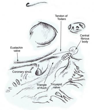
Diagrams / tables
Images hosted on other servers:

Purkinje network
Compact atrioventricular node

Conduction system
Microscopic (histologic) description
-
Sinus node:
- Cells include nodal cells, transitional cells and atrial muscle cells
- Nodal cells (P cells) are small ovoid, pale staining and poorly striated compared with the general myocardial cells
- The nodal cells are grouped together in interconnecting fascicles placed in a background of fibrous matrix
- Nodal cells are thought to be the source of normal impulse formation in the sinus node
- In the infant sinus node, the nodal cells predominate relative to the fibrous matrix; in contrast, in the adult sinus node, the fibrous tissue is predominant with the nodal cells scattered within the connective tissue
- Transitional cells (T cells) are elongated with characteristics intermediate between the packed nodal cells and the individual atrial myocardial cells
- Transitional cells are located at the margins of the node where the nodal cells become contiguous with atrial myocardium
Microscopic (histologic) images
Images hosted on other servers:


Sinoatrial (SA) node


Sinus node fibrosis
Additional references
