
Gallbladder & extrahepatic bile ducts
Gallbladder nonneoplastic
Adenomyomatous hyperplasia
Editorial Board Member: Catherine E. Hagen, M.D.
Deputy Editor-in-Chief: Raul S. Gonzalez, M.D.


Last author update: 15 April 2021
Last staff update: 14 September 2023
Copyright: 2003-2024, PathologyOutlines.com, Inc.
PubMed Search: Adenomyomatous hyperplasia gallbladder
Table of Contents
Definition / general | Essential features | Terminology | Epidemiology | Sites | Pathophysiology | Diagnosis | Radiology description | Radiology images | Case reports | Gross images | Microscopic (histologic) description | Microscopic (histologic) images | Sample pathology report | Differential diagnosis | Additional references | Board review style question #1 | Board review style answer #1 | Board review style question #2 | Board review style answer #2Cite this page: George AA, Garcia-Buitrago MT. Adenomyomatous hyperplasia. PathologyOutlines.com website. https://www.pathologyoutlines.com/topic/gallbladderadenomyomatous.html. Accessed April 19th, 2024.
Definition / general
- Benign reactive nonneoplastic lesion of the gallbladder
- Associated with chronic inflammation / injury (cholecystitis, cholelithiasis) (StatPearls: Adenomyomatosis [Accessed 26 January 2021])
Essential features
- Most common benign gallbladder polypoid lesion after cholesterol polyps (Euroasian J Hepatogastroenterol 2019;9:40)
- May form tumor-like lesions (adenomyomatous nodules) (Am J Surg Pathol 2020;44:1649)
- Most patients thought to be asymptomatic; right upper quadrant pain can be seen (StatPearls: Adenomyomatosis [Accessed 26 January 2021])
- Reported frequency: up to 10% of cholecystectomy specimens (Burt: MacSween's Pathology of the Liver, 7th Edition, 2017, StatPearls: Adenomyomatosis [Accessed 26 January 2021])
- May also represent diverticular change or hamartomatous malformation (Burt: MacSween's Pathology of the Liver, 7th Edition, 2017, Korean J Gastroenterol 2016;67:332)
- Usually found in the gallbladder but may rarely occur in extrahepatic bile ducts (Korean J Gastroenterol 2016;67:332)
- Rarely develops dysplasia and carcinoma (Am J Surg Pathol 2020;44:1649, Burt: MacSween's Pathology of the Liver, 7th Edition, 2017)
Terminology
- Also called adenomyoma, adenomyomatosis (Korean J Gastroenterol 2016;67:332)
- Generalized, segmental or localized types
- Generalized:
- Diffuse wall thickening (up to 5x normal) with intramural diverticula resembling cystic spaces within the wall
- Also called adenomyomatosis, adenomyosis
- Localized:
- Fundus has nodules from 0.5 to 2.5 cm with grayish white cut surface containing multiple cysts
- May cause gallbladder inversion
- Also called adenomyoma
- Generalized:
Epidemiology
- F:M = 3:1; some authors report similar prevalence between sexes (StatPearls: Adenomyomatosis [Accessed 26 January 2021])
- Wide age range of patients; most are diagnosed in their 50s (StatPearls: Adenomyomatosis [Accessed 26 January 2021])
- Rare in children (World J Clin Pediatr 2016;5:223)
Sites
- Gallbladder (fundus if localized), rarely extrahepatic bile ducts
Pathophysiology
- Associated with chronic inflammation / injury (cholecystitis, cholelithiasis) (StatPearls: Adenomyomatosis [Accessed 26 January 2021])
- May also represent diverticular change or hamartomatous malformation (Burt: MacSween's Pathology of the Liver, 7th Edition, 2017, Korean J Gastroenterol 2016;67:332)
Diagnosis
- Usually diagnosed by microscopic evaluation of hematoxylin and eosin stained slides
Radiology description
- Ultrasound is the most commonly used imaging modality; CT or MRI may also be used (HPB (Oxford) 2016;18:129)
Radiology images
Images hosted on other servers:


Ultrasound images of gallbladder adenomyoma
Case reports
- 17 year boy with adenomyomatosis of the gallbladder (Arch Argent Pediatr 2020;118:e43)
- 81 year old woman with right upper quadrant pain (BMC Gastroenterol 2010;10:41)
- 3 cases with fundal variant (J Clin Med Res 2010;2:150)
Gross images
Contributed by Alan A. George, D.O. and Monica T. Garcia-Buitrago, M.D.
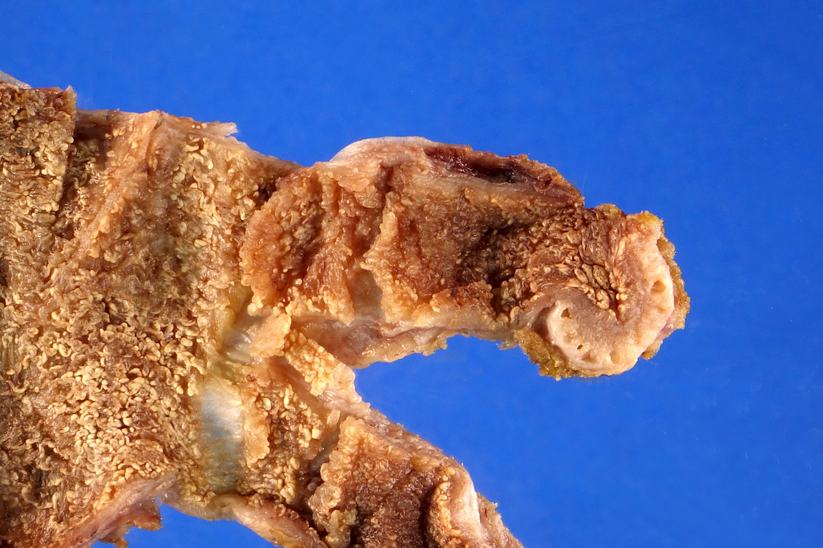
Localized adenomyoma
Images hosted on other servers:

Focal adenomyoma
Microscopic (histologic) description
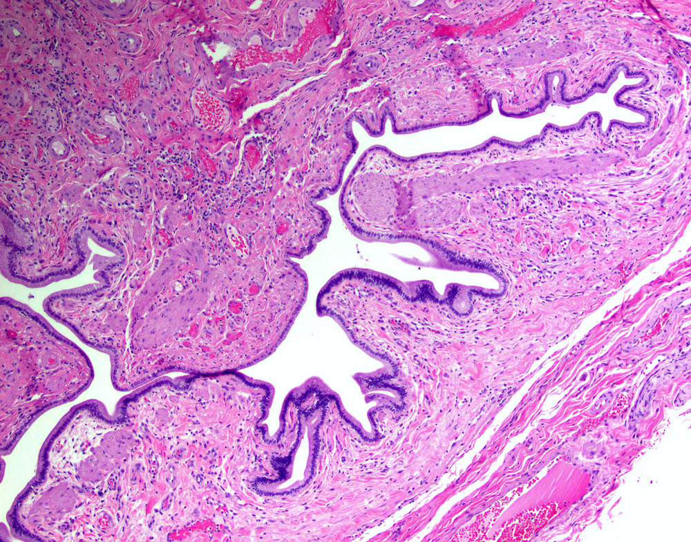
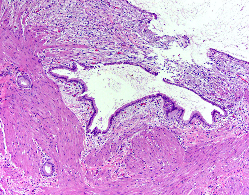
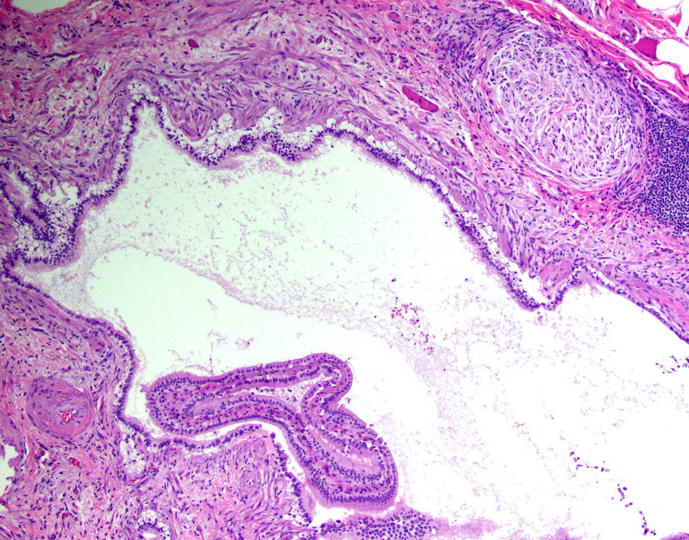
- Cystically dilated benign biliary glands accompanied by smooth muscle hypertrophy of gallbladder wall, thickened / fibrotic subserosa (StatPearls: Adenomyomatosis [Accessed 26 January 2021])
- Glands are distinct from the Rokitansky-Aschoff sinuses, which are epithelial diverticula, usually multifocal and occur throughout gallbladder secondary to injury, versus adenomyomatous nodule (distinct localized lesion of the gallbladder wall, 1 - 1.5 cm mural nodule in the fundus) (Am J Surg Pathol 2020;44:1649)
- May have reactive epithelial changes, papillary change and intestinal metaplasia (StatPearls: Adenomyomatosis [Accessed 26 January 2021])
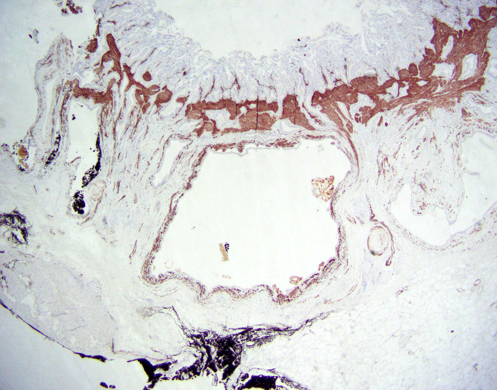
- Rarely, benign glands are seen in proximity to nerves, appearing as perineural and intraneural invasion only in the subserosal layer (benign gland-like structures may migrate into nerves due to chemotactic factors or signaling substances with activation of cell receptors) (Am J Surg Pathol 2007;31:1598)
- Adenomyomatous nodules may rarely show dysplastic / carcinomatous transformation, whereas dysplasia in Rokitansky-Aschoff sinuses appears to be more common; however, the true association between adenomyomatous nodules and neoplasia has not yet been determined (Am J Surg Pathol 2020;44:1649)
- Recently, papillary dysplastic lesions of adenomyomas have been identified (intracholecystic neoplasms of adenomyomas), demonstrating cystic and solid areas with papillary projections that show biliary, gastric and intestinal phenotypes, with low or high grade dysplasia (Am J Surg Pathol 2020;44:1649)
- In general, biliary dysplasia (biliary intraepithelial neoplasia [BilIN]) can be either low grade or high grade and is recognized by the abrupt transition from normal mucosa with nuclear hyperchromasia, overlapping and enlargement in low grade, with the inclusion of those features as well as loss of nuclear polarity/nuclear stratification seen in high grade dysplasia
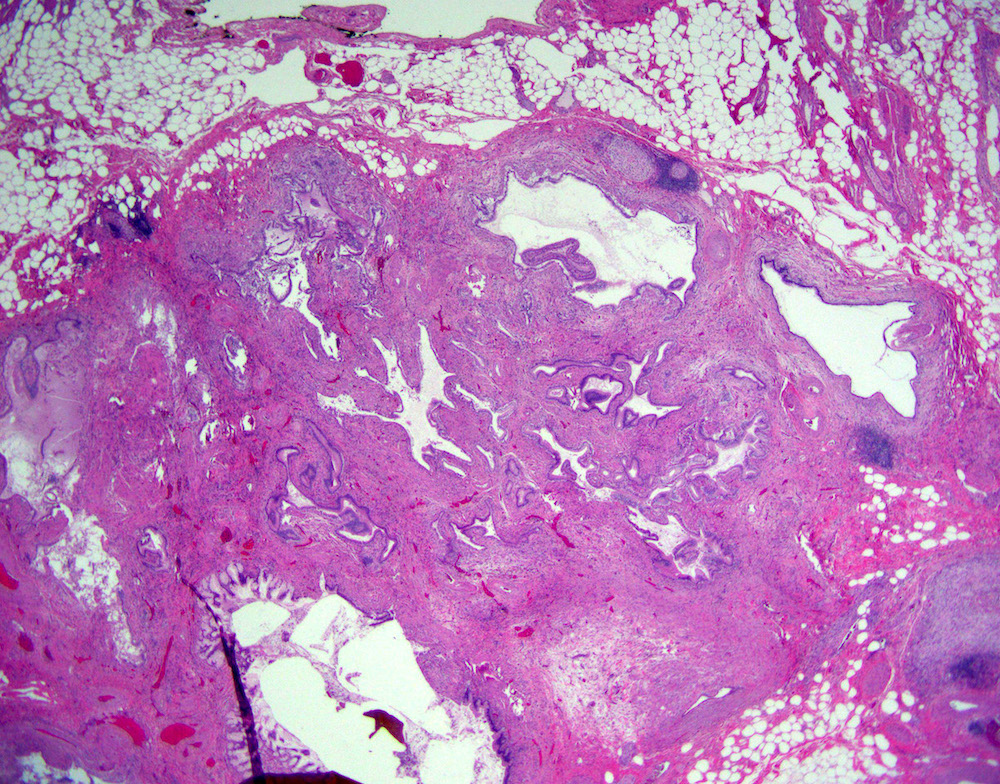
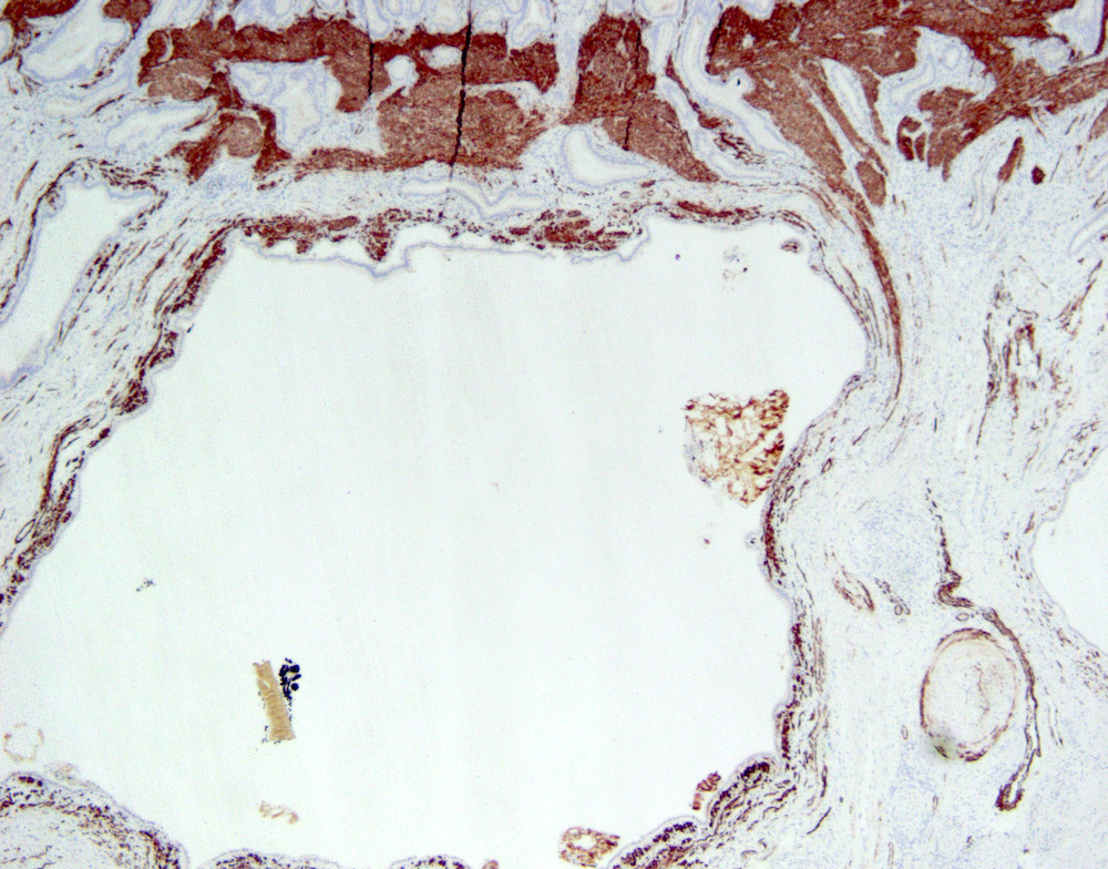
Microscopic (histologic) images
Contributed by Monica T. Garcia-Buitrago, M.D.

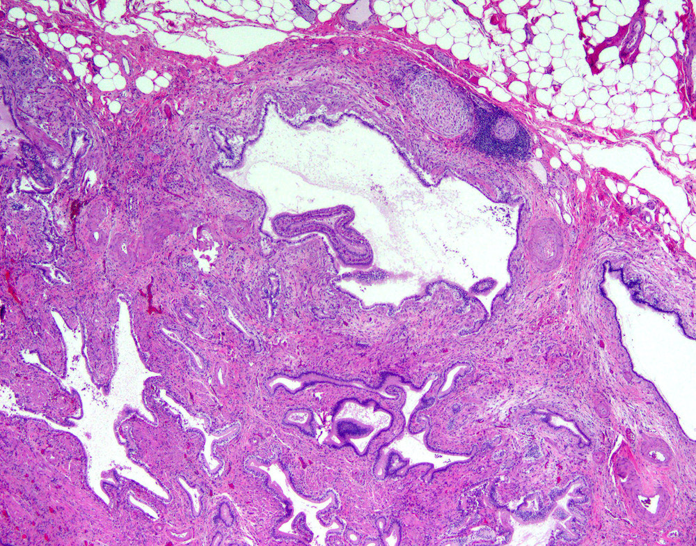
Cystically dilated biliary glands
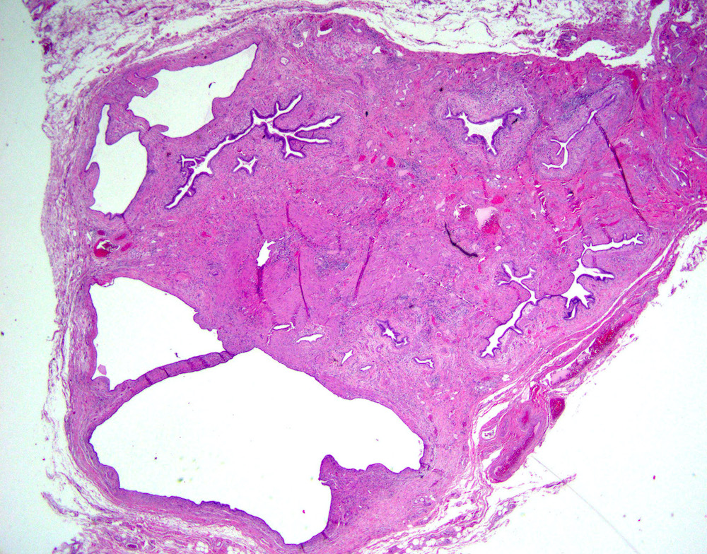
Adenomyomatous hyperplasia / adenomyomatous nodule
Dilated biliary gland, surrounding smooth muscle

Smooth muscle hyperplasia surrounding biliary glands
Thickened smooth muscle surrounding biliary gland
Cystically dilated biliary glands, adjacent nerve
Smooth muscle surrounding cystically dilated biliary glands
Sample pathology report
- Gallbladder, cholecystectomy:
- Chronic cholecystitis and cholelithiasis with adenomyomatous hyperplasia
Differential diagnosis
- Adenocarcinoma:
- Glands and tubules lined by cuboidal to columnar epithelium
- Cytologic atypia
- Prominent nucleoli
- Increased mitotic activity
- Desmoplastic stroma
- Chronic cholecystitis:
- Lymphoplasmacytic infiltrates in lamina propria, may extend deeper, occasionally form lymphoid aggregates / follicles, occasionally may contain eosinophils
- May contain Rokitansky-Aschoff sinuses: epithelial diverticula that can occur throughout gallbladder secondary to injury
- Thickened wall with mural fibrosis
- May have smooth muscle hyperplasia
Additional references
Board review style question #1
The above lesion is found in the gallbladder of a 60 year old woman with gallstones. What is the diagnosis?
- Acute cholecystitis
- Adenomyomatous hyperplasia
- Biliary intraepithelial neoplasia
- Follicular cholecystitis
- Well differentiated adenocarcinoma
Board review style answer #1
Board review style question #2
Which of the following is most commonly associated with adenomyomatous hyperplasia of the gallbladder?
- Acute cholecystitis
- Biliary intraepithelial neoplasia
- Chronic biliary epithelial inflammation / injury (chronic cholecystitis, cholelithiasis)
- Invasive adenocarcinoma
- Involvement of extrahepatic bile ducts
Board review style answer #2
C. Chronic biliary epithelial inflammation / injury (chronic cholecystitis, cholelithiasis)
Comment Here
Reference: Adenomyomatous hyperplasia
Comment Here
Reference: Adenomyomatous hyperplasia
