
Fallopian tubes & broad ligament
Broad ligament tumor-like lesions
Walthard cell nests
Author: Shabnam Zarei, M.D.
Editorial Board Member: Gulisa Turashvili, M.D., Ph.D.
Deputy Editor-in-Chief: Jennifer A. Bennett, M.D.

Last author update: 10 January 2022
Last staff update: 25 October 2022
Copyright: 2002-2025, PathologyOutlines.com, Inc.
PubMed Search: Walthard cell nests


Table of Contents
Definition / general | Essential features | ICD coding | Epidemiology | Sites | Pathophysiology | Etiology | Clinical features | Case reports | Gross description | Gross images | Microscopic (histologic) description | Microscopic (histologic) images | Immunofluorescence description | Positive stains | Negative stains | Sample pathology report | Differential diagnosis | Additional references | Board review style question #1 | Board review style answer #1 | Board review style question #2 | Board review style answer #2Cite this page: Zarei S. Walthard cell nests. PathologyOutlines.com website. https://www.pathologyoutlines.com/topic/fallopiantubeswalthard.html. Accessed April 1st, 2025.
Definition / general
- Nests, plaques or cysts (with eosinophilic luminal material) of metaplastic transitional epithelium involving the serosal surface of fallopian tube, mesosalpinx and mesovarium
- Benign common incidental finding
Essential features
- Benign incidental finding in the subserosa of tubal, paratubal and paraovarian soft tissue
- Bland transitional type epithelium forming small nests / cysts
- CK7+, GATA3+, P63+, PAX8-, CK20- and uroplakin-
ICD coding
Epidemiology
- Common incidental finding
Sites
- Serosa of fallopian tube, including fimbriated end, fimbrial peritoneal junction, mesosalpinx and meso-ovarium
Pathophysiology
- Nests or cysts, including mesothelial cells from the tubal and paratubal serosa, that undergo transitional cell metaplasia
Etiology
- Unknown
Clinical features
- Suggested association between Walthard cell nests and Brenner tumors or mucinous tumors of the ovary (stronger association with the former, though unexplained) (Arch Pathol Lab Med 2008;132:1753)
- Walthard cell nests can be the origin for rare examples of Brenner tumors outside the ovarian parenchyma (Diagn Pathol 2020;15:22)
- Histologically, presence of nests of bland, metaplastic transitional type cells on the serosal surface is diagnostic of Walthard nests
- Pelvic tuberculosis is characterized by granulomatous peritonitis whereas endometriosis will show endometrial type glands and stroma within the serosal surface of adnexal tissue
- Disseminated carcinomatosis shows malignant cells with stromal reaction in the serosal nodules / plaques
- Clinically, Walthard cells are benign asymptomatic and incidental findings, whereas the other differential diagnosis usually presents with abdominal pain, mass, constitutional symptoms or ascites
- Clinically can be mistaken for pelvic tuberculosis (diagnosed based on the presence of granulomas on the tissue along with patient's clinical symptoms) or disseminated tumor (diagnosed based on the presence of malignant tumor cells along with patient's clinical presentation such as abdominal pain / mass, ascites or elevated serum tumor markers)
Case reports
- 10 year old girl with acute appendicitis and urothelial rests (Fetal Pediatr Pathol 2021 Aug 2 [Epub ahead of print])
- 53 year old postmenopausal woman with extraovarian Brenner tumor of the uterus (Diagn Pathol 2020;15:22)
Gross description
- Small 1 - 2 mm yellow-white nodules beneath the serosa of the fallopian tube and paratubal soft tissue
Gross images
AFIP images
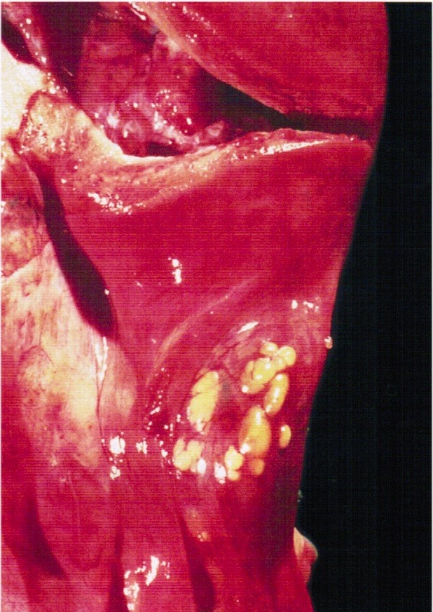
Several small yellow nodules
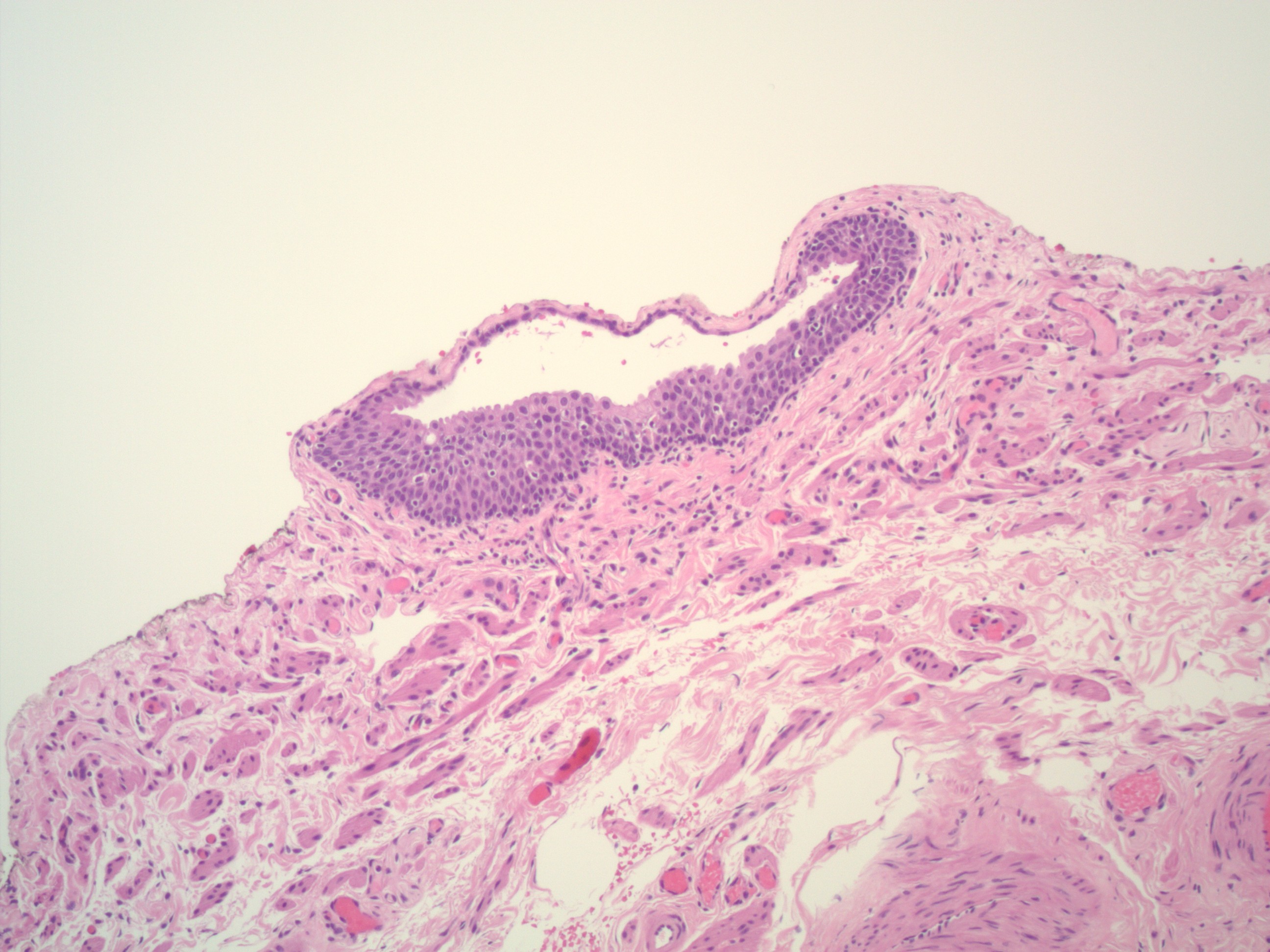
Microscopic (histologic) description
- Small solid nests or cysts of transitional type epithelium with relatively uniform nuclei with irregular borders and nuclear grooves, located beneath the serosa of tubal / paratubal / paraovarian tissue
- Cystic lesions may have eosinophilic luminal secretions
- Rare or no mitotic figures
Microscopic (histologic) images
Contributed by Shabnam Zarei, M.D. and AFIP images
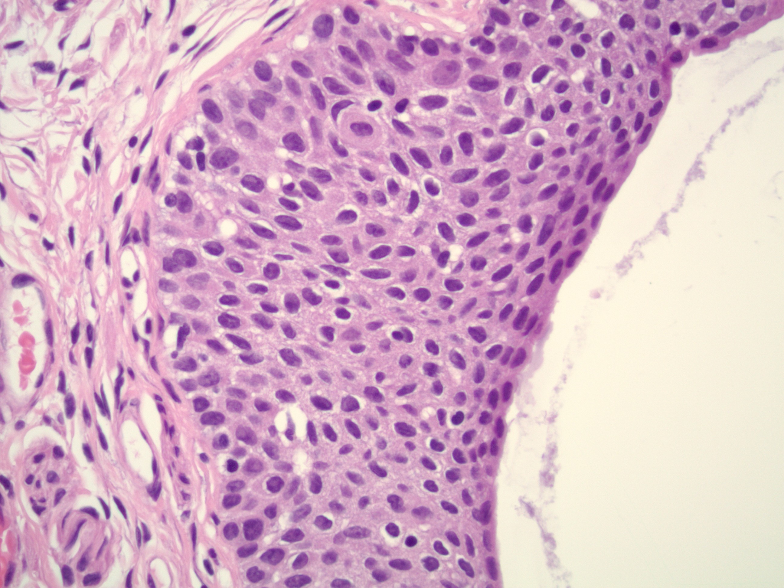
Transitional type epithelium
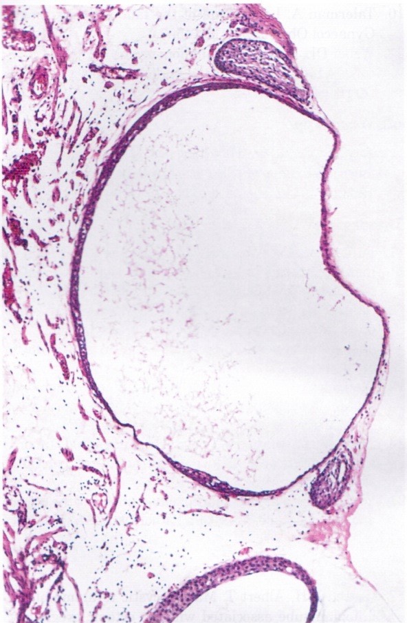
Cystic nests
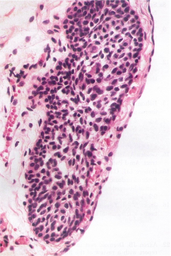
Bland appearing transitional cells
Immunofluorescence description
- Dual staining for α tubulin and γ tubulin shows attenuated and patchy distribution of cilia in the metaplastic cells of Walthard cell nests and Brenner tumor, suggesting a common origin from tubal epithelium (Eur J Cancer 2013;49:3839)
Positive stains
Negative stains
Sample pathology report
- These are benign, incidental findings and are typically not included within the pathology report
Differential diagnosis
- Benign Brenner tumors:
- Endometriosis:
- Presence of endometrial glands, endometrial stroma or hemosiderin laden macrophages
- 2 out of 3 required for the diagnosis
- Paratubal cysts:
- No solid component
- Lined by ciliated tubal type epithelium
- Endosalpingiosis:
- Glands lined by ciliated tubal type epithelium
Additional references
Board review style question #1
How does the immunohistochemical profile of Walthard cell rests differ from benign Brenner tumor?
- Benign Brenner tumors are P63+, GATA3+, uroplakin+, whereas Walthard cell nests are P63+, GATA+ and uroplakin-
- They share the same IHC phenotype
- Walthard cell nests are PAX2 / PAX8- but benign Brenner tumors show diffuse and strong staining with PAX2 / PAX8+
- Walthard cell nests are positive for uroplakin and GATA3; Brenner tumors are positive for GATA3 but negative for uroplakin
Board review style answer #1
A. Walthard cell nests are positive with P63, GATA3, CK7 and are negative with CK20, uroplakin and PAX2 / PAX8 (focal weak)
Comment Here
Reference: Walthard cell nests
Comment Here
Reference: Walthard cell nests
Board review style question #2
Which of the following is true about the pathologic finding in the image shown above?
- If bilateral, they are associated with higher risk for malignant Brenner tumor
- They are usually GATA3- and PAX8+
- They can mimic peritoneal carcinomatosis by imaging and intraoperatively
- They can only be found on the fallopian tube serosal surface
Board review style answer #2
C. Walthard cell nests are benign incidental findings and can mimic peritoneal carcinomatosis by both imaging and intraoperative examination
Comment Here
Reference: Walthard cell nests
Comment Here
Reference: Walthard cell nests
